Diagnosis and Evaluation of Bone Cysts
Diagnosis and evaluation of bone cysts involves a combination of clinical evaluation, imaging, and occasionally biopsy to confirm the diagnosis and rule out other bone lesions or tumours.
Most bone cysts are incidental findings, discovered on X-rays taken after a fall or sports injury. In some cases, children may present with mild pain or a limp if the cyst affects movement or causes structural weakness.
1. Clinical assessment:
- Doctors ask about:
- Pain: Is it constant or triggered by movement?
- Trauma: Any recent falls or fractures?
- Function: Any difficulty using the limb?
2. Imaging tests:
A. X-ray
- First-line diagnostic tool.
- A simple bone cyst usually appears as a well-defined, clear (radiolucent) area inside a long bone.
- Aneurysmal bone cysts may show expansive or multi-chambered spaces.
B. MRI (Magnetic Resonance Imaging)
- Provides detailed images of soft tissues and fluid levels.
- Particularly useful for aneurysmal bone cysts, which may appear aggressive.
C. CT scan (Computed Tomography)
- Used for complex or deep cysts, such as those in the pelvis or spine.
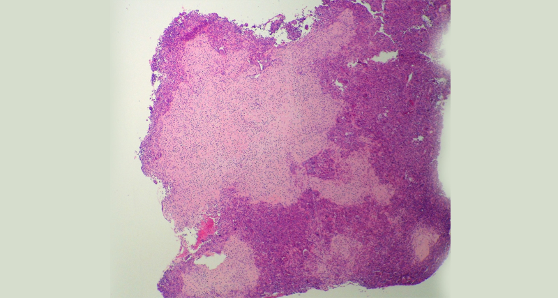
3. Bone biopsy
- Occasionally needed if the imaging is inconclusive.
- Involves removing a small sample of bone tissue for lab analysis.
- Helps rule out malignant bone tumours, which may resemble cysts.
4. Blood tests
- Not usually necessary unless other systemic conditions are suspected.
In South Africa, public hospitals such as Red Cross, Groote Schuur, or Chris Hani Baragwanath have paediatric orthopaedic units equipped to diagnose bone cysts. Private imaging centres may offer faster access to MRI or CT scans.
Diagnosis and Evaluation of Bone Cysts
Early diagnosis helps avoid complications such as fractures or bone deformity, especially if the cyst is near a growth plate.
👉 [Next: Treatment Options for Bone Cysts]


