
Diagnosing Hodgkin lymphoma involves a combination of clinical examination, imaging, laboratory testing, and tissue biopsy. Because its symptoms often overlap with common infections or autoimmune conditions, reaching a definitive diagnosis can take time. Early and accurate detection is critical to determining the disease stage, planning treatment, and improving outcomes.
Initial Presentation and Clinical Examination
Patients typically present with painless swelling of lymph nodes, unexplained fevers, night sweats, or persistent fatigue. Physicians conduct a thorough physical exam, palpating lymph nodes in the neck, armpits, and groin. If internal lymphadenopathy is suspected (in the chest or abdomen), imaging is used to evaluate deeper nodes.
A detailed medical history—including recent infections, family cancer history, exposure to viruses like Epstein-Barr virus (EBV), weight loss, and systemic symptoms—helps distinguish Hodgkin lymphoma from other causes.
Laboratory Testing
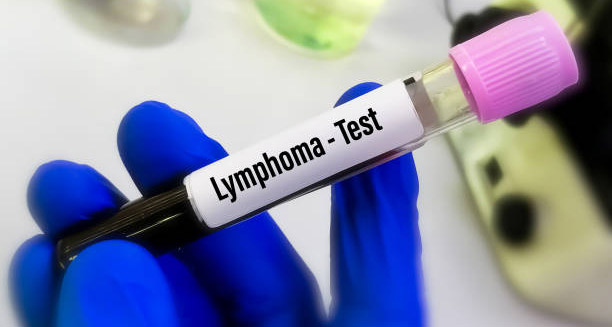
Blood tests are an early diagnostic step. A complete blood count (CBC) may reveal anemia, elevated white blood cells, or low lymphocytes. Though non-specific, these findings raise suspicion. Additional tests like erythrocyte sedimentation rate (ESR) and C-reactive protein (CRP) measure inflammation and are often elevated in Hodgkin lymphoma.
Lymph Node Biopsy
Definitive diagnosis requires biopsy of an affected lymph node. Typically, an excisional biopsy removes the entire node for pathological examination. In cases where the node is inaccessible, needle biopsies guided by imaging may be performed.
The hallmark of classical Hodgkin lymphoma is the presence of Reed-Sternberg cells—large, abnormal B cells with a distinctive “owl’s eye” appearance. Immunohistochemistry helps differentiate Hodgkin lymphoma from other diseases.
Imaging and Staging
After diagnosis, imaging determines disease extent. The preferred method is a PET-CT scan, which provides detailed anatomical and metabolic information, identifying involved lymph nodes and organs. This guides staging from Stage I (single lymph node region) to Stage IV (multiple organs involved).
Additional imaging like MRI may be used if the spinal cord or brain are suspected to be involved. A chest X-ray can detect large mediastinal masses early in the process.
Bone Marrow Biopsy
A bone marrow biopsy is sometimes performed, especially if there is suspicion of marrow involvement. This involves sampling marrow from the pelvic bone to check for malignant cells, which would indicate advanced disease and may affect treatment.
Molecular and Genetic Testing
While not always necessary, molecular and genetic testing of biopsy samples can identify specific subtypes, prognostic markers, and help personalise treatment. For example, nodular lymphocyte-predominant Hodgkin lymphoma (NLPHL) has unique genetic and immunological features influencing management.
Staging System
The Ann Arbor staging system classifies Hodgkin lymphoma into four stages (I to IV), with “A” indicating absence and “B” indicating presence of systemic symptoms (fever, night sweats, weight loss). For example, Stage IIIB indicates widespread lymph node involvement with symptoms.
Staging guides treatment decisions and prognosis.
Additional Assessments
Physicians evaluate performance status and organ function through routine blood chemistry and tests of liver and kidney function. These assessments determine if patients can tolerate specific chemotherapy agents or require dose adjustments. Discussions about fertility preservation are important, especially in younger patients.
Special Considerations in Children and Adolescents
Diagnosing Hodgkin lymphoma in children can be challenging because symptoms mimic infections such as mononucleosis. Paediatric oncologists use minimally invasive biopsies and tailored imaging protocols to reduce discomfort and radiation exposure.
Challenges and Multidisciplinary Approach
Diagnosis delays are common when symptoms are vague or misattributed. Early clinical suspicion and prompt referral to specialists improve outcomes. General practitioners play a crucial role in identifying warning signs.
A multidisciplinary team—including haematologists, radiologists, pathologists, and oncologists—typically reviews diagnosis, staging, and treatment planning to ensure accuracy and consensus.
Summary:
Diagnosing Hodgkin lymphoma requires a systematic approach: careful physical examination, blood tests, imaging studies, and lymph node biopsy. Advanced techniques such as PET-CT and immunohistochemistry refine diagnosis and staging, enabling personalised treatment plans. Early and precise diagnosis leads to the best possible outcomes.