This image displays four brain illustrations, each showing red inflamed regions, symbolising the brain swelling seen in cases of Japanese encephalitis. Diagnosis typically involves evaluating neurological symptoms, performing a lumbar puncture (spinal tap) to detect viral markers in cerebrospinal fluid, and using neuroimaging (MRI or CT scans) to visualise brain inflammation. Laboratory tests may also identify the Japanese encephalitis virus (JEV) through serological or PCR methods. The image offers a visual representation of how clinicians identify the location and extent of encephalitis, guiding appropriate treatment and care.
Diagnosing Japanese Encephalitis by Detecting Brain Inflammation
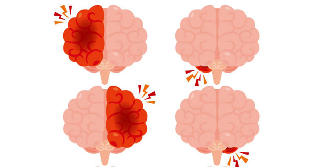
Illustrated brains with highlighted red areas indicating inflammation—representing how Japanese encephalitis is diagnosed through clinical and imaging techniques