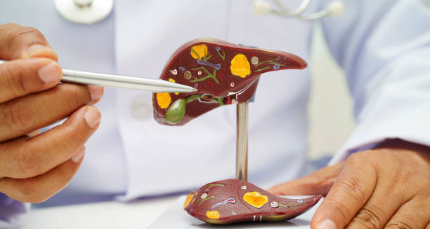

Jaundice is a visible symptom that typically signals an underlying medical condition affecting the liver, bile ducts, or red blood cells. While the yellow discolouration of the skin and eyes may be easily observed, determining the cause of jaundice requires a structured diagnostic approach. Early and accurate diagnosis is vital in identifying potentially serious or life-threatening conditions such as bile duct obstruction, hepatitis, or liver cancer. Clinicians rely on a combination of medical history, physical examination, laboratory tests, and imaging studies to evaluate the origin and severity of jaundice. Understanding the mechanisms behind bilirubin metabolism is crucial to interpreting diagnostic results correctly. Bilirubin, the substance responsible for the yellowing seen in jaundice, is produced when red blood cells are broken down. It is processed in the liver and excreted through the bile ducts. A disruption at any point in this pathway may lead to bilirubin accumulation and, subsequently, jaundice.
Initial Clinical Evaluation
Medical History
The first and most important step is a thorough medical history. Doctors ask about when the yellowing started and how long it has lasted. They check for recent travel, especially to places where hepatitis or malaria is common. They review all medicines used, including herbal remedies and over-the-counter drugs. Alcohol use is also important. Family history of liver or blood problems matters too. They ask about diet, lifestyle, and job risks, especially in healthcare, food handling, or chemical work. This information helps doctors guess the likely causes, such as viral hepatitis, alcohol damage, or blood disorders.
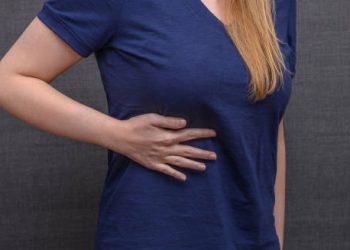
Physical Examination
Doctors perform a detailed physical exam to find clues. Yellowing of the whites of the eyes (scleral icterus) often appears before the skin changes. They feel the liver to check its size and firmness; a big, hard liver can mean cirrhosis or cancer. Tenderness in the right upper belly suggests inflammation or blockage. Signs like spider veins on the skin, red palms, and fluid in the belly point to long-term liver disease. An enlarged spleen may happen in blood disorders or portal hypertension.
Laboratory Investigations
Bilirubin Tests
Blood tests measure total bilirubin, the overall level in the blood. Doctors check unconjugated (indirect) bilirubin, which rises in cases like haemolysis before the liver. They also measure conjugated (direct) bilirubin, which increases when the liver or bile ducts fail to remove bilirubin properly. Comparing these two types helps pinpoint the problem’s location.
Liver Function Tests (LFTs)
Doctors test enzymes like ALT and AST, which rise when liver cells get hurt, for example in hepatitis. ALP and GGT levels increase if bile flow is blocked. They also check albumin and clotting time (PT). Low albumin or long PT means the liver is not working well.
Full Blood Count (FBC)
A low haemoglobin with a high number of young red cells (reticulocytes) suggests red blood cells are breaking down. Raised white blood cells can mean infection or inflammation.
Viral Hepatitis Panel
Doctors test for hepatitis A, B, C, D, and E, especially if jaundice comes with tiredness, fever, or belly pain. Chronic hepatitis B and C raise the risk of cirrhosis and liver cancer.
Autoimmune and Metabolic Markers
Tests for anti-smooth muscle antibodies or anti-nuclear antibodies can suggest autoimmune hepatitis. Low ceruloplasmin points to Wilson’s disease. Iron tests check for haemochromatosis. Alpha-1 antitrypsin levels also help find rare causes. These tests help find treatable but uncommon liver problems.
Urinalysis
Urine tests are quick and useful. Dark urine with bilirubin confirms conjugated jaundice. Urobilinogen is often missing if bile flow is blocked but high if red cells break down too fast. This helps decide if the problem is in or after the liver.
Stool Colour and Bile Pigments
Patients may notice pale or clay-coloured stools when bile does not reach the intestines. Although this is a subjective sign, it points to obstructive jaundice and calls for more imaging tests.
Imaging Techniques
Ultrasound Scan
Doctors usually start with an abdominal ultrasound. It is safe and easy to do. Ultrasound can spot gallstones, bile duct widening, liver lumps or cysts, and signs of cirrhosis.
CT Scan (Computed Tomography)
A CT scan gives detailed images of the body. It can find pancreatic cancer, bile duct tumours, and swollen lymph nodes. It helps plan surgery if needed.
MRI and MRCP (Magnetic Resonance Cholangiopancreatography)
MRI, especially MRCP, shows the bile ducts and pancreas clearly. Doctors use it when ultrasound results are unclear.
Endoscopic Retrograde Cholangiopancreatography (ERCP)
ERCP uses a flexible tube with a camera passed through the mouth to the bile ducts. It helps see blockages, remove stones, place stents, or take tissue samples. ERCP has risks and is used mainly when treatment is planned.
Liver Biopsy
If blood tests and scans do not show the cause, doctors may take a small piece of liver tissue. They look at it under a microscope to check inflammation, scarring, tumours, or storage diseases like Wilson’s or haemochromatosis. This test helps with unclear liver problems or chronic disease.
Specialised Tests in Neonates
Newborns with jaundice need specific tests like transcutaneous bilirubin measurements and blood bilirubin levels. A Coombs test checks for red cell breakdown. Blood typing of mother and baby helps find incompatibility. Early detection is key to prevent brain damage called kernicterus.
Conclusion
Diagnosing jaundice needs a careful mix of exam, lab tests, and imaging. By figuring out if the problem is before, in, or after the liver, doctors can find the cause and start treatment fast. Each step—from noticing yellow skin to biopsies—plays a key role. Quick diagnosis improves results and avoids problems like liver failure, infection, or brain damage in babies. Patients with jaundice must get thorough checks, especially if they have fever, confusion, or rapid weight loss.