Diagnosis of Bullous Pemphigoid
Diagnosis of bullous pemphigoid involves a combination of clinical examination, skin biopsy, and laboratory testing. A confirmed diagnosis of bullous pemphigoid ensures appropriate treatment. Additionally, helps rule out other similar skin conditions, such as pemphigus vulgaris or dermatitis herpetiformis.
The diagnostic process begins with a full medical history and visual inspection of the skin. A dermatologist will look for the pattern, size, and location of the blisters. As well as the condition of the surrounding skin. The presence of firm, fluid-filled blisters on red, itchy skin — particularly in older adults — raises suspicion.
To confirm the diagnosis, a skin biopsy is performed. A small sample of blistered or inflamed skin is removed and examined under a microscope. This helps determine the level at which the skin layers are separating and whether immune cells are present.
Direct immunofluorescence testing is also done on skin tissue. This test detects deposits of antibodies (IgG) and complement proteins (C3) at the junction between the epidermis and dermis. The presence of a linear pattern of these immune proteins is highly characteristic of bullous pemphigoid.
Diagnosis of Bullous Pemphigoid
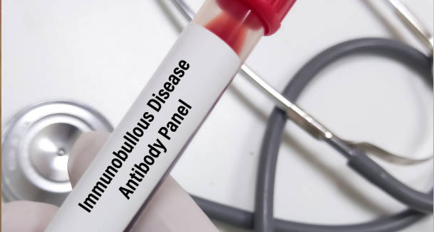
Blood tests can also be helpful. An ELISA test may be used to detect circulating autoantibodies to BP180 and BP230 — the proteins targeted by the immune system. Elevated levels of these antibodies support the diagnosis and can also be used to monitor disease activity over time.
In cases where the diagnosis remains unclear. Indirect immunofluorescence or salt-split skin testing may be used to distinguish bullous pemphigoid from other autoimmune blistering disorders.
Early and accurate diagnosis of bullous pemphigoid is crucial because starting the correct treatment early can significantly reduce symptoms, prevent complications, and improve quality of life.
[Next: Treatment of Bullous Pemphigoid →]


