
Diagnosis of Empyema
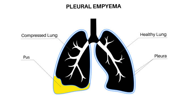
The diagnosis of empyema starts with a clinical evaluation, especially in patients who have recently had pneumonia or other lung infections. Since empyema involves pus collecting in the pleural space, doctors must confirm the presence of fluid and determine whether it’s infected. Early and accurate diagnosis is key to preventing serious complications.
The doctor begins by asking about symptoms such as chest pain, shortness of breath, cough, and fever. A physical examination may reveal reduced breath sounds, dullness to percussion, or decreased chest expansion on one side—all signs of fluid build-up.
Imaging Tests | Diagnosis of Empyema
Chest X-ray – May show pleural effusion (fluid around the lung), but not whether it’s infected.
Ultrasound – Useful for detecting fluid and guiding drainage. It can differentiate between clear fluid and thick pus.
CT scan – Provides a detailed view of the chest, helping identify pockets of pus, loculated fluid, or lung collapse. This is especially helpful when empyema is advanced or complex.
Diagnostic Thoracentesis
This is a key step. The doctor uses a needle to remove a sample of pleural fluid from the chest. The fluid is then analysed in the lab for:
Appearance – cloudy or foul-smelling fluid suggests empyema
Cell count – high white blood cells indicate infection
Protein and glucose levels – help distinguish between types of effusions
Culture – to identify the bacteria responsible, allowing targeted antibiotic treatment
In TB-endemic areas like South Africa, fluid may also be tested for Mycobacterium tuberculosis using specialised tests like GeneXpert or TB culture.
Diagnosis of Empyema
If empyema is suspected in a child, imaging and thoracentesis are often done at the same time, under sedation or anaesthesia.
In South Africa, delays in diagnosis are often due to limited access to imaging or specialist care in rural settings. Mobile X-ray units and ultrasound machines in clinics are helping to bridge this gap.
The diagnosis of empyema involves combining clinical signs, imaging, and fluid analysis. Once confirmed, treatment can begin immediately—reducing the risk of long-term lung damage and life-threatening complications.
[Next: Treatment of Empyema →]