
Diagnosis of Glaucoma
The diagnosis of glaucoma relies on a thorough clinical evaluation, including eye pressure measurement, optic nerve examination, and visual field testing. Since glaucoma often develops without early symptoms, routine eye checks are the only way to detect the condition before irreversible damage occurs. A timely diagnosis enables early treatment that can preserve vision and slow disease progression.
Modern diagnostic methods make it possible to detect glaucoma years before a patient notices any vision changes. The key is combining several tests to build a complete picture of eye health.
Comprehensive Eye Examination
A standard glaucoma assessment includes:
Patient history: Risk factors such as family history, diabetes, steroid use, or ethnicity
Visual acuity test: Measures how well a person can see at different distances
Slit-lamp examination: Assesses the front of the eye for inflammation, damage, or abnormalities
These initial steps guide whether further glaucoma-specific testing is required.
Tonometry: Measuring Eye Pressure
Tonometry measures intraocular pressure (IOP), which is one of the most important indicators of glaucoma. Elevated pressure suggests a higher risk, though not all glaucoma patients have high readings.
There are several types of tonometry:
Applanation tonometry: Gold standard, uses a device that gently flattens the cornea
Non-contact tonometry (puff test): Uses a brief burst of air
Rebound tonometry: Portable and often used for children or home monitoring
Normal IOP ranges between 10–21 mmHg, but glaucoma can occur at lower or higher values depending on other factors.
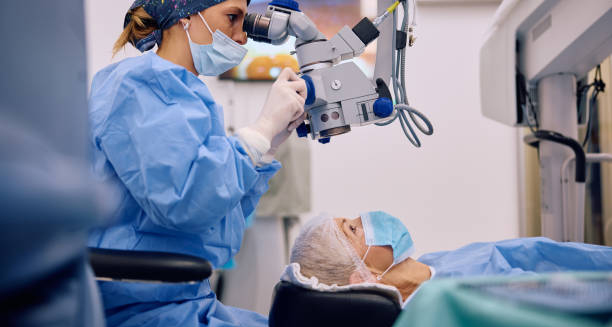
Ophthalmoscopy: Examining the Optic Nerve
Using a special lens and microscope, the doctor examines the optic disc at the back of the eye. Features suggestive of glaucoma include:
Cupping of the optic nerve
Thinning of the neuroretinal rim
Asymmetry between the two eyes
Progressive cupping is a sign of optic nerve damage, often visualised in sequential photos or scans.
Visual Field Testing
This is a key test to detect functional vision loss:
Uses perimetry machines to map out the person’s peripheral vision
Patients press a button when they see a light spot in various locations
Results show areas of reduced sensitivity or blind spots
In glaucoma, visual field loss often starts in the nasal periphery and progresses slowly.
Optical Coherence Tomography (OCT)
OCT is an advanced imaging technique that provides cross-sectional images of the retina and optic nerve. It helps measure:
Thickness of the retinal nerve fibre layer (RNFL)
Changes over time, even before functional loss is apparent
OCT is especially useful for diagnosing early glaucoma and monitoring disease progression.
Gonioscopy: Assessing the Drainage Angle
This test evaluates whether the drainage angle between the cornea and iris is open or blocked. It helps distinguish:
Open-angle glaucoma
Angle-closure glaucoma
A special lens is placed on the eye to allow visualisation of the angle. It is particularly important when acute angle-closure glaucoma is suspected.
Pachymetry: Measuring Corneal Thickness
Central corneal thickness affects IOP readings and is an independent risk factor for glaucoma. Thinner corneas:
May lead to underestimation of IOP
Are associated with higher risk of optic nerve damage
Pachymetry is a quick, painless test using ultrasound or optical methods.
Differential Diagnosis
Other conditions can mimic glaucoma, such as:
Optic neuritis
Ischaemic optic neuropathy
Compressive lesions (e.g. tumours)
Retinal diseases
A comprehensive examination helps rule out these alternatives and confirm a glaucoma diagnosis.
Screening and Follow-Up
Routine screening is recommended for:
All adults over 40, every 2 years
People with risk factors, annually
Children with family history of congenital glaucoma, as advised by specialists
Once diagnosed, glaucoma patients require:
Lifelong monitoring
Regular visual field and OCT testing
Adjustments to treatment based on progression
Importance of Early Diagnosis
Early detection can:
Prevent significant vision loss
Reduce reliance on more invasive treatments
Maintain independence and quality of life
Because damage is irreversible, diagnosis must occur before symptoms develop—another reason why eye pressure alone is not enough to rule out glaucoma.
Conclusion | Diagnosis of Glaucoma
The diagnosis of glaucoma requires a combination of clinical observation, pressure measurement, imaging, and functional testing. With early detection and regular monitoring, patients can preserve their vision and manage the condition effectively for life.