
Diagnosis of Gout
The diagnosis of gout is primarily clinical but can be confirmed with laboratory tests and imaging when necessary. Early and accurate diagnosis of gout is critical for initiating appropriate treatment, managing flare-ups, and preventing joint and kidney complications. Because the symptoms can mimic other types of arthritis, such as rheumatoid arthritis or septic arthritis, a thorough evaluation is essential.
Gout occurs due to the deposition of uric acid crystals in joints and tissues, triggered by high levels of uric acid in the bloodstream. Identifying these crystals or the conditions that lead to their formation helps differentiate gout from similar conditions and informs long-term management strategies.
Clinical Evaluation
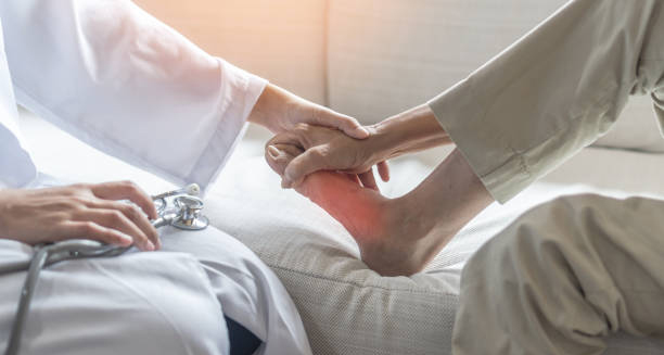
The first step in diagnosing gout involves a comprehensive history and physical examination. Key questions include:
When did the symptoms begin?
Is the pain intermittent or persistent?
Are specific triggers (food, alcohol, injury) involved?
Is there a family history of gout or kidney disease?
During the physical exam, clinicians assess:
Joint redness, swelling, and tenderness
Presence of tophi (chalky nodules under the skin)
Range of motion in affected joints
The combination of sudden joint pain, swelling, warmth, and rapid onset—especially in the big toe—is highly suggestive of gout.
Joint Fluid Analysis
The gold standard for confirming gout is joint aspiration:
A small needle is used to extract synovial fluid from the inflamed joint
The fluid is examined under a polarised light microscope
Needle-shaped, negatively birefringent uric acid crystals confirm the diagnosis
This test is especially important when the diagnosis is uncertain, or if septic arthritis is a possible alternative.
Blood Tests
Several blood tests support the diagnosis:
Serum uric acid levels: Elevated levels (>6.8 mg/dL or >400 µmol/L) suggest gout but may be normal during an acute attack
C-reactive protein (CRP) and erythrocyte sedimentation rate (ESR): Markers of inflammation
White blood cell count: May be elevated during flares
Kidney function tests, including creatinine and eGFR, to assess uric acid clearance
Repeated testing is often needed, as levels can fluctuate based on hydration, diet, and medication use.
Imaging
When joint aspiration is not feasible or gout has progressed to chronic stages, imaging can be helpful.
X-rays
May show joint erosion, particularly in chronic or tophaceous gout
Limited in early-stage disease
Ultrasound
Can detect urate crystal deposits
Shows the “double contour sign,” indicating crystal coating on cartilage
Useful for visualising tophi and guiding joint aspiration
Dual-energy CT (DECT)
Highly sensitive in detecting urate crystals
Useful in atypical or difficult-to-diagnose cases
Not widely available in all healthcare settings
Differential Diagnosis
Gout can resemble several other conditions:
Septic arthritis (joint infection): Requires urgent antibiotic treatment
Pseudogout: Caused by calcium pyrophosphate crystals
Rheumatoid arthritis: Usually involves multiple joints symmetrically
Reactive arthritis: Associated with prior infection
Joint fluid analysis and imaging help distinguish gout from these mimics.
Gout in Women and Elderly
In postmenopausal women and older adults:
Symptoms may be less typical
Flare-ups may involve knees or hands rather than toes
Other forms of arthritis may coexist, complicating diagnosis
This group often requires more detailed testing to confirm gout.
When to Refer
Referral to a rheumatologist is advised when:
The diagnosis is uncertain
Gout is resistant to standard therapies
There are multiple or complex comorbidities
Advanced joint damage or tophaceous deposits are present
Specialist input may also be necessary for long-term urate-lowering therapy management.
Conclusion | Diagnosis of Gout
The diagnosis of gout is based on clinical presentation, supported by synovial fluid analysis, blood tests, and imaging when needed. An accurate diagnosis of gout allows for appropriate treatment, helps rule out more serious conditions like infection, and sets the stage for long-term management to prevent flares and joint damage. Early diagnosis not only improves outcomes but also reduces the likelihood of chronic disability.