
The diagnosis of hydronephrosis requires a combination of clinical evaluation and diagnostic testing to determine the presence, severity, and cause of kidney swelling. The key to an effective diagnosis of hydronephrosis lies in promptly identifying the underlying obstruction or dysfunction responsible for the impaired flow of urine. Timely diagnosis helps to protect kidney function and prevent complications such as infection, scarring, or even irreversible renal damage.
Doctors see patients with many different symptoms—or sometimes no symptoms at all. Because of this, they must stay alert and think about hydronephrosis when checking urinary problems, belly pain, or signs of kidney trouble. Today’s diagnostic tools give clear, non-invasive ways to look at the urinary system and check how well the kidneys work.
Initial Clinical Assessment
First, doctors take a full history and do a physical exam to start diagnosing hydronephrosis.
- Patient History: The doctor asks about recent urine problems, pain, fever, how often one urinates, or changes in urine colour. For older adults, questions may include prostate health, medications, or nerve problems. For children, birth and development details matter.
- Physical Examination: The doctor may press on the belly or side to find pain or a lump. Babies may have a swollen belly or a kidney that can be felt. Fever, fast heartbeat, or signs of dehydration may show infection or serious illness.
This information helps doctors decide if more tests are needed.
Urine Tests
These urine tests give quick clues about urinary tract health and help diagnose hydronephrosis.
- Dipstick Test: Detects blood, white blood cells, nitrites, or protein—signs of infection, stones, or swelling.
- Microscopic Exam: Confirms infection and checks for kidney stones or crystals.
- Urine Culture: If infection is likely, this test finds the exact bacteria to guide antibiotics.
Urine problems are common when hydronephrosis results from infection, blockage, or stones.
Blood Tests
Blood tests tell doctors about kidney function and general health.
- Creatinine and Urea: High levels may show kidney problems, especially if both kidneys swell.
- Estimated Glomerular Filtration Rate (eGFR): Measures how well kidneys filter blood.
- Inflammation Markers (CRP or ESR): These rise when infection or swelling is present.
- Complete Blood Count (CBC): Checks for anemia, high white cells, or other signs of infection.
These results support imaging findings and help assess urgency.
Ultrasound Imaging
Doctors often use ultrasound first because it is safe, quick, and does not use radiation.
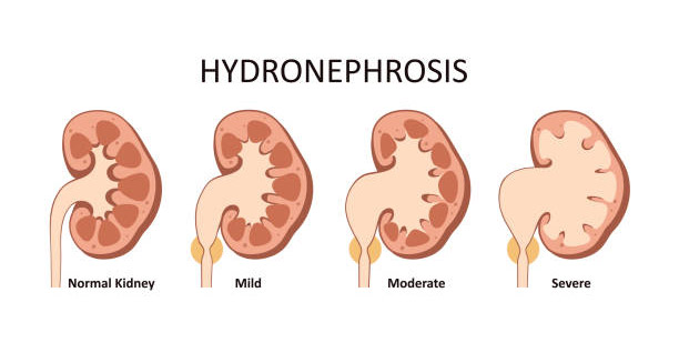
- Kidney Size and Swelling: Ultrasound shows how much the kidneys have enlarged and if one or both are affected.
- Detects Blockages: It can find stones, narrow areas, or lumps in the urinary tract.
- Checks Ureter and Bladder: Ultrasound spots bladder swelling, leftover urine, or ureter problems.
For unborn babies, routine ultrasounds in the second trimester often catch hydronephrosis early, helping plan care after birth.
Computed Tomography Scan (CT scan)
CT scans help when ultrasound is unclear or stones are suspected.
- Detects Stones: CT finds stones that ultrasound might miss.
- Detailed Images: Shows kidneys, ureters, and nearby organs clearly to find tumours or unusual anatomy.
- Contrast CT: Sometimes used to check blood flow but avoided if kidneys do not work well.
CT is vital in emergencies to find the cause and how serious the swelling is.
Magnetic Resonance Imaging (MRI)
MRI is useful when avoiding radiation is important, like in pregnancy or children needing many scans.
- MR Urography: Produces detailed pictures of the urinary system without X-rays.
- Good for Birth Defects: Helps find congenital issues, especially in children.
- Checks Kidney Function: Some MRI methods can measure how well each kidney works if only one is affected.
MRI is less common than CT but valuable in specific cases.
Voiding Cystourethrogram (VCUG)
This test mainly helps children and checks for urine flowing backward from the bladder to kidneys (VUR).
- Procedure: A catheter fills the bladder with contrast, then X-rays are taken while the child urinates.
- Detects Blockages: Finds problems like valves blocking urine flow in baby boys.
- Checks Urine Flow: Shows if urine leaves the bladder normally or gets stuck.
Doctors order VCUG when children have repeated urinary infections or unclear hydronephrosis.
Nuclear Renal Scan (Renography)
In complex or ongoing cases, nuclear scans measure kidney function and urine drainage.
- Diuretic Renography: Tests how well each kidney works and empties after medicine that increases urine flow.
- Split Kidney Function: Helps decide if a weak kidney should be removed or saved.
- Checks Blockage Severity: Useful when symptoms and images do not match.
Though specialized, this scan guides treatment and diagnosis decisions.
Summary
The diagnosis of hydronephrosis involves many steps, from clinical checks to lab tests and imaging scans. It starts with suspicion based on symptoms, then uses ultrasounds and more detailed scans to find the cause and check kidney health. Ultrasound remains the main first step, but CT, MRI, and nuclear scans add clarity in difficult cases.
Making a quick and accurate diagnosis helps doctors decide on the best treatment—whether medicine, surgery, or watchful waiting. Early diagnosis allows many people with hydronephrosis to get proper care before problems start.