
Diagnosis of leukoplakia is vital to tell this condition apart from other oral health issues, some of which may be more serious. It usually starts with a detailed examination of the mouth by a dentist, doctor, or oral specialist. Since leukoplakia can sometimes be an early sign of oral cancer or other precancerous problems, diagnosing it early and correctly is crucial for proper treatment and monitoring.
Clinical Examination and Risk Assessment
The healthcare provider begins with a visual check of the mouth and lips, looking for thick, white, or grey patches that can’t be wiped away. These patches often appear on the inside cheeks, gums, tongue, or floor of the mouth. The provider also asks about habits that raise risk, such as smoking, heavy drinking, or poor oral hygiene. Diagnosis of leukoplakia depends heavily on spotting these risk factors and matching them with the lesions’ appearance and location.
Cytology and Biopsy: Confirming the Diagnosis
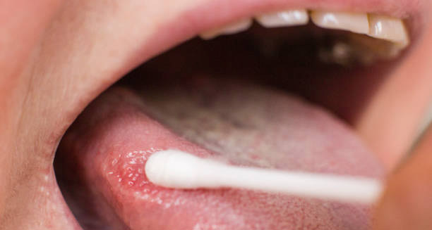
If the clinical exam points to leukoplakia, further testing may follow to confirm the diagnosis and exclude cancer. Oral exfoliative cytology uses a brush or spatula to collect cells from the patch surface. These cells are checked under a microscope for precancerous changes, but this method can sometimes miss deeper problems.
The most reliable test is a biopsy, where a small tissue sample is taken from the lesion. Types of biopsy include incisional (part of the lesion), excisional (whole lesion), or punch biopsy (a core of tissue). The choice depends on the lesion’s size, site, and clinical suspicion. A biopsy determines if the cells are normal, dysplastic (abnormal but not cancer), or malignant, making it essential in leukoplakia diagnosis.
Assessing Lesion Types and Malignancy Risk
Some leukoplakia patches are smooth and uniform (homogeneous), which carry a lower cancer risk. Others—speckled, nodular, or verrucous types—are non-homogeneous and need closer investigation. These higher-risk patches often require biopsy or more frequent follow-ups. Thus, diagnosis of leukoplakia involves not just finding the lesion but classifying it by appearance to assess cancer risk.
Additional Diagnostic Tools
To aid diagnosis, doctors may use special tests like toluidine blue staining, which highlights abnormal cells and helps pick biopsy sites. Autofluorescence devices can spot tissue changes by detecting differences in light reflection, signaling possible early cancer changes. These tools support but do not replace biopsy.
Patient History and Symptom Review
A thorough patient history is important. Doctors ask how long the lesion has been present, if it has changed, and about symptoms like pain, bleeding, or trouble eating. Though leukoplakia often causes no symptoms, any signs can guide the urgency and nature of further testing. Clinicians also check overall health, since immune problems or nutritional deficiencies can contribute to oral lesions.
Molecular Testing in High-Risk Cases
For suspicious or recurrent lesions, molecular or genetic tests on biopsy samples may be done. These can find chromosomal changes linked to oral cancer. Such advanced diagnostics may become more common, especially for high-risk or complicated leukoplakia cases.
Follow-Up and Monitoring
After diagnosis, patients usually need regular check-ups, especially if dysplasia is present. Leukoplakia can come back or develop into cancer, so ongoing surveillance with exams and sometimes repeat biopsies is key. Diagnosis of leukoplakia is a continuous process requiring careful monitoring.
Special Considerations: Proliferative Verrucous Leukoplakia
A rare, aggressive type called proliferative verrucous leukoplakia (PVL) demands close, long-term monitoring. PVL has a higher chance of turning cancerous and often needs care from a team including dental experts, oncologists, and pathologists.
Summary of Leukoplakia Diagnosis
In short, diagnosis of leukoplakia involves a thorough oral exam, risk factor review, cytology, biopsy, and sometimes advanced tests. Early and accurate diagnosis is critical because of the risk of cancer. Combining clinical skills, technology, and patient cooperation ensures leukoplakia is managed effectively.