
Accurate diagnosis of malignant brain tumour is crucial for starting treatment quickly and improving outcomes. These tumours grow fast and can threaten life. Because symptoms often overlap with other conditions, doctors use a careful step-by-step approach that combines clinical exams and advanced imaging.
This section explains the full process of diagnosing malignant brain tumour. We cover symptoms, tests, and tools doctors use to confirm the diagnosis.
Initial Clinical Evaluation
The diagnosis of malignant brain tumour begins with a detailed patient history and physical exam. Doctors ask about:
- When headaches started and how long they last
- Changes in behavior, memory, or personality
- Type and frequency of seizures
- Vision, hearing, or speech problems
Next, the neurological exam checks:
- Reflexes
- Muscle strength
- Balance and coordination
- Sensory responses
- Eye movements and pupil reactions
- Thinking and speech skills
If doctors find any abnormalities, they use this information to localize the tumour and decide how urgently to do more tests.
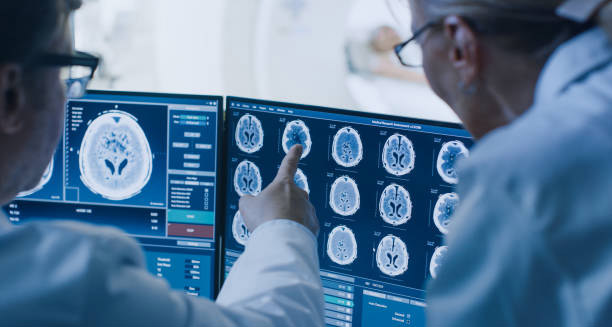
Imaging Tests
Brain imaging forms the backbone of the diagnosis of malignant brain tumour. Two main types are used:
Magnetic Resonance Imaging (MRI)
MRI provides the most detailed images of brain soft tissues. It uses magnets and radio waves to show:
- Tumour size, shape, and exact location
- Surrounding swelling or inflammation
- Clear differences between tumour and healthy tissue
Using contrast dye (gadolinium) improves image clarity. MRI can also map brain activity (fMRI) or blood flow (perfusion MRI) to help grade the tumour.
Computed Tomography (CT) Scan
CT scans are faster and used in emergencies like sudden seizures or stroke-like symptoms. They detect:
- Large masses, bleeding, or brain swelling
- Calcifications in some tumour types
- Skull bone involvement
CT is less detailed than MRI and usually acts as a first screening tool.
Advanced Imaging Techniques
To better understand tumour behavior and guide treatment, doctors may order:
Magnetic Resonance Spectroscopy (MRS)
This test analyzes brain chemicals. Malignant tumours show high levels of choline and low N-acetylaspartate (NAA), helping distinguish tumour types.
Positron Emission Tomography (PET)
PET scans reveal tumour metabolism. Malignant tumours consume more glucose, which PET detects. PET helps:
- Identify tumour grade
- Spot recurrence
- Detect spread to other sites
Biopsy
A biopsy gives a definite diagnosis by examining tumour cells.
Stereotactic Needle Biopsy
Doctors insert a thin needle into the brain using MRI or CT guidance. This is best for tumours deep in sensitive areas. Advantages include:
- Lower risk than open surgery
- Faster recovery
- High accuracy when done correctly
Open Surgical Biopsy (Craniotomy)
For accessible tumours, surgeons remove a part of the skull to take a larger sample. This often happens alongside tumour removal surgery.
Pathologists analyze biopsy samples to determine:
- Cell type (e.g., glioblastoma, astrocytoma)
- Grade (how aggressive the tumour cells look)
- Molecular markers (like IDH mutation, MGMT methylation)
This information guides chemotherapy, radiotherapy, and prognosis.
Lumbar Puncture (Spinal Tap)
Sometimes doctors perform a lumbar puncture to test cerebrospinal fluid (CSF), especially if the tumour affects the meninges or spinal cord. This test detects:
- Cancer cells in the CSF
- Raised brain pressure
- Signs of infection or inflammation
However, this test is avoided if there is high pressure inside the skull because of the risk of brain herniation.
Blood Tests
Blood tests cannot detect brain tumours directly. Still, they help:
- Rule out infections (e.g., high white blood cells)
- Check tumour markers in germ cell tumours (like AFP, HCG)
- Identify hormone problems from pituitary tumours
- Assess organ function before chemotherapy
Genetic and Molecular Testing
Modern diagnosis relies heavily on molecular profiling. Testing tumour samples for gene mutations and protein markers helps:
- Predict prognosis
- Tailor treatment plans
- Identify candidates for targeted therapies
Examples include:
- IDH mutations in gliomas suggest better outcomes
- EGFR amplification may respond to specific drugs
- 1p/19q co-deletion predicts better chemo response
Molecular testing is now essential for personalized care.
Differential Diagnosis
Before confirming malignant brain tumour, doctors consider other conditions with similar symptoms:
- Brain abscess
- Stroke or transient ischemic attacks (TIAs)
- Multiple sclerosis
- Epilepsy
- Brain metastases from cancers elsewhere
Accurate imaging and biopsy help tell these apart.
Multidisciplinary Review
After all tests, a tumour board of specialists—neurosurgeons, neurologists, oncologists, radiologists, and pathologists—reviews the case. Together, they confirm the diagnosis and plan the best treatment.
Summary
The diagnosis of malignant brain tumour involves many steps. It starts with clinical evaluation, followed by advanced imaging, biopsy, and molecular testing. Early and precise diagnosis allows doctors to plan effective treatment and improve patient outcomes. Advances in technology and genetics make diagnosis more accurate than ever.