
Diagnosis of Mallet Finger
The diagnosis of mallet finger is primarily clinical, based on history, physical exam, and when needed, imaging. Finding this condition early is critical. If diagnosis is missed or delayed, the injury can cause lasting deformity, stiffness, or arthritis. Because mallet finger affects the tendon or bone at the fingertip, doctors must separate it from other finger injuries that may look similar.
In this section, we look step by step at how doctors make a reliable diagnosis of mallet finger. This includes the questions they ask, the signs they check, and the scans they may order.
History and Patient Interview
Every accurate diagnosis of mallet finger begins with a careful history. Doctors usually ask:
- How and when the injury occurred: Mallet finger often follows direct trauma to an extended fingertip. Typical causes include sports like cricket, baseball, or netball, as well as catching a ball awkwardly or jamming a finger in a door.
- Which finger is affected: The middle and ring fingers are most often injured, but any finger can be involved.
- Pain details: Pain may be mild, but its location and timing help rule out fractures or dislocations.
- Finger function: Many patients notice the fingertip hanging or find they cannot straighten it.
- Previous injuries: Past arthritis, tendon problems, or trauma can affect both diagnosis and treatment.
This interview helps the doctor confirm whether the injury matches the typical pattern of mallet finger.
Visual Inspection
Looking at the finger gives strong clues in the diagnosis of mallet finger. Typical findings include:
- Finger droop: The fingertip bends down while the rest of the finger stays normal.
- Swelling or bruising: Often around the DIP joint.
- Redness or warmth: A sign of tissue injury or inflammation.
- Wound or cut: Suggests an open mallet injury, which carries more risk and often needs urgent care.
Doctors also compare both hands. Asking the patient to hold their hands flat or upright makes the droop more obvious.
Palpation and Functional Tests
Next, doctors gently press the joint and check movement. They use:
- Active extension test: The patient tries to straighten the fingertip. In mallet finger, this is not possible, although the middle joint works fine.
- Passive extension test: The doctor straightens the tip. If it stays straight when supported but droops again when unsupported, the tendon is damaged.
- Tenderness check: Pinpoints pain to tendon or bone.
- Joint stability check: Helps find any ligament injury.
These simple tests form the foundation for a solid diagnosis of mallet finger.
Imaging Studies
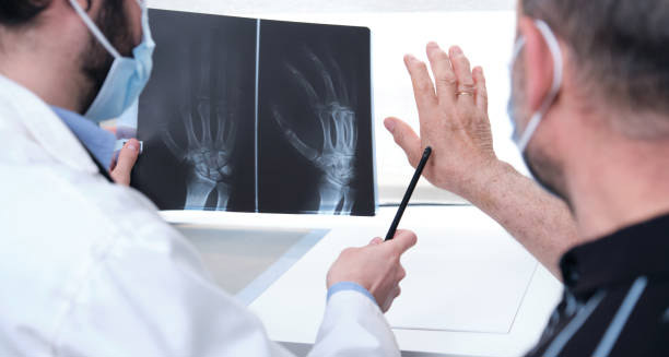
While the diagnosis of mallet finger is often clear without scans, imaging can confirm the extent of injury or rule out other problems.
- X-ray: The most common tool. It shows small bone fragments, dislocation, or signs of arthritis. A bony mallet injury is confirmed when the X-ray shows a fragment where the tendon attaches at the fingertip.
- Ultrasound: Useful for tendon tears, especially in children or when avoiding radiation.
- MRI: Rarely used, but helpful for complex or long-standing cases. It shows tendons, ligaments, and cartilage in detail.
Imaging is not always needed, but it strengthens diagnosis and helps when surgery is being considered.
Differential Diagnosis
Not every drooping finger is mallet finger. Doctors must check for other conditions:
- Jersey finger: Involves loss of flexion rather than extension.
- Tendon cuts: From sharp injuries, sometimes involving both flexor and extensor tendons.
- Arthritis or gout: These cause droop and stiffness but usually affect more than one joint.
- Boutonnière deformity: Involves the middle joint, not the tip.
Sorting through these conditions ensures treatment is correct.
Classifying the Injury
Once confirmed, the diagnosis of mallet finger is classified into two main types:
- Tendinous mallet finger: Caused by tendon rupture with no fracture.
- Bony mallet finger: Caused by a bone fragment pulled off with the tendon.
Bony injuries are further described by fragment size, joint involvement, and any dislocation. This classification guides the choice between splinting and surgery.
Importance of Early Diagnosis of Mallet Finger
Early diagnosis of mallet finger is vital. If left untreated, even a small tendon tear may cause:
- Permanent fingertip droop.
- Stress on nearby joints.
- Progressive instability or arthritis.
Starting splinting within one week gives the best chance for recovery without surgery. Waiting longer than 3–4 weeks often reduces success.
Follow-Up and Reassessment
Doctors reassess the finger after a few weeks of splinting. They check healing, adjust the splint, and look for improvement. Sometimes a repeat X-ray ensures bone fragments remain in place.
Summary
The diagnosis of mallet finger combines history, exam, and sometimes imaging. Spotting the classic fingertip droop, testing tendon function, and ruling out similar injuries guide treatment. Early recognition matters most. With quick diagnosis, patients can heal well and avoid long-term loss of function.