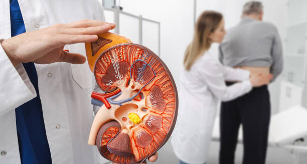
Hydronephrosis is a condition where one or both kidneys swell because urine cannot drain properly into the bladder. This happens when something blocks or slows the flow of urine, causing it to back up into the kidneys. As a result, the kidneys enlarge and come under pressure. The condition can affect just one kidney (unilateral hydronephrosis) or both (bilateral hydronephrosis) and can range from mild to severe.
Hydronephrosis is not a disease on its own. It usually signals another problem, such as a kidney stone, urinary tract obstruction, or birth defect. If left untreated, it can lead to permanent kidney damage, infections, and loss of kidney function. Early diagnosis and treatment are critical. Hydronephrosis can happen at any age and is often detected during pregnancy on routine ultrasounds.
Anatomy of the Urinary Tract
To understand hydronephrosis, it’s helpful to know the basic parts of the urinary system:
- Kidneys: Filter waste from the blood and produce urine
- Ureters: Tubes that carry urine from the kidneys to the bladder
- Bladder: Stores urine until it is released
- Urethra: The tube that carries urine out of the body
Hydronephrosis happens when urine flow is blocked anywhere from the kidneys to the urethra, creating pressure inside the kidneys.
Types of Hydronephrosis
Hydronephrosis can be classified by how long it lasts and what causes it:
- Acute Hydronephrosis: Sudden onset, often caused by a kidney stone or infection. It usually clears up when the blockage is removed.
- Chronic Hydronephrosis: Develops slowly, often due to structural problems or long-standing conditions.
- Obstructive Hydronephrosis: Caused by a physical blockage in the urinary tract.
- Non-Obstructive Hydronephrosis: Due to functional issues, such as weak bladder muscles or nerve problems.
Knowing the type helps doctors choose the right treatment plan.
Who Is Affected?
Hydronephrosis can affect anyone, but some groups are more at risk:
- Newborns and Infants: Often diagnosed during prenatal scans. Usually caused by congenital issues like ureteropelvic junction obstruction.
- Adults: Often due to kidney stones, tumours, or enlarged prostate in men.
- Pregnant Women: Hormonal changes and the growing uterus can temporarily block urine flow.
- Older Adults: Higher risk from prostate enlargement and age-related bladder issues.
Symptoms and Presentation
Hydronephrosis doesn’t always cause symptoms, especially early on. When it does, common signs include:
- Sharp or dull flank pain (side or back pain)
- Frequent or painful urination
- Nausea or vomiting
- Fever and chills (if infection is present)
- Blood in the urine (haematuria)
- Trouble urinating or feeling like the bladder isn’t empty
In babies, symptoms may include an enlarged belly, poor feeding, or frequent urinary infections.
Diagnosis of Hydronephrosis
Doctors use several tests to diagnose hydronephrosis and its cause:
- Physical Exam: Checking for a swollen kidney or abdominal mass.
- Ultrasound: First-choice imaging to confirm kidney swelling.
- CT Scan or MRI: Provides a detailed view of blockages or tumours.
- Urine Tests: Detect infection, blood, or crystals.
- Blood Tests: Measure kidney function (creatinine, BUN).
- Voiding Cystourethrogram (VCUG): Often for children to check urine reflux.
Risks if Left Untreated
Untreated hydronephrosis can lead to:
- Chronic kidney disease (CKD)
- Acute kidney injury (AKI)
- Recurrent urinary tract infections (UTIs)
- Urosepsis (life-threatening infection)
- Permanent kidney scarring
The longer the obstruction lasts, the higher the risk of permanent damage.
Treatment Goals
Treatment focuses on:
- Clearing the blockage or fixing the underlying cause
- Protecting kidney function
- Preventing infection
- Monitoring for recurrence
Some cases only need observation, especially mild prenatal hydronephrosis. Moderate or severe cases may need medication, drainage, or surgery.


