
Overview of Macular Hole
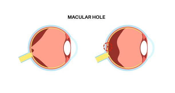
A macular hole is a small break in the centre of the retina, specifically in the macula. The macula is responsible for sharp, central vision—the vision needed for reading, driving, recognizing faces, and detailed tasks. When a macular hole forms, it damages this critical part of the eye, causing blurry or distorted central vision and sometimes a blind spot right in the center.
This condition is different from macular degeneration. While both affect the macula and impair central vision, a macular hole is a physical opening in the retinal tissue. In contrast, macular degeneration is a degenerative or inflammatory condition. Macular holes mainly occur in people over 60 and affect women more often than men. With timely treatment, especially surgery, vision can often be stabilized or partly restored.
Understanding the Macula and Retinal Anatomy
The retina is a thin, light-sensitive layer at the back of the eye. It captures images and sends them to the brain. At its centre lies the macula, a small but vital area packed with photoreceptor cells that provide detailed central vision.
A macular hole happens when the macula develops a full-thickness defect—meaning a gap through all retinal layers. This gap disrupts how light is processed in that spot, harming central vision.
How Macular Holes Develop
Macular holes usually develop slowly. Often, they result from ageing. As we age, the vitreous gel inside the eye shrinks and pulls away from the retina. If the pull is strong or focused on the macula, it can create a small tear or hole.
There are three stages of macular hole formation:
- Stage I: Foveal detachment (impending hole). The retina loses its normal shape but no hole yet.
- Stage II: Partial-thickness hole. A small break forms in the macula.
- Stage III: Full-thickness hole. The hole goes through all retinal layers, causing major vision loss.
Early detection improves chances of successful treatment and vision recovery.
Who Is at Risk?
Several factors raise the risk of developing a macular hole:
- Age over 60
- Female gender
- High myopia (severe nearsightedness)
- Previous eye surgery, especially cataract removal
- Eye trauma or injury
- Macular pucker or retinal detachment
- Diabetic eye disease and inflammatory retinal conditions
Sometimes, macular holes develop without any clear risk factors.
Symptoms and Functional Impact
The main symptom is a gradual loss of central vision. Peripheral vision usually stays normal. Early signs include:
- Blurry or distorted central vision (straight lines may look wavy)
- Trouble reading or recognizing faces
- A dark or grey spot in the center of vision
- Reduced color perception or brightness
If untreated, the hole can grow, causing permanent central vision loss.
How It Differs from Other Macular Disorders
It is important to distinguish macular hole from other macular problems:
- Macular degeneration: Gradual thinning and breakdown of the macula due to ageing.
- Macular pucker: Scar tissue on the retina surface causing wrinkling or distortion.
- Macular hole: A full-thickness opening in the macula.
Each condition needs different treatment, so correct diagnosis is crucial.
Importance of Early Detection and Treatment
Detecting a macular hole early offers the best chance to save or improve vision. Surgery called vitrectomy can close the hole and restore central vision. Surgery works best in Stage II or early Stage III holes.
Delaying diagnosis or treatment lowers the chance of success and increases permanent damage risk.
Summary
A macular hole is a small but serious break in the retina’s centre that affects vision. It mostly affects older adults and starts subtly. Without treatment, it can cause significant vision loss. Advances in surgery now allow many people to recover vision when treated early. If you notice symptoms like distorted central vision or a dark spot, see an eye specialist promptly.