
Keloid scars are usually diagnosed through visual examination and a detailed patient history. Due to their distinct appearance, dermatologists or general practitioners can often identify them by sight. However, a thorough assessment is important to confirm the diagnosis and rule out other skin conditions.
Doctors assess the scar’s size, shape, texture, location, and how it has changed over time. They also consider the patient’s medical background and family history. Correct diagnosis is essential, as keloids require specific treatments and have a high chance of returning if not managed properly.
1. Medical History
Diagnosis starts with a comprehensive medical history. The doctor will ask about past skin injuries, surgeries, burns, acne, vaccinations, or piercings. These events often lead to keloid formation.
Keloids typically appear weeks or months after the skin has healed and may continue growing without new trauma. Family history is also important. Keloids often run in families, and people of African, Asian, or Latin American descent are more prone to them. Knowing this can help support the diagnosis.
2. Clinical Examination
A hands-on clinical exam is the most important step in diagnosing a keloid. The doctor will examine several features:
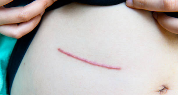
- Appearance: Keloids are raised, shiny, smooth, and firm. They often feel rubbery or dense.
- Borders: Unlike hypertrophic scars, keloids grow beyond the original wound.
- Location: Common sites include the chest, shoulders, earlobes, jawline, and neck.
- Growth: Keloids may keep growing long after the wound has healed.
- Colour: The scar may appear red, purple, brown, or dark, depending on skin tone.
The doctor may gently press the area to check for tenderness, warmth, or symptoms like pain and itching. These sensations are common in keloid scars and can help confirm the diagnosis.
3. Differentiating Keloids from Other Conditions
It’s crucial to distinguish keloids from similar conditions. The most common confusion is with hypertrophic scars. These are also raised but stay within the wound’s original borders and often flatten over time.
Other skin conditions that may mimic keloids include:
- Dermatofibromas – firm skin nodules that can feel similar.
- Keloid-like tumours – rare growths that resemble keloids.
- Hypertrophic lichen planus or sarcoidosis – skin conditions that cause thickened lesions.
Correct diagnosis helps avoid unnecessary treatments, such as surgery for a dermatofibroma or medications meant for inflammatory skin diseases.
4. Dermatoscopy and Imaging
Most of the time, doctors diagnose keloids by sight and touch. However, in complex cases, they may use tools like dermatoscopy. This handheld device magnifies the skin, revealing patterns of blood vessels, pigmentation, and texture.
Ultrasound imaging is sometimes used to check how deep the scar goes. This is useful when planning injections, cryotherapy, or radiation therapy. It can also help distinguish a keloid from a deep cyst or tumor.
5. Skin Biopsy: When Needed
A skin biopsy is rarely required for diagnosing keloids. But if the scar looks unusual or keeps growing despite treatment, a biopsy may be done to rule out cancer or other conditions.
In this procedure, a small piece of the scar is removed under local anaesthesia. A pathologist then examines the tissue under a microscope. Keloids typically show thick collagen bundles in disorganised patterns and a dense dermis.
Biopsies should be used with caution, especially on keloid-prone skin. The incision itself can trigger more scarring.
6. Patient Symptoms and Functional Assessment
The patient’s experience plays a key role in diagnosis. Keloids often cause itching, pain, or sensitivity, which help differentiate them from asymptomatic skin conditions.
If the scar is near a joint or moves with body motion, the doctor will assess whether it limits mobility or causes friction. These factors are important for planning treatment and monitoring progress.
7. Psychosocial Impact
Keloids can affect more than just the skin. People with visible scars may feel embarrassed or anxious. This is especially common in teenagers and young adults.
To assess this, doctors may use questionnaires such as the Dermatology Life Quality Index (DLQI). This helps measure how much the scar affects the patient’s emotional well-being and daily life. A full diagnosis should always include mental health as part of the picture.
8. Genetic Testing and Research
While not yet routine, genetic testing for keloid risk is being studied. Some genes linked to collagen production and wound healing may explain why some people are more prone to keloids.
In the future, genetic testing may help predict who is at risk before elective surgery or cosmetic procedures. For now, doctors rely on family history and ethnic background to assess genetic predisposition.
Final Thoughts
Diagnosing keloid scars relies mainly on clinical expertise and patient history. Visual inspection and physical examination are usually enough. In rare or unclear cases, tools like imaging or biopsy may help.
Early and accurate diagnosis allows for faster treatment and can reduce the risk of further scarring or emotional distress. By considering physical symptoms and mental health together, doctors can create a more complete care plan.
Recognising a keloid early is the first—and most important—step toward effective management.