Mental Matters

Health & Wellness

Causes of Leukoplakia

Causes of Leukoplakia

Causes of leukoplakia are often linked to chronic irritation of the mucous membranes in the mouth, most commonly from tobacco use or poor dental hygiene. Main Causes of Leukoplakia The main causes of leukoplakia include long-term tobacco use, especially smoking or chewing tobacco, and drinking alcohol. Both can irritate the lining inside the mouth. People who use tobacco daily for a long time have a much higher risk of getting leukoplakia. Chemicals in cigarettes, cigars, pipe tobacco, and chewing tobacco create a harmful environment in the mouth. This can cause abnormal cell growth. How Alcohol and Tobacco Work Together Alcohol use is another key cause of leukoplakia. Drinking alcohol, especially when combined with tobacco use, makes the damage to the mouth lining worse. This increases the chance of abnormal thickening. Together, tobacco and alcohol raise the risk of leukoplakia much more than either one alone. Dental trauma and constant irritation also cause leukoplakia. Dentures that do not fit well, broken or sharp teeth, or rough spots on dental tools can rub against the mouth lining. This rubbing triggers the body to respond by forming thick, white patches. If this irritation keeps going, leukoplakia may develop. People who bite the inside of their cheeks or use their teeth to open things may also cause this irritation. Poor Hygiene and Nutritional Deficiencies as Causes of Leukoplakia Poor mouth hygiene adds to the causes of leukoplakia. When people do not clean their mouth regularly, plaque and bacteria build up. This can lead to swelling and damage to soft tissues. Long exposure to bacteria and irritants can change cells and cause leukoplakia. People with gum disease or frequent mouth infections face higher risks if they do not treat these issues quickly. Lack of certain nutrients is another cause. Not getting enough vitamin A and B vitamins can weaken the lining of the mouth. This makes it easier for irritants to cause problems and raises the chance of leukoplakia. Infection and Immune System Factors in Causes of Leukoplakia Sometimes, chronic infections play a role. The Epstein-Barr virus (EBV) links to a type called hairy leukoplakia. This mostly affects people with weak immune systems, like those with HIV/AIDS. The virus causes changes in mouth cells, creating the white patches typical of this condition. Weak immune systems also increase the chance of leukoplakia. People with certain illnesses, or those taking drugs that lower immunity, face higher risks. This includes patients on chemotherapy, organ transplant recipients, and those using immunosuppressants for autoimmune diseases. Other Causes and Considerations of Leukoplakia Some leukoplakia cases have no clear cause. People who do not smoke or drink alcohol sometimes develop it. These unexplained cases still need careful checks to rule out serious issues and watch for cancer signs. Rarely, genetics may play a part. Some families show more leukoplakia cases, hinting that inherited factors affect risk. However, lifestyle and environmental factors cause most cases. Workers exposed to industrial chemicals, like polycyclic aromatic hydrocarbons (PAHs), might also develop leukoplakia. This can happen in jobs such as construction, mining, or chemical manufacturing. If people in these jobs do not use proper protection, repeated mouth lining irritation can occur. Summary of Causes of Leukoplakia In summary, the causes of leukoplakia come from many sources. Lifestyle choices, environmental factors, and health problems all play a role. Tobacco, alcohol, irritation, poor hygiene, vitamin shortages, and weak immunity are common causes. Knowing these risk factors helps prevent leukoplakia and catch it early. People at risk should visit their dentist regularly and see a doctor if white patches stay in the mouth. [Next: Symptoms of Leukoplakia →]


Causes of Leukoplakia Read More »

Symptoms of Leukoplakia

Symptoms of Leukoplakia

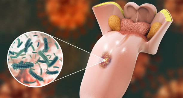
Symptoms of leukoplakia can be subtle in the early stages but may develop into more noticeable signs as the condition progresses. What Symptoms of Leukoplakia Look Like Symptoms of leukoplakia usually show as white or greyish patches inside the mouth or on the tongue. These patches often grow slowly over weeks or months. They usually do not hurt and can go unnoticed, especially if they form in hard-to-see spots, like inside the cheeks or under the tongue. Even though these patches do not cause pain at first, you should never ignore them. Symptoms of leukoplakia can sometimes signal more serious mouth problems, including oral cancer. The most common sign among symptoms of leukoplakia is thick, white plaques. These patches stick firmly and cannot be rubbed off easily. Their surface may look raised, ridged, or rough. Unlike oral thrush — caused by a yeast infection and wiped away easily — leukoplakia patches stay in place and last a long time. They may appear alone or in groups, and their size and shape can change a lot. Texture Changes and Other Symptoms of Leukoplakia Another typical symptom of leukoplakia is that the affected mouth area changes in feel. It may feel rough or leathery when touched. Some people also feel tightness or like something is stuck in their mouth when patches get bigger. Sometimes, the patches become sore or uncomfortable, especially when eating spicy or acidic foods, but this is rare. Sometimes, red patches appear with the white ones. This is called erythroleukoplakia and is more serious because it has a higher risk of turning into cancer. Any red spots in a leukoplakia patch need immediate medical check. Mixed red and white patches should alert you and your doctor to do more tests. Why Symptoms of Leukoplakia Can Go Unnoticed One sneaky part about symptoms of leukoplakia is they usually do not cause pain. Many people do not notice the patches until a dentist or doctor points them out during a check-up. Because these patches do not hurt, people delay treatment. This delay can raise the chance of serious problems later. Symptoms of leukoplakia may also include changes in speech or swallowing. This happens if the patches grow in places that block normal mouth use. When patches develop on the sides of the tongue or floor of the mouth, they may irritate speech or chewing. In severe cases, the tongue might move less, causing slurred speech or trouble saying some sounds. Variations and Warning Signs in Symptoms of Leukoplakia In hairy leukoplakia — a form linked to weak immune systems, especially in people with HIV/AIDS — symptoms may show fuzzy, hairy-looking white patches on the tongue’s sides. Hairy leukoplakia is caused by the Epstein-Barr virus. It usually does not carry the same cancer risk but still signals serious immune problems. Sometimes, patients notice that patches change in look over time. For example, thin patches may become thicker, uneven, or bumpy. Rapid changes in color, size, or surface texture of leukoplakia patches are warning signs. You should get them checked right away because these changes might mean early cancer. Why Professional Diagnosis Matters Symptoms of leukoplakia can look like other mouth problems. White patches might come from lichen planus, candidiasis, or burns from chemicals. Because of this, do not try to diagnose yourself. Only a trained healthcare worker or dental expert can tell leukoplakia apart by checking clinically and doing a biopsy if needed. Lifestyle Factors and Monitoring Symptoms of Leukoplakia Certain habits can make symptoms of leukoplakia worse. Smoking or chewing tobacco and drinking alcohol regularly link strongly to leukoplakia development and persistence. These habits not only worsen symptoms but also raise the chance of the patches turning cancerous. It is very important to watch symptoms of leukoplakia closely. People diagnosed with it should see their dentist or mouth specialist often to check for any signs of change. Sometimes, patches shrink or go away if harmful habits, like smoking or bad dental care, stop. But if symptoms stay or get worse, more tests or treatment are usually needed. Summary of Symptoms of Leukoplakia Recognising the symptoms of leukoplakia helps catch it early and avoid problems. Look out for white, thick patches that cannot be scraped off, occasional pain or irritation, and sometimes red spots in the patches. Since these symptoms often do not hurt and can be hard to notice, regular mouth exams are vital. This is especially true for people who smoke, drink alcohol a lot, or have weak immune systems. [Next: Diagnosis of Leukoplakia →]

Symptoms of Leukoplakia Read More »

Diagnosis of Leukoplakia

Diagnosis of Leukoplakia

Diagnosis of leukoplakia is vital to tell this condition apart from other oral health issues, some of which may be more serious. It usually starts with a detailed examination of the mouth by a dentist, doctor, or oral specialist. Since leukoplakia can sometimes be an early sign of oral cancer or other precancerous problems, diagnosing it early and correctly is crucial for proper treatment and monitoring. Clinical Examination and Risk Assessment The healthcare provider begins with a visual check of the mouth and lips, looking for thick, white, or grey patches that can’t be wiped away. These patches often appear on the inside cheeks, gums, tongue, or floor of the mouth. The provider also asks about habits that raise risk, such as smoking, heavy drinking, or poor oral hygiene. Diagnosis of leukoplakia depends heavily on spotting these risk factors and matching them with the lesions’ appearance and location. Cytology and Biopsy: Confirming the Diagnosis If the clinical exam points to leukoplakia, further testing may follow to confirm the diagnosis and exclude cancer. Oral exfoliative cytology uses a brush or spatula to collect cells from the patch surface. These cells are checked under a microscope for precancerous changes, but this method can sometimes miss deeper problems. The most reliable test is a biopsy, where a small tissue sample is taken from the lesion. Types of biopsy include incisional (part of the lesion), excisional (whole lesion), or punch biopsy (a core of tissue). The choice depends on the lesion’s size, site, and clinical suspicion. A biopsy determines if the cells are normal, dysplastic (abnormal but not cancer), or malignant, making it essential in leukoplakia diagnosis. Assessing Lesion Types and Malignancy Risk Some leukoplakia patches are smooth and uniform (homogeneous), which carry a lower cancer risk. Others—speckled, nodular, or verrucous types—are non-homogeneous and need closer investigation. These higher-risk patches often require biopsy or more frequent follow-ups. Thus, diagnosis of leukoplakia involves not just finding the lesion but classifying it by appearance to assess cancer risk. Additional Diagnostic Tools To aid diagnosis, doctors may use special tests like toluidine blue staining, which highlights abnormal cells and helps pick biopsy sites. Autofluorescence devices can spot tissue changes by detecting differences in light reflection, signaling possible early cancer changes. These tools support but do not replace biopsy. Patient History and Symptom Review A thorough patient history is important. Doctors ask how long the lesion has been present, if it has changed, and about symptoms like pain, bleeding, or trouble eating. Though leukoplakia often causes no symptoms, any signs can guide the urgency and nature of further testing. Clinicians also check overall health, since immune problems or nutritional deficiencies can contribute to oral lesions. Molecular Testing in High-Risk Cases For suspicious or recurrent lesions, molecular or genetic tests on biopsy samples may be done. These can find chromosomal changes linked to oral cancer. Such advanced diagnostics may become more common, especially for high-risk or complicated leukoplakia cases. Follow-Up and Monitoring After diagnosis, patients usually need regular check-ups, especially if dysplasia is present. Leukoplakia can come back or develop into cancer, so ongoing surveillance with exams and sometimes repeat biopsies is key. Diagnosis of leukoplakia is a continuous process requiring careful monitoring. Special Considerations: Proliferative Verrucous Leukoplakia A rare, aggressive type called proliferative verrucous leukoplakia (PVL) demands close, long-term monitoring. PVL has a higher chance of turning cancerous and often needs care from a team including dental experts, oncologists, and pathologists. Summary of Leukoplakia Diagnosis In short, diagnosis of leukoplakia involves a thorough oral exam, risk factor review, cytology, biopsy, and sometimes advanced tests. Early and accurate diagnosis is critical because of the risk of cancer. Combining clinical skills, technology, and patient cooperation ensures leukoplakia is managed effectively. [Next: Treatment of Leukoplakia →]

Diagnosis of Leukoplakia Read More »

Treatment of Leukoplakia

Treatment of Leukoplakia

The treatment of leukoplakia combines lifestyle changes, medical care, and regular check-ups to stop it from becoming oral cancer. If caught early, treatment is usually simple. However, the exact plan depends on how severe the patches are, where they are, and if they show any signs of being precancerous (dysplasia). Working closely with a dentist or doctor helps create a plan tailored to each person’s needs. Stopping the Cause: Tobacco and Alcohol The first step in treating leukoplakia is finding and removing what’s causing it. Smoking or chewing tobacco is the main trigger in many cases. Drinking alcohol, especially with tobacco use, also raises the risk. Quitting both tobacco and alcohol often helps the white patches shrink or disappear. For some people, these lifestyle changes alone can clear up the problem, especially if the patches are not showing any dysplasia. When More Treatment Is Needed If leukoplakia stays after quitting irritants or if the patches look thick, bumpy, or are in risky spots like the tongue or floor of the mouth, further treatment may be needed. Doctors usually do a biopsy to check for dysplasia. If precancerous changes are found, surgery may be recommended to remove the patches and prevent cancer. Surgical options include traditional cutting, laser treatment, or freezing (cryotherapy). Laser treatment is often chosen because it is precise, causes less bleeding, and helps patients heal faster. Medications for Leukoplakia Sometimes, doctors use creams or gels to treat leukoplakia. Retinoids, which come from vitamin A, can help reverse early changes in the cells but may have side effects and aren’t good for long-term use. Anti-inflammatory or antifungal creams might be used if there is swelling or infection along with leukoplakia. Regular Monitoring and Follow-Up Regular check-ups are very important in managing leukoplakia. Patients usually need oral exams every three to six months, depending on their risk. Follow-ups help catch any new patches, return of old ones, or changes in existing lesions early. Sometimes, doctors use special imaging or repeat biopsies if the patches look different or cause new symptoms. Nutrition and Leukoplakia Eating a healthy diet with plenty of fruits and vegetables may support the mouth’s health. These foods contain antioxidants that help protect cells from damage. Some studies suggest that supplements like beta-carotene or lycopene might help reduce lesions, but more research is needed before these can be regularly recommended. Treatment for Oral Hairy Leukoplakia Oral hairy leukoplakia, linked to the Epstein-Barr virus, usually affects people with weak immune systems like those with HIV. This form often does not need treatment unless it causes symptoms or cosmetic concerns. If treatment is needed, antiviral drugs such as acyclovir may be given. Managing the underlying immune problem is also important. Handling Difficult or Recurrent Cases In rare cases, leukoplakia does not respond to usual treatments or keeps coming back. Specialists may suggest stronger treatments or refer the patient to an oral surgeon or cancer specialist. This teamwork is especially important if the patches have high-grade dysplasia or early cancer changes. Educating Patients for Better Outcomes Education is key to successful treatment. Patients must understand the need to avoid irritants, follow medical advice, and attend regular check-ups. Doctors and dentists play a major role in supporting patients and providing complete care. Summary of Leukoplakia Treatment In summary, treating leukoplakia involves many steps based on the lesion and risk factors. Quitting tobacco and alcohol can be enough in some cases, but others need surgery or medicines. Ongoing monitoring is critical to spot any early signs of cancer. With proper care, treatment of leukoplakia can lower the risk of oral cancer and improve oral health. [Next: Complications of Leukoplakia →]

Treatment of Leukoplakia Read More »

Complications of Leukoplakia

Complications of Leukoplakia

Complications of leukoplakia can range from minor mouth discomfort to serious, life-threatening problems like oral cancer. Understanding the complications of leukoplakia is important for catching it early and managing long-term oral health. Most cases are harmless, but some have a higher chance of turning into cancer. This risk is greater in people who smoke or have weak immune systems. Risk of Cancer in Leukoplakia One major concern is that leukoplakia can turn into cancer. It is seen as a precancerous condition. Research shows that about 3% to 17.5% of leukoplakia cases may turn into squamous cell carcinoma over time. This risk is much higher with non-homogenous leukoplakia, especially if the patches look speckled or bumpy. If a biopsy shows changes in the cells, the danger increases, and doctors may suggest surgery or close monitoring. Recurrence and Persistent Lesions Another issue is that leukoplakia can come back after treatment. Even after removing the patches with surgery, lasers, or freezing, they may reappear. This happens more often if the person keeps using tobacco or drinking alcohol. These recurring patches can cause ongoing pain, especially while eating, talking, or brushing. Long-term cases also mean more doctor visits and more biopsies. This can feel emotionally draining and cost a lot over time. Psychosocial and Emotional Impact Leukoplakia can also affect a person’s mental and emotional well-being. People with visible or large patches in the mouth often feel embarrassed or anxious. This is worse if the spots are on the lips or front of the tongue. As a result, they may avoid social situations. In serious cases, the fear of cancer or feeling judged can even lead to depression or pulling away from others. Functional Problems in the Mouth Some complications of leukoplakia involve problems with how the mouth works. If the patches are near important areas like salivary glands or the sides of the tongue, they can cause pain or stiffness. This makes it hard to chew or speak clearly. If the condition turns into cancer and needs surgery, it may lead to lasting issues with swallowing or talking. Complications of Leukoplakia in Immunocompromised People People with weak immune systems are more likely to develop severe forms of leukoplakia. For instance, those with HIV/AIDS may get a type called hairy leukoplakia. This form is strongly linked to Epstein-Barr virus. While it usually does not become cancer, it may signal that the person’s immune health is getting worse. In these cases, leukoplakia is part of a bigger health problem, which might also include other infections and poor healing. Diagnostic Challenges and Delays Even finding the right diagnosis can be a problem. Not all white patches in the mouth are leukoplakia. Conditions like lichen planus, fungal infections, or chemical burns can look very similar. If doctors don’t take a biopsy or misread the signs, cancer may go unnoticed—especially in places with few dental specialists. That’s why careful exams and follow-up are so important for anyone with suspicious patches. From Leukoplakia to Oral Cancer When complications of leukoplakia lead to oral cancer, the results can be serious or even deadly. Advanced oral cancer often needs strong treatment, like surgery, radiation, or chemotherapy. These treatments can cause side effects, including disfigurement and trouble with speech or swallowing. The chances of surviving are much better when cancer is found early. That’s why any case of leukoplakia must be taken seriously from the start. Cost and Access to Care Money problems can also be a complication. Regular check-ups, tests, surgery, and follow-ups all add up. In many places, dental care isn’t fully covered by insurance. This makes it hard for some people to get the care they need. In lower-income areas, there may not even be access to the right specialists. This delay can lead to worse outcomes and more advanced disease. Preventing Complications of Leukoplakia The good news is that many of these problems can be prevented. Quitting smoking and drinking less alcohol are key steps. Keeping your mouth clean and seeing the dentist regularly also helps. In some cases, doctors may remove high-risk patches early to stop them from becoming cancerous later. The Importance of Early Action To sum up, the complications of leukoplakia affect not just the body, but also the mind and daily life. From cancer risk to emotional stress, this condition needs careful attention and regular follow-up. Doctors must help patients understand the risks and take early steps to avoid serious problems. New tools like biomarkers and easier tests may improve care in the future. Until then, staying informed and proactive is the best way to manage this condition. [Next: Back to Overview →]

Complications of Leukoplakia Read More »

Overview of Leukaemia

Leukaemia

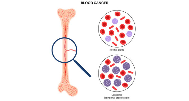
Overview of Leukaemia The overview of Leukaemia refers to a comprehensive understanding of the disease known as leukaemia, which is a type of blood cancer that starts in the bone marrow and affects how white blood cells are made. Overview of Leukaemia is essential to help patients, caregivers, and doctors understand how the disease develops, spreads, and affects the body. Leukaemia happens when the bone marrow makes too many abnormal white blood cells. These faulty cells can’t do their job and get in the way of making red blood cells and platelets. Types and Classifications of Leukaemia Leukaemia isn’t just one disease. It’s a group of blood cancers, each with different subtypes, outcomes, and treatments. Doctors group them by how fast they grow—acute or chronic—and the type of blood cell they affect—lymphoid or myeloid. Acute types like Acute Lymphoblastic Leukaemia (ALL) and Acute Myeloid Leukaemia (AML) grow quickly and need fast treatment. Chronic types like Chronic Lymphocytic Leukaemia (CLL) and Chronic Myeloid Leukaemia (CML) grow more slowly. Doctors may choose to watch them for a while before starting treatment. The Overview of Leukaemia also shows how it affects people differently. For example, ALL is more common in children, while CLL is usually found in older adults. We still don’t know the exact cause, but some risk factors increase the chance of getting it. These include inherited traits, radiation, smoking, contact with certain chemicals, and past cancer treatments. Knowing these risks helps in spotting leukaemia early and maybe even lowering the chances of getting it. Recognising Symptoms of Leukaemia Symptoms vary based on the type and stage of leukaemia. Common signs include feeling very tired, frequent infections, losing weight without trying, anaemia, bruising or bleeding easily, and swollen lymph nodes. Because these signs can look like other less serious illnesses, diagnosis often takes time. That’s why knowing the signs and seeing a doctor early is so important. Doctors usually use blood tests, bone marrow samples, and scans to find and confirm leukaemia. Looking at blood and marrow under a microscope and doing lab tests helps identify the exact type. New genetic tests give doctors even better tools to understand the disease. These tools also help them choose treatments that match the patient’s specific cancer features. Treatment and Advances in Care Treatment depends on the type of leukaemia, the patient’s age, health, and genetic markers of the cancer. Chemotherapy is still one of the main treatments. Doctors may also use radiation or targeted drugs. A bone marrow transplant can be life-saving, especially for patients who relapse or have high-risk disease. New treatments like CAR-T cell therapy and monoclonal antibodies are becoming more common, especially when the disease doesn’t respond to standard treatments. The outlook for people with leukaemia has improved over the years. Early diagnosis, better treatments, and supportive care all help. Survival rates now vary by type, with some chronic forms being controlled for years. At the same time, doctors focus more on quality of life. Treatments now aim to reduce side effects and help people stay active and well. Public Awareness and Support in Leukaemia Care Public health plays a big part in the Overview of Leukaemia. Education, early testing, and research funding all help people understand and manage this disease better. Clinical trials are also key to finding new treatments. These trials give hope to patients who don’t get better with standard care and help doctors improve how they treat others in the future. Support systems are also very important. Patients often need emotional help to deal with their diagnosis and treatment. They may also need nutrition advice, physical therapy, and help managing side effects. Getting family, social workers, and support groups involved gives a more complete type of care. Campaigns and teamwork across countries have helped us learn more about leukaemia. Global databases and research studies help track patterns and see how the disease affects people in different places. These efforts also make sure that people in low- and middle-income countries get better care too. Why the Overview of Leukaemia Matters In summary, the Overview of Leukaemia covers many parts of this disease—from where it starts in the body to how it shows up, how we diagnose it, and how we treat it. It shows that leukaemia is complex and can affect anyone. Education, early awareness, and new ideas are key to making sure patients get the best and kindest care. By learning the Overview of Leukaemia, people and communities can take steps to stay informed, seek early help, and support cancer research around the world. [Next: Symptoms of Leukaemia →]

Leukaemia Read More »

Symptoms of Leukaemia

Symptoms of Leukaemia

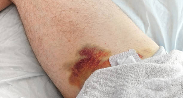
Symptoms of leukaemia often present subtly in the early stages, making the disease difficult to detect without medical tests. As the disease progresses, these symptoms become more noticeable and disruptive. They vary depending on the type—acute or chronic—and whether it affects lymphoid or myeloid cells. Still, recognising and understanding the most common and serious symptoms of leukaemia helps patients and healthcare professionals act quickly and begin the right treatment. This early awareness is vital because some forms of leukaemia can progress rapidly and need urgent care. The most reported symptoms of leukaemia include feeling very tired, sudden weight loss, frequent infections, and easy bruising or bleeding. Fatigue happens when leukaemia cells push out healthy red blood cells, causing anaemia. This kind of tiredness does not improve with rest. It can affect daily life and lower quality of living. Sudden weight loss and low appetite happen because the body uses a lot of energy to fight the rise of cancerous white blood cells. These broad signs often lead people to seek medical help. Early Physical Signs of Leukaemia A clear warning among symptoms of leukaemia is easy bruising or heavy bleeding. Patients might have frequent nosebleeds, bleeding gums, or heavy menstrual periods. This happens because leukaemia stops the bone marrow from making enough platelets, which are needed for blood to clot. In some people, small red or purple spots called petechiae appear on the skin, especially on the legs. These spots, though they look like small rashes, are signs of low platelet counts and could point to a blood problem. Frequent infections are also major symptoms of leukaemia. This is due to the immune system not working properly. Leukaemia causes the bone marrow to make faulty white blood cells that can’t fight infections. As a result, people become more prone to illnesses like bronchitis, pneumonia, or urinary infections. These illnesses might take longer to heal or keep coming back, which signals trouble for both patients and doctors. Swollen lymph nodes—in the neck, armpits, or groin—are other common symptoms of leukaemia. These swollen areas are often painless, so many people don’t notice them. Leukaemia cells can also build up in the spleen or liver, making them larger. This can cause a feeling of fullness or bloating, especially in the upper left side of the belly. Sometimes it leads to discomfort or pain if the disease is more advanced. Bone Pain and Severe Symptoms of Leukaemia Bone or joint pain is another issue some people face. It comes from the build-up of leukaemia cells inside the bone marrow, which puts pressure on the bones. The pain may feel sharp or dull and can affect just one area or several. It often shows up in the arms and legs, especially in children. In acute forms of leukaemia, symptoms can appear very quickly. They may include high fevers, night sweats, and pale skin because of fewer red blood cells. Chronic leukaemia moves more slowly. Its symptoms might be mild at first and mistaken for ageing or other health problems. But even chronic leukaemia can speed up and cause stronger symptoms during a phase called blast crisis. Some symptoms of leukaemia are less clear. These include headaches, blurry vision, or shortness of breath, which can happen if anaemia becomes severe. In rare cases, when the disease reaches the brain or spinal cord, it may cause seizures or confusion. This is more common in a type called acute lymphoblastic leukaemia. Some people might also see greenish skin lumps called chloromas, though this is rare. How Leukaemia Symptoms Differ in Children Children with leukaemia often show different symptoms than adults. They still experience tiredness, infections, and bruising, but may also stop growing normally, eat poorly, or become more irritable. Parents might see their child lose interest in playing or fall behind in reaching milestones. These quiet changes in behaviour can be some of the earliest signs of paediatric leukaemia. Because symptoms of leukaemia can look like other illnesses, misdiagnosis is common, especially early on. Doctors might confuse these signs with flu, low iron, or hormone problems. That’s why it’s important to get checked if multiple symptoms show up or get worse over time. Blood tests often catch unusual white cell, red cell, or platelet levels, leading to more tests like a bone marrow biopsy. Key Symptoms of Leukaemia to Watch For To sum up, the main symptoms of leukaemia include: These symptoms are easy to miss because they also happen with common illnesses. Still, knowing the full list of symptoms of leukaemia can lead to early diagnosis and better treatment. Whether the symptoms come on slowly or quickly, it’s always best to talk to a doctor if they persist or don’t have a clear cause. Early care makes a big difference, especially for fast-moving forms of the disease. [Next: Diagnosis of Leukaemia →]

Symptoms of Leukaemia Read More »

Diagnosis of Leukaemia

Diagnosis of Leukaemia

Diagnosis of leukaemia is a critical process that involves a combination of clinical evaluation, laboratory testing, and imaging techniques to confirm the presence and type of this complex blood cancer. Since the diagnosis of leukaemia affects treatment choices and long-term outcomes, early and accurate testing matters. Many patients first visit a doctor with vague symptoms like fatigue, fever, or frequent bruising. These signs push doctors to do further tests. A clear process helps ensure the disease is found and classified correctly. Initial Clinical Assessment The process begins with a full medical history and physical check-up. Doctors ask about symptoms, past illnesses, and any family history of cancer. Symptoms such as repeated infections, bleeding gums, night sweats, or swollen lymph nodes may raise red flags. During the physical exam, the doctor may notice signs like pale skin or an enlarged liver or spleen. Although these symptoms may appear in other conditions, they help guide next steps. If leukaemia is suspected, the general practitioner sends the patient to a blood specialist—a haematologist. This doctor then orders tests to confirm the presence of abnormal cells and rule out other illnesses. Blood Tests and Cell Examination One of the first tools used in the diagnosis of leukaemia is the full blood count (FBC). This test measures the levels of red cells, white cells, and platelets. A result showing low red cells, unusual white cell counts, or fewer platelets may point to leukaemia. Along with the FBC, doctors use a blood smear. This test checks the shape and stage of cells under a microscope. In acute leukaemia, early signs include abnormal white cells called blasts. In chronic leukaemia, more mature but still abnormal cells might appear. If these are present, doctors move on to more detailed testing. Bone Marrow Testing in Diagnosis of Leukaemia A firm diagnosis often comes from a bone marrow biopsy and aspiration. In this test, a small sample of bone marrow is taken—usually from the hip bone—using a needle under local anaesthetic. Doctors check this sample for the number and type of blood cells, especially the number of blast cells. If the blast count is 20% or more, acute leukaemia is usually confirmed. The sample also goes through further testing. Two key methods—cytogenetic analysis and flow cytometry—help spot gene changes and confirm the type of leukaemia. This information is vital for choosing the right treatment and understanding how the disease might progress. Genetic and Molecular Testing To better understand the diagnosis of leukaemia, doctors also use tests that study chromosomes and genes. Cytogenetic tests like karyotyping look at changes in the structure of chromosomes. These may show key clues—like the Philadelphia chromosome, which appears in CML, or other genetic shifts seen in ALL and AML. Doctors also use PCR and FISH tests. These spot very small changes in DNA that help confirm leukaemia and plan treatment. Some of these changes also guide the use of special drugs that target the cancer more precisely. Flow Cytometry and Immunophenotyping Another key test is flow cytometry, used for immunophenotyping. This method tags cells with fluorescent dyes and helps tell one type of leukaemia from another. For example, it can show whether the cancer involves B-cells or T-cells. Knowing this helps doctors design a plan that fits the exact disease type. This test also helps detect any leftover cancer after treatment—called minimal residual disease—which is important for tracking recovery and adjusting care. Lumbar Puncture and Imaging Sometimes, leukaemia spreads to the brain and spinal cord. This is more common in ALL. If doctors think this might be happening, they perform a lumbar puncture. This test collects fluid from the spine to check for cancer cells. It’s done if patients have symptoms like headaches, vision changes, or seizures. Imaging tools like X-rays, CT scans, or ultrasound may also help. They show if organs like the spleen or lymph nodes are larger than normal. These tests do not confirm the diagnosis but offer helpful information about how far the disease has spread. Staging and Risk Stratification Once the diagnosis is confirmed, doctors check how advanced the disease is. This step, called staging, looks at how much cancer is in the body and how fast it may grow. Doctors also consider risk factors like age, white cell count, and gene changes. For example, in ALL, patients are placed into standard-risk or high-risk groups. This helps guide how strong the treatment should be. In CLL, doctors use systems like Rai or Binet staging to measure disease level and decide when to start therapy. Grouping patients by risk makes treatments more personal and effective. Paediatric and Adult Diagnosis: Key Differences Children go through the same basic tests, but doctors take extra care during diagnosis. Kids may react more strongly to illness or feel more fear. Since children often get ALL, doctors move quickly to start treatment. Special paediatric teams use equipment and steps suited to younger bodies. Emotional support also plays a major role. Psychologists and counsellors often join the care team early to help families cope. They guide parents and children through what can be a confusing and scary process. Ongoing Tests and Monitoring After the first diagnosis of leukaemia, the journey continues with regular testing. Doctors keep track of blood counts, bone marrow changes, and gene activity to see how well treatment is working. These follow-up tests show whether the patient is in remission or if changes to treatment are needed. By keeping a close watch, doctors can spot a relapse early and act fast. For patients receiving bone marrow transplants or targeted drugs, ongoing testing helps doctors manage long-term results and avoid future problems. Final Thoughts on Diagnosis of Leukaemia The diagnosis of leukaemia is not a simple task. It involves many tests and expert review to make sure every detail is clear. Finding the exact type of leukaemia early makes it possible to start the right treatment and improve results. Thanks to new tools and advanced lab work, doctors can now find even tiny disease

Diagnosis of Leukaemia Read More »

Treatment of Leukaemia

Treatment of Leukaemia

Treatment of leukaemia is a multifaceted and highly individualised process that varies depending on the type of leukaemia, its stage, the patient’s age, overall health, and genetic factors. Because this blood cancer starts in the bone marrow and affects the blood and immune system, doctors must use strong and carefully planned treatments. The main goals in treating leukaemia are to remove cancer cells, bring back healthy blood cell production, stop the disease from returning, and help patients live well during and after treatment. Modern Approaches to Treatment of Leukaemia Today’s treatment of leukaemia often combines several methods. These include chemotherapy, targeted drugs, radiation, stem cell transplants, and new immune-based therapies. Patients also need supportive care to handle side effects, fight off infections, and manage emotional stress. Different types of leukaemia need different treatment plans. Acute leukaemia and chronic leukaemia do not respond the same way. Subtypes like ALL, AML, CLL, and CML each need unique approaches. Chemotherapy as the Foundation of Leukaemia Treatment Chemotherapy is the base of treatment for most leukaemia types, especially acute ones. These drugs attack fast-growing cells, including cancerous white blood cells. Doctors give chemotherapy in cycles. These breaks give the body time to recover. For Acute Lymphoblastic Leukaemia (ALL), chemotherapy has three stages. First is induction, which aims to kill most of the cancer cells. Then comes consolidation, which removes any cells left behind. Finally, the maintenance phase keeps the disease under control and often lasts for two to three years. Acute Myeloid Leukaemia (AML) also starts with induction chemotherapy. Doctors usually use drugs like cytarabine and anthracyclines. Sometimes, a second round is needed to reach full remission. After this, patients receive high-dose chemotherapy or stem cell transplants, depending on their risk level. In chronic leukaemia, like CLL and CML, chemotherapy isn’t always the first choice. Instead, targeted therapies are often used. But in some cases—like when other treatments don’t work or aren’t available—chemotherapy with monoclonal antibodies like rituximab is still used. Targeted Therapy: A Major Shift in Treatment of Leukaemia Targeted therapy has changed the way doctors treat leukaemia, especially in chronic forms. These drugs go after specific proteins or genes that help cancer grow. For example, in Chronic Myeloid Leukaemia (CML), drugs called tyrosine kinase inhibitors (TKIs)—like imatinib, dasatinib, or nilotinib—block a protein linked to the disease. This protein is caused by a genetic change called the Philadelphia chromosome. In Acute Promyelocytic Leukaemia (APL), doctors often use drugs like all-trans retinoic acid (ATRA) and arsenic trioxide instead of chemotherapy. These targeted treatments have very high success rates. Other targeted therapies include FLT3, IDH1/2, and BCL-2 inhibitors such as venetoclax, often used in CLL. These options help patients stay in remission longer and usually come with fewer side effects than traditional chemotherapy. Stem Cell Transplants and Radiation Stem cell transplantation, also called bone marrow transplant, is important for some high-risk or relapsed cases. It involves replacing damaged bone marrow with healthy cells from a donor or from the patient. Allogeneic transplants, using donor cells, are more common in serious cases. Before the transplant, doctors give strong chemotherapy or radiation to remove the cancer. Then, donor cells are added. This can cure the disease, but also brings serious risks like graft-versus-host disease (GVHD), infections, and organ problems. Autologous transplants use the patient’s own healthy cells. This is more often used when the disease is under control and mostly in certain lymphomas or clinical trials. Radiation is less common but still useful in some situations. It can shrink large organs, like the spleen or lymph nodes, or treat cancer that has reached the brain or spinal fluid. Total body irradiation (TBI) is also used before stem cell transplants to kill any remaining cancer and lower the risk of rejection. Immunotherapy and CAR-T in Treatment of Leukaemia One of the most exciting changes in the treatment of leukaemia is immunotherapy. These treatments use the body’s own immune system to fight cancer. For example, monoclonal antibodies like rituximab (used in CLL) and blinatumomab (used in ALL) help the immune system find and kill cancer cells. CAR-T cell therapy is a powerful new option. Doctors collect a patient’s T-cells, change them in a lab to attack leukaemia cells, and then put them back in the body. This treatment is used when other options have failed, especially in ALL and some B-cell lymphomas. While CAR-T therapy can be very effective, it can also cause serious side effects. These include cytokine release syndrome and brain-related problems. That’s why patients need close care in special treatment centres. Supportive, Palliative, and Age-Specific Care Supportive care is a key part of treatment. It includes blood transfusions to treat anaemia and low platelets, antibiotics to prevent infections, and drugs to boost blood cell production. This helps patients feel better and stay strong during treatment. Emotional care is just as important. Many patients feel anxiety or depression. Counselling, peer groups, and services like nutrition or physical therapy help improve mental and physical health. Palliative care is offered early for people who can’t have aggressive treatment or have incurable leukaemia. It focuses on easing pain and discomfort, giving emotional support, and helping patients live as fully as possible. Treatment of Leukaemia in Children and Older Adults Children need special care when being treated for leukaemia. Their bodies react differently to drugs, and doctors must think about long-term effects. Children with ALL often do very well because of fast diagnosis and strong treatment plans. Still, cancer treatment affects children emotionally and mentally. Paediatric teams include psychologists, play therapists, and school support workers to help children stay on track with their development. Older adults face other challenges. Many have other health problems, and their bodies may not handle strong treatments. In these cases, doctors may choose gentler treatments or suggest clinical trials that offer new, lower-risk options. Clinical Trials and the Future of Treatment Clinical trials are shaping the future of leukaemia care. These research studies test new drugs, better combinations, and advanced technologies. They offer hope for

Treatment of Leukaemia Read More »

Complications of Leukaemia

Complications of Leukaemia

Complications of leukaemia can significantly affect the quality of life, response to treatment, and overall prognosis for individuals diagnosed with this complex group of blood cancers. The disease harms the body’s ability to make and use healthy blood cells. Because of this, many problems come from both the illness itself and the treatments used, like chemotherapy or radiation. These treatments, along with the weakened immune system, increase the risk of infections and organ problems. So, it’s very important to understand all the possible issues that can happen. This helps patients and their care teams manage the disease better and lower the risks. Infections: A Serious Threat One of the most common and dangerous complications of leukaemia is infection. The disease affects how white blood cells are made and how they work. As a result, the immune system becomes weak and struggles to protect the body. This makes infections more likely and more severe. These infections can come from bacteria, viruses, or fungi. Sometimes, they become life-threatening. People who are getting chemotherapy or have had a bone marrow transplant are at an even higher risk. Their immune system is already low, so they must be extra careful. Infections may show up in strange ways or get worse quickly. That’s why quick treatment is often needed, including hospital care. Bleeding and Anaemia Another common issue is bleeding or easy bruising. This happens when the body doesn’t make enough platelets. Platelets help the blood to clot, so without enough, even small injuries can cause serious bleeding. Nosebleeds, gum bleeding, or dark bruises for no reason may appear early. In very bad cases, bleeding inside the brain or stomach may happen. Doctors often give platelet transfusions and monitor levels closely to prevent this. Anaemia is also a big concern. The bone marrow may stop making enough red blood cells, which carry oxygen. This can lead to feeling tired all the time, trouble breathing, pale skin, and dizziness. Some people need regular blood transfusions to feel well. Their treatment plan may also change to help manage this problem better. Organ and Nerve Problems in Complications of Leukaemia In more serious cases, the disease can damage organs. The liver, spleen, and lymph nodes might get larger due to the build-up of leukaemia cells. This can cause stomach pain or make a person feel full quickly. In types like chronic lymphocytic leukaemia (CLL), swelling in these organs may get so bad that they stop working properly. Kidney and heart problems can also appear. Sometimes the cancer causes this, and other times it’s because of side effects from strong drugs. Regular scans and blood tests help catch these problems early. Nervous system problems can develop too, especially if the cancer spreads to the brain or spinal cord. People might have headaches, confusion, blurry vision, or even seizures. In children, this is more common. Doctors often treat it with special chemotherapy given directly to the spine. But this treatment can also affect learning or development, especially if given during important growth stages. Treatment Side Effects and Emotional Strain Some people with leukaemia need a stem cell or bone marrow transplant. A serious problem that can happen after this is graft-versus-host disease (GVHD). In GVHD, the new cells from the donor attack the patient’s body. This can cause rashes, stomach problems, liver damage, and a weak immune system. It can be mild or very dangerous. To treat it, doctors often use drugs that lower the immune system, which brings its own risks. Living with leukaemia brings a heavy emotional load. The stress of facing a life-threatening illness, ongoing hospital visits, and tough treatments can lead to anxiety or depression. Some people may even develop post-traumatic stress. These mental health struggles can make it hard to follow treatment plans. That’s why support through therapy or counselling is so important in long-term care. Long-Term Complications of Leukaemia Another risk that may appear much later is getting a second type of cancer. Strong treatments like chemotherapy or radiation can harm healthy cells and increase the chance of other cancers. This may include skin cancer, breast cancer, or even another form of leukaemia. Fertility problems can happen too, especially in younger patients. These treatments may hurt the organs that control reproduction. Some people may become infertile for a short time or even forever. Hormones that control things like the thyroid or adrenal glands might also get out of balance. In those cases, hormone testing and treatment might be needed. Over time, some patients find that their treatment stops working. The cancer cells change and no longer respond to drugs that once helped. When this happens, doctors may try new approaches. These include targeted therapies, immunotherapy, or joining clinical trials for new treatments. Challenges for Children and Families In children, the complications of leukaemia can affect growth and learning. Brain treatments, missed school, and being away from friends can slow down development. Care teams often include teachers and therapists who help children stay on track and recover better. Families also face financial and social problems. The cost of treatment, travel, time off work, and caregiving can be a big burden. Some people struggle to get the latest treatment or support services due to where they live, money issues, or lack of insurance. Managing Complications of Leukaemia for Better Outcomes In the end, the complications of leukaemia reach far beyond the disease itself. They include many physical, emotional, and social problems. To treat these well, patients need a team of specialists who look at the whole picture. Preventing and handling these problems means staying alert, using helpful therapies, and focusing on the person as a whole. Whether it’s stopping infections, protecting the organs, or offering emotional help, dealing with the complications of leukaemia plays a key role in helping people live better and longer lives. [Next: Back to Overview →]

Complications of Leukaemia Read More »

Scroll to Top