
Diagnosis of a Broken Tailbone
Diagnosis of a Broken Tailbone is typically based on clinical symptoms and physical examination. When needed, imaging tests confirm the extent and nature of the injury. An accurate diagnosis of a broken tailbone ensures effective treatment and helps prevent the development of chronic pain conditions such as coccydynia.
A healthcare provider will begin by asking detailed questions about the incident, symptoms, and how long the pain has lasted. Information about difficulty sitting, bowel movement discomfort, or pain during specific activities helps narrow down the diagnosis.
During the physical exam, the doctor may palpate the area near the coccyx to check for tenderness, swelling, or deformity. In some cases, a rectal examination may be done to feel for abnormal movement of the coccyx or detect dislocation.
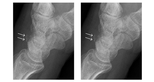
X-rays are the most common imaging method used to confirm a fracture. Lateral (side) views of the coccyx allow doctors to see misalignment or bone fragments. However, small cracks may be hard to detect if swelling obscures the view.
CT scans may be used in more complex cases or when multiple injuries are suspected. They provide a more detailed image of the bones and can help determine whether the injury involves surrounding structures.
Diagnosis of a Broken Tailbone
MRI is useful when soft tissue damage, inflammation, or infection is suspected. It can reveal issues like bruised ligaments or bone marrow swelling that may not be visible on X-ray or CT.
Diagnosis also includes ruling out other causes of lower back or pelvic pain, such as herniated discs, sciatica, or sacroiliac joint dysfunction. A thorough review of posture, lifestyle, and past injuries may also be conducted.
An early and accurate diagnosis of a broken tailbone allows healthcare providers to develop a recovery plan tailored to the patient’s lifestyle, activity level, and overall health.
[Next: Treatment of a Broken Tailbone →]