Mental Matters

Author name: admin

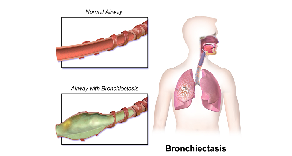
Medical illustration comparing normal airway to an airway affected by bronchiectasis

Bronchiectasis

Bronchiectasis – Overview Bronchiectasis is a chronic lung condition where the airways become permanently widened and damaged. This structural change makes it difficult to clear mucus, leading to repeated infections and long-term breathing difficulties. This condition affects the bronchi, the tubes that carry air in and out of the lungs. Over time, trapped mucus builds up, creating an environment that allows bacteria to thrive, triggering a cycle of inflammation, infection, and further airway damage. The condition can affect anyone but is more commonly found in older adults and people with a history of lung infections or underlying conditions such as cystic fibrosis or immune system disorders. In South Africa, respiratory infections like tuberculosis are a leading contributor to developing it. Bronchiectasis Although bronchiectasis is a progressive disease, early diagnosis and consistent treatment can help control symptoms, slow its progression, and improve overall quality of life. It’s often classified as either localised (affecting one area of the lung) or widespread (affecting multiple lung segments). Treatment aims to keep the lungs clear of mucus, prevent infections, and minimise further lung damage. There is no cure, but regular chest physiotherapy, prescribed antibiotics, and in some cases, bronchodilators or surgery can significantly improve symptom control and lung function. With appropriate care, many patients live full, active lives despite their diagnosis. [Next: Causes of Bronchiectasis →]

Bronchiectasis Read More »

Causes of Bronchiectasis

Causes of Bronchiectasis Causes of Bronchiectasis vary, but all lead to inflammation and damage of the airway walls. The most common causes of bronchiectasis include severe lung infections, immune system problems, allergic reactions, congenital conditions, and obstruction of the airways. In many cases, especially in developing countries, tuberculosis is a major contributor. One of the primary causes is past infection. Lung infections such as pneumonia, whooping cough, or tuberculosis can cause enough inflammation to damage the bronchial walls permanently. This damage leads to scarring and the development of abnormally widened airways where mucus collects. Congenital conditions like cystic fibrosis are a well-known cause. In cystic fibrosis, thick, sticky mucus builds up in the lungs from birth, leading to chronic infection and bronchiectasis. While rare in South Africa, it is a major factor in high-income countries. Immunodeficiency conditions — where the body has trouble fighting infections — also increase the risk. People with HIV, primary immunodeficiencies, or those on immune-suppressing treatments (such as chemotherapy or corticosteroids) may develop frequent lung infections that lead to bronchiectasis. Allergic bronchopulmonary aspergillosis (ABPA) is another cause. This condition results from an allergic reaction to a common fungus called Aspergillus, and it causes airway inflammation and mucus plugging, eventually leading to airway dilation. Causes of Bronchiectasis Obstruction of the airways can also trigger bronchiectasis. Inhaled foreign bodies, tumours, or lymph node pressure can block airflow and create a breeding ground for infection. Over time, these infections can cause lasting damage. In some cases, the exact cause remains unknown — this is referred to as idiopathic bronchiectasis. Understanding the causes of bronchiectasis is essential for effective management. When the root cause is identified early, targeted treatment can reduce progression and prevent recurrent infections that worsen lung damage. [Next: Symptoms of Bronchiectasis →]

Causes of Bronchiectasis Read More »

Symptoms of Bronchiectasis

Symptoms of Bronchiectasis Symptoms of bronchiectasis develop gradually and often mimic other chronic lung conditions, making early diagnosis a challenge. The most common symptoms of bronchiectasis include a persistent productive cough, thick mucus, shortness of breath, frequent chest infections, and general fatigue. These symptoms may worsen during flare-ups or after respiratory infections. The hallmark symptom is a chronic cough that brings up large amounts of sputum (phlegm). The mucus is often thick, sticky, and may be yellow or green — especially during infections. Patients may need to clear their throat and cough frequently throughout the day. Shortness of breath is another key symptom, particularly during physical activity. As the lungs become less efficient, patients may struggle to catch their breath even with moderate exertion. This can limit mobility and affect daily routines. Chest pain or tightness is sometimes reported. This is typically due to the constant coughing or inflammation in the lungs. Some people also experience wheezing, which can be confused with asthma. Recurrent respiratory infections are a significant issue. People with bronchiectasis tend to experience frequent bouts of bronchitis or pneumonia. These infections cause worsening symptoms, more mucus production, and sometimes fever or chills. Symptoms of bronchiectasis Clubbing — a change in the shape of the fingertips or nails — may develop over time in people with long-standing bronchiectasis. It’s a sign of chronic oxygen deficiency in the bloodstream. Fatigue is common and often underestimated. Constant coughing, disrupted sleep, and frequent infections take a toll on energy levels, affecting work, family life, and mental health. Symptoms may worsen during cold weather, after illness, or when exposed to pollutants or smoke. It’s important to monitor these patterns and work with a healthcare provider to manage flare-ups. Identifying symptoms of bronchiectasis early makes it possible to start treatment before lung damage becomes severe. If you or someone you know has a persistent cough with daily mucus, especially after previous lung infections, medical evaluation is strongly recommended. [Next: Diagnosis of Bronchiectasis →]

Symptoms of Bronchiectasis Read More »

Diagnosis of Bronchiectasis

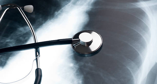
Diagnosis of Bronchiectasis Diagnosis of bronchiectasis involves a combination of medical history, physical examination, imaging, and lab tests. A clear diagnosis of bronchiectasis allows healthcare providers to determine the severity of the disease, identify underlying causes, and begin tailored treatment to reduce long-term damage. The process begins with a detailed history. Doctors will ask about the duration and pattern of symptoms — especially coughing, sputum production, breathlessness, and history of chest infections or childhood illnesses. A history of tuberculosis, pneumonia, or asthma is often relevant. A physical examination may reveal crackles or wheezing sounds when listening to the lungs with a stethoscope. Doctors also look for signs like finger clubbing, chronic infection, or general fatigue. High-resolution CT (HRCT) scans are the gold standard for diagnosis. Doctors use these detailed images to confirm whether the airways are abnormally widened, thickened, or filled with mucus—classic signs of bronchiectasis. Chest X-rays may be done initially but are less sensitive. Lung function tests (spirometry) assess how well the lungs are working. These tests help determine if airway obstruction is present and monitor progression over time. Diagnosis of bronchiectasis Sputum cultures identify which bacteria or fungi are present in the mucus. This guides antibiotic treatment, especially during infections. In some cases, doctors test sputum for tuberculosis or resistant organisms like Pseudomonas aeruginosa. Blood tests may check for signs of infection, immune system function, and underlying conditions like autoimmune diseases or allergic responses (e.g. IgE levels in allergic bronchopulmonary aspergillosis). In children or patients with suspected cystic fibrosis, doctors may perform a sweat chloride test or genetic testing to confirm the diagnosis. An accurate diagnosis of bronchiectasis allows doctors to create the right treatment plan, prevent repeated infections, preserve lung function, and improve quality of life. [Next: Treatment of Bronchiectasis →]

Diagnosis of Bronchiectasis Read More »

Treatment of Bronchiectasis

Treatment of Bronchiectasis Treatment of bronchiectasis focuses on controlling infections, clearing mucus from the lungs, reducing inflammation, and managing any underlying causes. There is no cure, but consistent treatment of bronchiectasis can dramatically improve symptoms and reduce flare-ups. Airway clearance is a central part of treatment. Chest physiotherapy — including postural drainage, percussion, and breathing exercises — helps remove mucus and prevent blockages. Devices such as flutter valves or oscillating PEP (positive expiratory pressure) tools may assist with this process. Antibiotics are commonly prescribed, especially during infections. Oral antibiotics are used for mild infections, while more severe or resistant cases may require intravenous (IV) antibiotics. Some patients benefit from long-term, low-dose antibiotic therapy to reduce flare-up frequency. Bronchodilators and corticosteroids may be used in people with overlapping asthma or chronic obstructive pulmonary disease (COPD). These medications help relax airway muscles and reduce inflammation, improving airflow. For people with thick mucus, mucolytic agents or saline nebulisers can thin the secretions and make it easier to cough them up. Vaccinations play an important role in prevention. Annual flu vaccines and pneumococcal vaccines help protect against respiratory infections that can worsen bronchiectasis. Surgery is rarely required but may be considered in patients with localised bronchiectasis who do not respond to medication. Lung resection may help if only one area of the lung is severely damaged. Treatment of bronchiectasis Pulmonary rehabilitation and regular exercise are also beneficial. They help improve lung function, increase endurance, and reduce shortness of breath. In severe or advanced cases, oxygen therapy or referral for lung transplantation may be considered — although these situations are uncommon. Self-management, including quitting smoking, maintaining hydration, avoiding air pollutants, and treating flare-ups early, is essential for long-term success in treatment of bronchiectasis. [Next: Complications and Management of Bronchiectasis →]

Treatment of Bronchiectasis Read More »

Complications and Management of Bronchiectasis

Complications and Management of Bronchiectasis Complications and management of bronchiectasis go hand in hand. If left untreated, the condition can lead to worsening lung damage, respiratory failure, or serious infections. However, with proactive care, most complications and management of bronchiectasis challenges can be avoided or controlled. One major complication is recurrent lung infections. These infections cause further damage to the airways and increase mucus production, making symptoms worse. Regular sputum monitoring, prompt antibiotics, and good airway hygiene help manage this risk. Pneumonia and respiratory failure are severe complications, particularly in people with widespread lung involvement. These cases may require hospitalisation, oxygen support, and aggressive treatment. Haemoptysis (coughing up blood) may occur due to fragile or damaged blood vessels in the lungs. Minor bleeding can be managed with rest and antibiotics, but heavy bleeding needs urgent medical care and possibly surgery or embolisation. Lung function decline is another concern. If bronchiectasis progresses, breathing becomes harder, oxygen levels drop, and physical activity is affected. Regular spirometry testing and early intervention can slow this process. Complications and management of bronchiectasis In rare cases, abscesses or Pseudomonas colonisation may develop, which are harder to treat and require long-term antibiotic therapy. Chronic fatigue, depression, and reduced quality of life are commonly reported by patients. Support groups, mental health support, and regular exercise play important roles in emotional wellbeing. Management of bronchiectasis involves a combination of medical care and lifestyle changes. Consistent follow-up with a respiratory specialist, adherence to treatment plans, vaccination, and healthy habits are key to long-term control. With proper complications and management of bronchiectasis, most individuals can lead fulfilling lives, minimise hospital visits, and maintain good lung health well into the future. Back to Overview

Complications and Management of Bronchiectasis Read More »

Bruised Ribs

Bruised Ribs

Bruised Ribs – Overview Bruised ribs is an injury where the muscles or cartilage around the ribcage are damaged. Without fracturing the bone itself. A bruised rib is typically the result of blunt trauma. Such as a fall, sports injury, or impact from a seatbelt — and although not as serious as a fracture, it can be extremely painful and take several weeks to fully heal. The ribcage is made up of 12 pairs of ribs that protect vital organs like the lungs and heart. When bruised, the soft tissue between and around these bones becomes inflamed, leading to pain, swelling, and tenderness. The injury may also affect the intercostal muscles, which help with breathing and movement. Bruised ribs are commonly confused with broken ribs due to similar symptoms. However, bruised ribs don’t involve a break in the bone and usually carry less risk of internal complications. That said, deep bruising can still cause difficulty breathing and discomfort during sleep or physical activity. Bruised Rib Typical causes include sports collisions, slipping and falling onto the side of the body, physical assaults. Additionally, even intense coughing fits. Diagnosis is made through physical examination and, if necessary, imaging like X-rays or CT scans to rule out fractures. Treatment focuses on pain management, rest, and gradual return to normal activity. Unlike some injuries, ribs can’t be immobilised with a cast, so healing requires careful avoidance of activities that aggravate the injury. A bruised rib generally takes 3 to 6 weeks to heal, but persistent symptoms may warrant further evaluation to exclude more serious conditions. [Next: Causes of a Bruised Rib →]

Bruised Ribs Read More »

Boxers exchanging punches during a fight, highlighting potential rib injuries

Causes of a Bruised Rib

Causes of a Bruised Rib Causes of a Bruised Rib typically involve blunt trauma to the chest or repeated strain on the ribcage. The most common causes of a bruised rib include falls, sports injuries, car accidents, and excessive coughing. Identifying the source of the injury is crucial for effective treatment and future prevention. Falls are a major cause, especially when someone lands directly on their side or back. Slipping on wet floors, falling off a bicycle, or tumbling down stairs can create enough force to bruise the ribs without breaking them. Sports injuries are also frequent culprits. Contact sports like rugby, hockey, or football often result in collisions or tackles that impact the chest. High-speed sports like cycling and skateboarding also carry risk when a rider falls or crashes into a hard surface. Car accidents are another significant source. A seatbelt can compress the chest during a sudden stop or crash, causing bruising to the ribs and surrounding tissues. While seatbelts save lives, the pressure they apply during impact can still result in soft tissue injury. Repeated physical strain, such as heavy lifting, intense gym training, or repetitive twisting motions, can also bruise the intercostal muscles and soft tissue between the ribs. Rowers, dancers, and weightlifters are especially prone to these types of injuries. Causes of a Bruised Rib Severe or prolonged coughing fits can bruise the ribs too. This is especially common in people with respiratory illnesses like bronchitis, pneumonia, or chronic obstructive pulmonary disease (COPD). The repeated contraction of chest muscles puts pressure on the ribcage, which can lead to bruising — particularly in older adults or those with fragile bones. In rare cases, minor rib injuries may result from underlying medical conditions like osteoporosis or bone cancer, where the ribs are more susceptible to damage. Understanding the causes of a bruised rib can help prevent recurrence. Protective gear during sport, proper lifting technique, treating persistent coughs early, and improving home safety are all helpful preventative strategies. [Next: Symptoms of a Bruised Rib →]

Causes of a Bruised Rib Read More »

Man holding his lower back with spine highlighted in red, indicating pain or injury

Symptoms of a Bruised Rib

Symptoms of a Bruised Rib Symptoms of a Bruised Rib usually develop quickly after injury and are often similar to those of a broken rib, which is why a professional evaluation is important. The most common symptoms of a bruised rib include localised chest pain, tenderness, swelling, and discomfort when breathing or moving the upper body. These symptoms can range from mild to severe, depending on the impact. Pain is the most noticeable symptom. It usually occurs in one specific area of the ribcage and may feel sharp or achy. The pain tends to worsen with movement, deep breathing, coughing, sneezing, or laughing. Some people also experience a burning or throbbing sensation, especially at night or when lying on the affected side. Tenderness over the injured area is common. Light touch, pressure, or wearing tight clothing can trigger pain. Bruising may develop externally as a visible blue or purple discolouration on the skin above the ribs. In deeper injuries, bruising may not be visible, but discomfort remains significant. Swelling around the ribs may appear within hours of the injury. In some cases, the area feels warm or tight. Applying pressure or lying on the bruised side can intensify the discomfort. Symptoms of a Bruised Rib Difficulty breathing deeply is another typical symptom. Although the ribs are not broken, inflammation of the soft tissue limits expansion of the chest, making full breaths painful. This may lead to shallow breathing, which increases the risk of lung infections like pneumonia if left unmanaged. Muscle spasms in the surrounding area may also occur as the body tries to protect the injury. These spasms can cause additional stiffness and reduced mobility in the upper torso. People often mistake symptoms of a bruised rib for pulled muscles or internal organ issues. However, rib bruises tend to produce very localised and consistent pain during specific movements, which helps differentiate them from other conditions. If symptoms persist beyond a few days, worsen, or are accompanied by shortness of breath, blood in coughs, or dizziness, immediate medical evaluation is needed to rule out more serious problems. [Next: Diagnosis of a Bruised Rib →]

Symptoms of a Bruised Rib Read More »

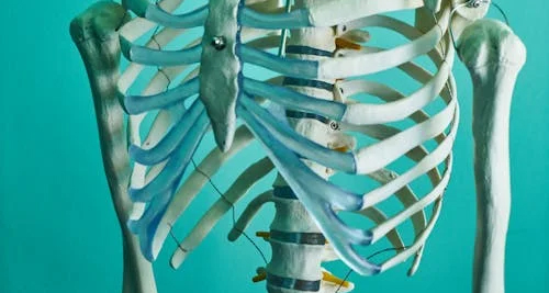
Medical rib cage model showing upper torso bones against a teal background

Diagnosis of a Bruised Rib

Diagnosis of a Bruised Rib Diagnosis of a Bruised Rib relies primarily on a clinical examination, followed by imaging tests if necessary to rule out fractures or complications. A proper diagnosis of a bruised rib helps ensure the correct treatment approach. Additionally, prevents overlooking more serious injuries like lung damage or broken ribs. The doctor begins by asking about the injury. How it happened, when it started hurting, and whether other symptoms such as difficulty breathing or chest tightness are present. A physical examination follows, where the doctor gently presses along the ribcage to find tender or swollen areas. Most bruised ribs don’t require imaging, especially if the injury is mild and there are no signs of complications. However, if the patient has severe pain, signs of breathing difficulty, or is involved in high-impact trauma (like a car accident), further tests may be needed. X-rays are commonly used to rule out broken ribs. Although they do not always detect bruising of soft tissues or muscles, X-rays are useful in eliminating more serious skeletal injuries. CT scans may be ordered in cases involving multiple trauma or if lung or organ damage is suspected. These provide a more detailed look at the chest cavity and can reveal internal bleeding or other complications. Diagnosis of a Bruised Rib An ultrasound may also be used to evaluate bruising of the intercostal muscles or check for fluid build-up around the lungs (pleural effusion). In respiratory cases, oxygen saturation and lung function may be assessed to ensure breathing isn’t compromised. MRI scans are rarely required but may be used to evaluate soft tissue damage if symptoms are prolonged or unexplained. In patients with a history of osteoporosis, cancer, or bleeding disorders. Additionally, blood tests or bone scans may be requested to check for underlying causes or coexisting conditions. An early and accurate diagnosis of a bruised rib prevents unnecessary anxiety and supports a smoother recovery process, especially when the pain mimics that of a more serious internal injury. [Next: Treatment of a Bruised Rib →]

Diagnosis of a Bruised Rib Read More »

Scroll to Top