Mental Matters

Author name: admin

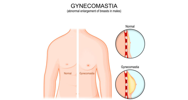
Medical illustration comparing normal male chest with gynaecomastia

Causes of Gynaecomastia

Causes of Gynaecomastia The causes of gynaecomastia are rooted in a hormonal imbalance, typically involving an increase in oestrogen or a decrease in testosterone. Although it may seem alarming, the causes of gynaecomastia are often benign and treatable, especially when identified early. Gynaecomastia, the enlargement of breast tissue in males, can affect newborns, adolescents, and adults, and can arise from a range of natural, medical, or environmental factors. Understanding what causes gynaecomastia is key to diagnosing and treating it properly. The condition happens when hormone levels—especially the balance between estrogen and testosterone—change. These changes may be due to natural aging, certain medications, or underlying health problems. In some cases, lifestyle factors or substance use can also play a role. Importantly, gynaecomastia is not the same as extra fat in the chest, which is called pseudogynaecomastia. However, both conditions can look similar, and sometimes the causes overlap. That’s why it’s important for doctors to look at the whole picture—including hormone levels, medical history, and overall health—when making a diagnosis. By identifying the root cause, healthcare providers can offer the most effective and personalised care. 1. Hormonal Imbalance The human body requires a delicate balance between oestrogen and testosterone. Men naturally produce oestrogen in small amounts, but when the balance tips in favour of oestrogen, breast tissue can develop. Common reasons for hormonal imbalance include: Puberty: Hormonal fluctuations during adolescence are the most frequent cause. Up to 70% of boys experience temporary gynaecomastia, which usually resolves within 6–24 months. Ageing: As men age, testosterone levels decline and fat tissue increases. Fat can convert testosterone into oestrogen via an enzyme called aromatase, leading to breast tissue growth. Obesity: Being overweight raises oestrogen levels and contributes to both true gynaecomastia and pseudogynaecomastia. In most cases, the hormonal imbalance is transient and not linked to serious health issues. However, persistent or painful enlargement should always be evaluated. 2. Medications Several medications are known to alter hormone levels or mimic oestrogen, which can lead to breast tissue growth in men. These include: a. Anti-androgens Used to treat prostate cancer, enlarged prostate, and some skin conditions, drugs like: Flutamide Spironolactone Finasteridecan block testosterone and increase the relative impact of oestrogen. b. Antipsychotics and Antidepressants Drugs such as risperidone, haloperidol, and tricyclic antidepressants may raise prolactin levels, which can stimulate breast tissue growth. c. Cardiovascular drugs Calcium channel blockers, digoxin, and ACE inhibitors have been implicated in some cases, though the mechanism is not always clear. d. Chemotherapy Cancer treatments can disrupt hormone regulation, especially when used in combination with hormone-blocking drugs. e. Ulcer and acid-reflux medications Cimetidine and ranitidine (now less commonly used) have weak oestrogenic effects and can contribute to gynaecomastia with long-term use. Always inform your healthcare provider about all medications and supplements, as switching drugs may help reverse the symptoms. 3. Substance Use Certain substances, legal and illicit, are associated with hormone disruption and breast enlargement: Alcohol: Chronic use affects liver function, reducing the liver’s ability to clear oestrogen from the body. Cannabis: Studies have linked long-term use to reduced testosterone levels and possible oestrogenic effects. Anabolic steroids: Bodybuilders and athletes who use synthetic testosterone may develop gynaecomastia during the “off-cycle” period, when natural testosterone drops but oestrogen levels remain elevated. Heroin and amphetamines: These may suppress testosterone production through central nervous system effects. Avoiding or discontinuing these substances often leads to improvement, although long-standing gynaecomastia may persist without surgical intervention. 4. Health Conditions Several medical conditions can disturb hormonal balance, leading to breast tissue development in males: a. Liver disease Chronic liver disorders, including cirrhosis, impair oestrogen breakdown and increase its levels in the bloodstream. Conditions such as non-alcoholic fatty liver disease also contribute indirectly via obesity. b. Kidney failure Long-term dialysis can cause hormonal shifts, particularly with reduced testosterone production and elevated prolactin. c. Hyperthyroidism An overactive thyroid gland increases sex hormone-binding globulin (SHBG), which binds testosterone and leaves more unbound oestrogen active in the body. d. Pituitary tumours Some tumours produce excess prolactin, a hormone that stimulates breast tissue development in both sexes. e. Hypogonadism Primary testicular failure, whether genetic (e.g., Klinefelter syndrome) or acquired, results in reduced testosterone and higher oestrogen effects. Damage from mumps, injury, or radiation can also lead to low testosterone and subsequent gynaecomastia. In all cases, treating the underlying health issue is crucial for resolving the breast enlargement and preventing recurrence. 5. Tumours and Hormone-Secreting Growths Although rare, tumours that secrete hormones may directly increase oestrogen or decrease testosterone: Testicular tumours (e.g., Sertoli or Leydig cell tumours) Adrenal gland tumours HCG-producing tumours (commonly seen in lung, liver, or stomach cancers) These require urgent assessment and imaging studies. A testicular ultrasound or abdominal scan may be ordered if a mass is suspected. 6. Herbal Products and Supplements Some plant-based products contain phytoestrogens, compounds that mimic oestrogen in the body. Common examples include: Tea tree oil and lavender oil (topical use has been linked to breast growth in boys) Soy-based products (usually in high, concentrated amounts) Herbal supplements, such as those used for body enhancement or testosterone boosting, may contain undeclared hormone-active ingredients The risk is higher when products are used frequently or over long periods. 7. Idiopathic Gynaecomastia In some cases, no clear cause is found — this is termed idiopathic gynaecomastia. While frustrating, this form is often harmless and may stabilise or regress over time. Nonetheless, ongoing monitoring and exclusion of serious causes are essential, especially in adult males or post-pubertal boys with persistent symptoms. Conclusion | Causes of Gynaecomastia The causes of gynaecomastia come from many different sources. Often, it begins with natural changes in hormone levels—especially during puberty or older age. However, other factors can also play a role. For example, certain medications, such as those for high blood pressure or ulcers, may lead to breast tissue growth. In addition, long-term health problems like liver or kidney disease can affect hormone balance. Substance use, including alcohol, anabolic steroids, or marijuana, can also increase the risk. Fortunately, in most cases, gynaecomastia goes away once

Causes of Gynaecomastia Read More »

Man covering chest due to breast swelling or tenderness

Symptoms of Gynaecomastia

Symptoms of Gynaecomastia The symptoms of gynaecomastia can vary from mild breast enlargement to significant swelling and tenderness, affecting one or both breasts. While the physical appearance is the most noticeable feature, the symptoms of gynaecomastia can also include pain, emotional discomfort, and psychological distress—especially in adolescents and adult men. Gynaecomastia is defined as the benign enlargement of male breast tissue caused by hormonal imbalances, typically involving increased oestrogen activity or decreased androgens. It can affect newborns, adolescents, and older men, and its symptoms may range from barely perceptible to significantly distressing, both physically and emotionally. Common Physical Symptoms The physical signs of gynaecomastia are usually the most obvious, and often include: Firm or rubbery tissue underneath the nipple area Symmetrical or asymmetrical breast enlargement (one-sided or both) Tenderness or mild pain in the affected breast A disc-like mound of tissue beneath the areola (the dark area around the nipple) In some cases, visible swelling that resembles female breast development Unlike fat tissue associated with obesity, the glandular tissue in gynaecomastia is firmer and more nodular to the touch. The tissue is usually located centrally under the nipple and does not extend broadly like adipose tissue in pseudogynaecomastia. Unilateral vs Bilateral Symptoms Unilateral gynaecomastia (one breast only) occurs in about one-third of cases Bilateral gynaecomastia (both breasts) is more common and may occur evenly or unevenly Asymmetry often causes additional anxiety, as men may fear cancer or another underlying disease While breast cancer in men is rare, any lump that is hard, fixed, or associated with nipple discharge warrants immediate medical evaluation. Pain and Tenderness Pain is not always present, but when it occurs, it may include: A dull ache or sensitivity Discomfort when pressure is applied, such as when lying on the chest or wearing tight clothing Tender nipples, which may be irritated by contact with fabrics Pain is typically more noticeable during the growth phase of gynaecomastia, when hormonal fluctuations are most active. Emotional and Psychological Effects The symptoms of gynaecomastia go beyond the physical. Many men and boys experience: Body image concerns Shame or embarrassment, particularly during social or intimate situations Avoidance of activities like swimming or changing in public locker rooms Low self-esteem, especially during adolescence Depression or anxiety, in severe or long-standing cases In teenagers, these psychological symptoms may significantly impact quality of life, school participation, and peer relationships. Nipple Changes Although rare, some men notice: Nipple protrusion or change in shape Increased pigmentation or sensitivity In isolated cases, clear or milky discharge, which should be evaluated further Any bloody or spontaneous nipple discharge must be promptly assessed to rule out malignancy. Distinguishing Features of True Gynaecomastia To confirm that symptoms are consistent with true gynaecomastia and not fat-related enlargement (pseudogynaecomastia), key signs include: A firm mass under the nipple Tissue that does not change significantly with weight loss No signs of infection or inflammation In contrast, pseudogynaecomastia (caused by excess body fat) tends to be soft, evenly distributed, and responsive to changes in diet and exercise. Progression and Duration of Symptoms Gynaecomastia can be: Transient, especially in newborns and teenagers Persistent, in cases related to medications, liver disease, or ageing Progressive, if underlying causes are not addressed Symptoms often resolve spontaneously within 6 to 24 months in adolescents but may persist into adulthood if not evaluated. When to Seek Medical Advice It is important to consult a healthcare provider if: The swelling is rapid or painful There is nipple discharge or skin dimpling Only one side is affected Symptoms cause emotional distress There is a family history of breast cancer or testicular disease These features help differentiate benign gynaecomastia from more serious conditions such as tumours or hormonal disorders. Conclusion | Symptoms of Gynaecomastia The symptoms of gynaecomastia include not only visible breast enlargement but also tenderness, nipple sensitivity, and psychological discomfort. Understanding the full range of symptoms of gynaecomastia is essential for early recognition, appropriate diagnosis, and compassionate management. For many men, timely reassurance and evaluation can ease physical discomfort and restore confidence. [Next: Diagnosis of Gynaecomastia →]

Symptoms of Gynaecomastia Read More »

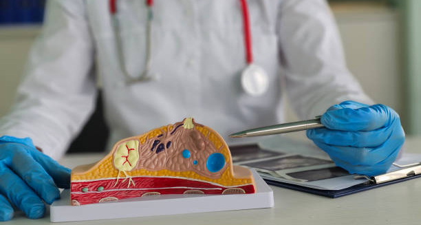
Doctor pointing at anatomical model of male breast tissue

Diagnosis of Gynaecomastia

Diagnosis of Gynaecomastia The diagnosis of gynaecomastia involves a combination of clinical examination, patient history, and targeted testing to rule out underlying causes and confirm the presence of glandular breast tissue. Because the diagnosis of gynaecomastia can sometimes overlap with other conditions, especially pseudogynaecomastia or male breast cancer, a thorough and careful assessment is essential. Gynaecomastia is a clinical diagnosis in most cases, but tests may be needed to determine the underlying cause, especially in persistent, asymmetric, or painful cases. Understanding the diagnostic process can help reassure patients and guide appropriate treatment decisions. 1. Clinical History A detailed patient history provides valuable insight into the potential causes of gynaecomastia. Doctors will typically ask about: Age of onset and duration of symptoms Rate of progression (sudden vs slow development) Presence of pain or tenderness Medication history, including prescription and recreational drugs Use of supplements or anabolic steroids Alcohol consumption History of chronic illness, such as liver, kidney, or thyroid disorders Sexual health and fertility issues, including erectile dysfunction or testicular size The goal is to identify hormonal imbalances or external factors contributing to glandular breast development. 2. Physical Examination A focused physical exam is essential. It should assess: Size and consistency of breast tissue Whether the tissue is firm, glandular, or soft and fatty Location of enlargement (true gynaecomastia is central under the nipple) Signs of inflammation, infection, or discharge Testicular examination to check for masses or asymmetry Assessment of secondary sexual characteristics, such as body hair, muscle mass, and genital development True gynaecomastia is distinguished from pseudogynaecomastia by the presence of firm, disc-like tissue beneath the nipple, while fat-related enlargement tends to be soft and diffuse. 3. Blood Tests If the diagnosis is uncertain or if there are signs of hormonal imbalance, blood tests may be performed to evaluate: Oestrogen and testosterone levels Liver function tests (to check for cirrhosis or hepatic disease) Kidney function tests Thyroid function (TSH and T4) Luteinising hormone (LH) and follicle-stimulating hormone (FSH) Prolactin Beta-hCG and AFP, which may be elevated in testicular or adrenal tumours These tests help uncover systemic or endocrine causes, such as hypogonadism, tumours, or hyperthyroidism. 4. Imaging Studies a. Breast Ultrasound Helps distinguish gynaecomastia from male breast cancer Identifies glandular tissue vs cysts or masses Useful in cases of unilateral enlargement or hard masses b. Mammography Used if malignancy is suspected (especially in older men) Detects irregular, spiculated lesions characteristic of cancer c. Testicular Ultrasound Ordered if physical exam reveals testicular irregularities Screens for germ cell tumours, which may produce hormones that affect breast tissue 5. Staging and Classification Gynaecomastia can be classified based on severity: Staging helps determine the need for surgery or other interventions. 6. Differential Diagnosis The diagnosis of gynaecomastia must rule out other causes of breast enlargement or masses in men: Condition Key Distinction Pseudogynaecomastia Fatty tissue only, no firm glandular mass Male breast cancer Hard, unilateral lump, possible discharge Mastitis or abscess Painful, red, inflamed swelling Lipoma Soft, mobile lump away from nipple Dermatological lesions Localised skin thickening or cysts In cases where a malignant tumour is suspected, referral to a breast or oncology specialist is essential. 7. Adolescent Gynaecomastia In teenage boys, no testing is usually required unless: The tissue is persisting longer than 2 years There is significant pain or asymmetry The breast tissue is rapidly growing Other signs of endocrine disruption are present Most adolescent cases resolve spontaneously and do not require further investigation. When to Refer Referral to an endocrinologist, breast specialist, or surgeon is appropriate if: There is a suspicion of cancer Blood tests reveal hormonal abnormalities The patient desires surgical correction for cosmetic or psychological reasons Gynaecomastia is causing severe emotional distress Conclusion | Diagnosis of Gynaecomastia The diagnosis of gynaecomastia begins with a detailed medical history. First, the doctor asks about symptoms, medications, and any underlying health issues. Next, they perform a physical exam to check for breast tissue growth and rule out other causes. In some cases, blood tests help identify hormone imbalances. Imaging, such as ultrasound or mammography, may also be used to look more closely at the breast tissue. It is important to tell true gynaecomastia apart from conditions like pseudogynaecomastia, which is caused by fat buildup, or rare but serious issues like breast cancer. By making a clear diagnosis, doctors can give the right advice and choose the best treatment. With an accurate diagnosis, patients receive not only medical care but also reassurance. As a result, they can better understand their condition and feel more confident moving forward. [Next: Treatment of Gynaecomastia→]

Diagnosis of Gynaecomastia Read More »

Doctor preparing for male breast reduction surgery

Treatment of Gynaecomastia

Treatment of Gynaecomastia The treatment of gynaecomastia depends on the cause, duration, severity, and impact on a person’s physical and emotional wellbeing. In many cases, the treatment of gynaecomastia is not required, as the condition may resolve on its own. However, persistent or distressing cases often warrant medical or surgical intervention to address symptoms and restore confidence. Gynaecomastia is the benign enlargement of male breast tissue due to hormonal imbalance, particularly an increased ratio of oestrogen to testosterone. It can affect males of all ages — from newborns to older men — and may be unilateral or bilateral. While not life-threatening, the psychological impact can be significant. 1. Observation and Reassurance In many adolescents and some adults, gynaecomastia is: Physiological (a normal part of development) Transient, resolving within 6 to 12 months Associated with no underlying disease In such cases, doctors may recommend: Monitoring without intervention Regular follow-up to ensure the condition is improving or stabilising Reassurance that it is common and often temporary This approach is especially appropriate for teenage boys during puberty, where as many as 60% may experience some degree of breast enlargement. 2. Addressing Underlying Causes Secondary gynaecomastia caused by medical conditions or medications requires treatment of the root issue. This includes: Discontinuing or replacing drugs known to cause gynaecomastia (e.g. spironolactone, anti-androgens, anabolic steroids) Managing hormonal disorders, such as hypogonadism or hyperthyroidism Treating liver or kidney disease that may alter hormone metabolism Stopping the use of recreational drugs such as cannabis or alcohol Doctors will often perform blood tests to assess hormone levels and organ function when secondary causes are suspected. 3. Medical Therapy Medical treatment is most effective in the early, active phase of gynaecomastia, when breast tissue is still proliferating (within 6–12 months of onset). a. Selective Oestrogen Receptor Modulators (SERMs) Tamoxifen is commonly used to reduce breast tissue size and tenderness Works by blocking oestrogen receptors in breast tissue Often used off-label with encouraging results Generally well tolerated but may cause fatigue or hot flushes b. Aromatase Inhibitors Anastrozole or letrozole reduce oestrogen production Less effective than tamoxifen for established gynaecomastia Reserved for specific cases with hormonal abnormalities c. Androgens or Testosterone Therapy May help in cases of testosterone deficiency Only used if there is a clear diagnosis of hypogonadism Should be carefully monitored to avoid adverse effects Medical therapy is rarely effective in long-standing fibrotic gynaecomastia, where the tissue becomes firm and unresponsive to hormonal treatment. 4. Surgical Treatment Surgery is considered the definitive treatment of gynaecomastia, particularly in cases of: Persistent enlargement despite medical therapy Psychological distress or body image concerns Pain or tenderness Excess skin or large-volume tissue, especially in older adults or those with massive weight loss There are two main surgical options: a. Liposuction Ideal for cases with fat-dominant breast tissue Involves suctioning excess fat through a small incision Less invasive, quicker recovery Limited ability to remove glandular tissue b. Excisional Surgery (Mastectomy) Involves direct removal of glandular tissue via a small incision around the areola Sometimes combined with liposuction for optimal contouring More effective for firm, fibrous, or long-standing tissue Recovery from surgery typically involves: Wearing compression garments Managing swelling and bruising Returning to work within 1 to 2 weeks Scarring is minimal and usually fades over time. Most patients report high satisfaction post-surgery. 5. Psychological and Supportive Care The emotional effects of gynaecomastia should not be underestimated. Many affected individuals experience: Low self-esteem Social withdrawal Avoidance of sports or swimming Anxiety or depression Supportive strategies include: Counselling or cognitive behavioural therapy (CBT) Support groups for men with body image issues Education about the condition to reduce stigma Treating the psychological aspects can be just as important as addressing the physical symptoms. 6. Lifestyle Measures Certain lifestyle adjustments may support overall management: Weight loss and exercise can reduce chest fat and improve appearance, though they do not affect glandular tissue Avoiding alcohol and drugs known to impact hormone levels Healthy eating to support hormone balance and body confidence However, it’s important not to falsely promise resolution through diet and exercise alone when true glandular gynaecomastia is present. Conclusion | Treatment of Gynaecomastia The treatment of gynaecomastia can range from simple observation to medical and surgical interventions, depending on the cause and severity. While many cases resolve naturally, persistent or emotionally distressing gynaecomastia may require hormone therapy or surgery. A holistic approach to the treatment of gynaecomastia, addressing both physical and psychological aspects, offers the best outcomes for patients seeking relief and restoration of confidence. [Next: Complications of Gynaecomastia →]

Treatment of Gynaecomastia Read More »

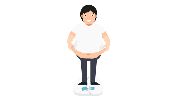
Overweight man standing on cracked scale

Complications of Gynaecomastia

Complications of Gynaecomastia The complications of gynaecomastia are primarily psychological, but physical and clinical concerns can also arise in persistent or misdiagnosed cases. Although gynaecomastia is not a life-threatening condition, the complications of gynaecomastia can significantly affect quality of life, especially when symptoms are severe, long-standing, or overlooked. Understanding these potential consequences is essential for clinicians and patients alike to guide management decisions and avoid unnecessary distress. 1. Psychological and Social Complications Perhaps the most common — and often underestimated — complications are emotional in nature. Men and boys with gynaecomastia frequently report: Embarrassment and shame, especially in public changing areas Body image dissatisfaction, often leading to social withdrawal Bullying or teasing, particularly during adolescence Avoidance of activities like swimming, sport, or intimacy Depression, anxiety, or social phobia These psychological effects may persist long after the physical condition resolves and can interfere with education, relationships, and personal development. 2. Chronic Pain or Tenderness Some individuals experience: Persistent breast pain or sensitivity Discomfort when lying on the chest or wearing fitted clothing Nipple tenderness or itching This is more common when gynaecomastia is due to hormone fluctuations or medications, and may fluctuate in severity. Pain can disrupt sleep, concentration, or exercise routines, adding to emotional strain. 3. Skin and Tissue Changes Long-standing or significant breast enlargement can lead to: Stretching of skin and areolae Sagging or drooping (ptosis) of the breast tissue Excess skin that may remain even after weight loss or regression These cosmetic issues may persist even if the underlying glandular tissue shrinks, prompting some men to seek surgical correction. 4. Surgical Complications (When Applicable) Although surgery is generally safe, as with any procedure, risks include: Bleeding or haematoma formation Infection Scarring, including hypertrophic or keloid scars Asymmetry or contour irregularities Changes in nipple sensation (temporary or permanent) Complications can be minimised by choosing a qualified surgeon, following postoperative instructions, and attending follow-up visits. 5. Medication Side Effects For those using pharmacological treatments like tamoxifen or aromatase inhibitors: Side effects may include fatigue, hot flushes, headaches, or libido changes Long-term safety of off-label medications is still under study Monitoring and communication with a healthcare provider are essential Discontinuation of therapy due to side effects may also lead to a return of symptoms, particularly if the underlying cause is not addressed. 6. Missed or Delayed Diagnosis of Serious Conditions One of the most important complications is the potential for missed diagnosis of an underlying issue: Testicular tumours or adrenal tumours that secrete oestrogens Chronic liver disease, which alters hormone metabolism Kidney failure or thyroid disorders Rare cases of male breast cancer Assuming all gynaecomastia is benign can delay detection of these serious diseases. Red flags that require further investigation include: Unilateral breast enlargement Hard, fixed lumps Nipple discharge or skin dimpling Lymph node swelling Proper diagnostic work-up, including imaging and hormonal testing, is crucial when atypical features are present. 7. Reduced Physical Performance or Activity In athletes or those with active lifestyles, gynaecomastia can: Interfere with upper body movements due to discomfort Cause self-consciousness during team sports Lead to avoidance of gym or outdoor activities These issues can contribute to decreased fitness, which may further affect confidence and hormone regulation. 8. Ongoing Recurrence After Resolution In some cases: Gynaecomastia recurs even after initial resolution May follow a return to causative medications or substance use Hormonal imbalances may fluctuate, especially in older adults or those with underlying endocrine conditions This can create a cycle of repeated distress and treatment attempts, especially if the underlying trigger is not addressed fully. Conclusion | Complications of Gynaecomastia The complications of gynaecomastia extend beyond physical symptoms, encompassing a wide range of emotional, psychological, and medical challenges. While most cases are benign and treatable, untreated or misdiagnosed gynaecomastia can significantly impact quality of life. By recognising and addressing the complications of gynaecomastia, healthcare professionals can provide holistic care that prioritises both physical outcomes and emotional wellbeing. [Next: Back to Overview →]

Complications of Gynaecomastia Read More »

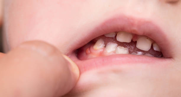
Close-up of inflamed gums in a child’s mouth

Gum Disease

Gum Disease Gum Disease is a common but preventable condition that affects the soft tissues surrounding the teeth. Often caused by poor oral hygiene, Gum Disease begins as inflammation of the gums (gingivitis) and can progress to more serious forms, such as periodontitis, if left untreated. Early detection and consistent care are essential for preserving both oral and overall health. While many people experience some level of gum irritation in their lifetime, advanced Gum Disease can lead to tooth loss, jawbone damage, and has even been linked to systemic conditions like heart disease and diabetes. Understanding its symptoms, causes, and treatment options is key to maintaining healthy gums and a strong smile. What Is Gum Disease? Gum Disease refers to inflammation and infection of the gingival tissue (gums) that support and surround the teeth. It progresses in stages: 1. Gingivitis The earliest and mildest form Caused by plaque build-up at the gumline Gums appear red, swollen, and may bleed during brushing or flossing Reversible with proper oral hygiene 2. Periodontitis Occurs when gingivitis is left untreated Plaque hardens into tartar (calculus), causing deeper infection Gums pull away from the teeth, forming periodontal pockets Involves loss of supporting bone and tissues Can result in loose teeth or tooth loss Periodontitis itself can be classified further into mild, moderate, and severe stages depending on the depth of infection and bone involvement. Who Is at Risk? Anyone can develop this disease, but certain risk factors increase susceptibility: Poor oral hygiene Smoking or tobacco use Diabetes, especially if uncontrolled Hormonal changes, including pregnancy or menopause Genetic predisposition Stress, which weakens immune function Poor nutrition, particularly deficiencies in vitamin C Medications that cause dry mouth or affect gum tissue (e.g. anti-epileptics, calcium channel blockers) Even people who brush daily can develop this disease if they neglect proper flossing or professional dental cleanings. Signs and Symptoms The most common signs of Gum Disease include: Red, swollen gums Bleeding when brushing or flossing Bad breath (halitosis) Receding gums Sensitive or loose teeth Pain when chewing Changes in bite or fit of dentures In its early stages, Gum Disease may be painless, which makes regular dental check-ups essential for early detection. How Gum Disease Affects Overall Health Recent research has linked this disease with various systemic health conditions: Heart disease: Chronic inflammation may contribute to arterial plaque and increase heart attack risk Diabetes: Gum infections can worsen blood sugar control, while diabetes increases the risk of periodontitis Pregnancy complications: Gum Disease has been associated with preterm birth and low birth weight Respiratory illness: Oral bacteria may be inhaled, increasing the risk of pneumonia or bronchitis Rheumatoid arthritis and cognitive decline have also been studied in relation to chronic gum inflammation These associations highlight the importance of treating this disease not just as a dental issue, but a whole-body health concern. Prevention Prevention is the most effective defence against Gum Disease. Daily habits should include: Brushing teeth twice a day using fluoride toothpaste Flossing or using interdental brushes once a day Regular dental check-ups and professional cleanings every 6 months Avoiding tobacco products Managing chronic conditions like diabetes Eating a balanced diet rich in vitamins and low in sugar For individuals at higher risk, more frequent dental visits and additional home care may be recommended. Conclusion | Gum Disease Gum Disease is both widespread and preventable, beginning as mild gum inflammation and potentially leading to serious oral and systemic health problems. Through daily oral hygiene, professional care, and early detection, individuals can manage or entirely avoid the complications of Gum Disease. Understanding its stages and risk factors empowers people to take proactive steps toward lifelong dental health. [Next: Causes of Gum Disease →]

Gum Disease Read More »

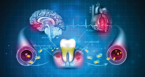
Illustration linking gum disease to brain, heart, and blood vessel health issues

Causes of Gum Disease

Causes of Gum Disease The causes of Gum Disease are primarily bacterial, though a variety of lifestyle, health, and genetic factors influence its development. At its core, the causes of Gum Disease begin with plaque — a sticky film of bacteria that forms on the teeth — but progression depends on how the body responds and how well oral hygiene is maintained. Understanding these causes allows for better prevention, early detection, and effective treatment of both gingivitis and more advanced periodontal disease. 1. Dental Plaque: The Root Cause The most direct cause of Gum Disease is plaque accumulation. Plaque is: A soft, colourless film of bacteria Formed when bacteria feed on sugars and starches in the mouth Easily removed with brushing and flossing If not removed, plaque: Hardens into tartar (calculus) within 24 to 72 hours Tartar cannot be removed with brushing and must be eliminated by a dentist Acts as a reservoir for bacteria, triggering gum inflammation This is the starting point of gingivitis, the earliest form of Gum Disease. 2. Smoking and Tobacco Use Tobacco is one of the strongest risk factors for Gum Disease. It: Reduces blood flow to the gums, impairing healing Weakens the body’s immune response Increases tartar build-up and bacterial colonisation Masks symptoms such as bleeding gums, delaying diagnosis Smokers are up to seven times more likely to develop periodontitis than non-smokers, and outcomes from treatment tend to be poorer. 3. Poor Oral Hygiene Habits Infrequent or incorrect brushing and flossing allow plaque to: Accumulate near the gumline Enter periodontal pockets Progress into deeper layers of gum and bone tissue Neglecting routine dental check-ups and cleanings further accelerates the problem, as tartar build-up goes unnoticed. 4. Hormonal Changes Certain hormonal shifts can increase gum sensitivity and inflammation: Puberty Pregnancy Menstruation Menopause During these times, gums may respond more strongly to plaque, resulting in temporary or chronic inflammation if not managed properly. 5. Diabetes and Other Chronic Illnesses Uncontrolled diabetes significantly increases the risk of Gum Disease due to: Impaired blood circulation Reduced resistance to infection Elevated glucose in saliva, feeding bacteria Other chronic conditions such as HIV/AIDS, leukaemia, and autoimmune diseases may also heighten vulnerability due to immune suppression. 6. Medications Several medications can contribute to Gum Disease, either by causing dry mouth or affecting gum tissue. These include: Antihistamines Antidepressants Calcium channel blockers (used for blood pressure) Anti-epileptics (like phenytoin) Reduced saliva flow increases the risk of plaque accumulation, while some drugs can cause gum overgrowth (gingival hyperplasia), making oral hygiene more difficult. 7. Nutritional Deficiencies A diet lacking in essential nutrients can impair gum health. Key nutrients include: Vitamin C: Deficiency may cause bleeding gums and slow healing Calcium and vitamin D: Important for bone and tooth strength Antioxidants: Help control inflammation Processed foods high in sugar and low in fibre contribute to plaque formation and bacterial imbalance in the mouth. 8. Stress and Immune Function Chronic stress affects gum health by: Weakening the immune system’s ability to fight infection Increasing levels of cortisol, which may worsen inflammation Leading to poor oral habits, like teeth grinding or neglecting hygiene People under prolonged stress may also delay dental visits, compounding the problem. 9. Genetics Genetics can influence a person’s susceptibility to Gum Disease. Some individuals have: A genetic predisposition to stronger inflammatory responses A family history of early tooth loss or aggressive periodontitis Variations in immune response genes Knowing this history allows dentists to monitor at-risk patients more closely. 10. Bruxism (Teeth Grinding) Bruxism does not cause Gum Disease directly, but: It puts excessive pressure on teeth and gums May exacerbate existing gum inflammation Increases the likelihood of gum recession and bone loss in periodontitis Custom mouthguards and stress management are often recommended for individuals who grind their teeth. Summary Table of Causes Cause Mechanism Plaque and tartar Direct bacterial invasion of gum tissue Smoking Reduces healing and immune defence Poor oral hygiene Allows progression from plaque to periodontitis Hormonal changes Increases gum sensitivity to plaque Diabetes Impairs healing, raises infection risk Medications Cause dry mouth or gum overgrowth Poor nutrition Delays recovery and weakens tissue integrity Stress Alters immune function and oral habits Genetics Increases inflammatory response Bruxism Worsens tissue breakdown in affected gums Conclusion | Causes of Gum Disease The causes of gum disease go far beyond poor brushing. Of course, plaque buildup is the main cause. However, many other factors can make the disease worse or speed up its progress. For example, smoking reduces blood flow to the gums, making it harder for them to heal. In addition, high stress levels can weaken the immune system, leaving the gums more vulnerable to infection. Medical conditions like diabetes also raise the risk, especially when blood sugar is not well controlled. Furthermore, some people may be more likely to get gum disease because of their genes. Even with good brushing habits, they may still need extra care. By learning about all the possible causes, people can take stronger steps to protect their gums. This includes not only good oral hygiene, but also quitting smoking, managing health conditions, and getting regular dental check-ups. In the end, understanding these risks helps you care for both your mouth and your overall health. [Next: Symptoms of Gum Disease→]

Causes of Gum Disease Read More »

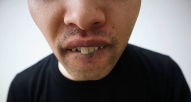
Man with visible gum issues and missing teeth showing symptoms of gum disease

Symptoms of Gum Disease

Symptoms of Gum Disease The symptoms of gum disease often begin subtly, with many individuals unaware of the condition until it becomes more advanced. In the early stages, the symptoms of gum disease may be limited to redness or mild bleeding of the gums, but if left untreated, they can progress to significant tissue damage, tooth loss, and even systemic health complications. Gum disease, medically known as periodontal disease, is a chronic inflammatory condition affecting the soft tissue and bone that support the teeth. It typically develops in stages, starting with gingivitis (reversible gum inflammation) and potentially advancing to periodontitis (irreversible damage to gum and bone structures). Recognising the signs early allows for more conservative and effective treatment. Early Symptoms (Gingivitis) In its initial stage, gum disease is usually painless, but several key warning signs may appear: Red or swollen gums Bleeding during brushing or flossing Persistent bad breath (halitosis) Mild gum tenderness Gums that appear shiny or overly smooth At this stage, the inflammation is limited to the gums and can usually be reversed with improved oral hygiene and professional cleaning. However, without intervention, gingivitis can escalate. Moderate to Severe Symptoms (Periodontitis) As gum disease progresses into periodontitis, more severe and noticeable symptoms emerge: Receding gums, making teeth appear longer Formation of deep pockets between teeth and gums Loose or shifting teeth Pus discharge from the gums Pain while chewing Sensitivity to hot or cold foods These signs indicate structural damage beneath the gumline, including the loss of connective tissue and alveolar bone that support teeth. Gum Recession One of the hallmark features of advancing gum disease is gum recession. The gums pull away from the teeth, exposing the roots and increasing sensitivity. Receding gums also: Compromise the seal around teeth Create deeper spaces for bacteria to thrive Make teeth more prone to decay and sensitivity In some cases, gum recession may be the first visible sign a person notices. Bleeding Gums While bleeding gums may seem minor, they are not normal and should never be ignored. This symptom often indicates: Chronic inflammation The presence of plaque or tartar at the gumline Inadequate flossing or brushing techniques Persistent bleeding is usually the body’s response to infection and should prompt dental evaluation. Halitosis (Chronic Bad Breath) Bad breath caused by gum disease stems from: Bacterial activity in plaque Decaying food particles trapped in gum pockets Infected or necrotic gum tissue Unlike transient halitosis due to food or dehydration, gum disease-related halitosis tends to be ongoing and resistant to mouthwash or mints. Tooth Mobility As the condition worsens: Bone supporting the teeth deteriorates Ligaments and connective tissues weaken Teeth may shift, wobble, or fall out Tooth mobility is a late-stage sign that indicates the disease has already caused significant damage. Systemic Signs and Associations Although the focus is on the mouth, some systemic symptoms may be linked to gum disease, especially in advanced cases: Low-grade fever (in severe infection) Fatigue or malaise due to chronic inflammation Increased risk for diabetes complications, heart disease, and pregnancy issues Research suggests a strong correlation between periodontitis and systemic health due to the constant low-level release of inflammatory markers and bacteria into the bloodstream. When to See a Dentist You should schedule an immediate dental check-up if you experience: Bleeding gums not caused by injury Gum recession or visible tooth roots Persistent bad breath despite hygiene Painful, swollen, or pus-filled gums Teeth feeling loose or changing position Early intervention can prevent irreversible damage and reduce the need for surgery or tooth replacement. Conclusion | Symptoms of Gum Disease The symptoms of gum disease begin subtly but can escalate quickly without appropriate care. From bleeding and swelling to receding gums and loose teeth, recognising the symptoms of gum disease in its early stages offers the best chance for full recovery. Routine dental visits and attentive oral hygiene are essential to detect and address problems before they become permanent. [Next: Diagnosis of Gum Disease →]

Symptoms of Gum Disease Read More »

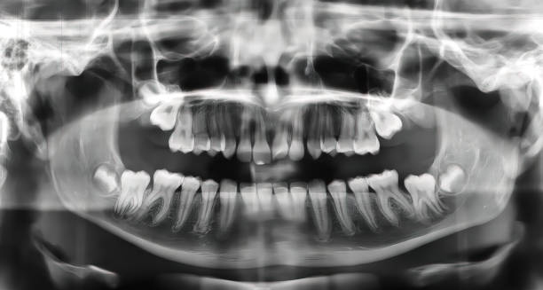
Dental X-ray showing bone loss and periodontal issues related to gum disease

Diagnosis of Gum Disease

Diagnosis of Gum Disease The diagnosis of gum disease relies on a combination of clinical evaluation, dental history, and imaging techniques. Since the early signs are often painless and easily missed, regular dental check-ups play a vital role in the timely diagnosis of gum disease, allowing for early treatment and prevention of further complications. Gum disease can be silent in its early stages, which is why many people are unaware they have it. Dentists use systematic assessments to identify inflammation, attachment loss, and bone damage — all crucial in evaluating how far the disease has progressed. Clinical Examination The first step in diagnosing gum disease is a detailed oral examination, during which the dentist or hygienist will: Assess the colour and contour of the gums Check for swelling, bleeding, or tenderness Look for recession of gum tissue Identify calculus (tartar) and plaque build-up These visible signs offer early clues to inflammation and infection. Periodontal Probing One of the most important diagnostic tools is the periodontal probe, a calibrated instrument used to measure the depth of the space between the tooth and surrounding gum tissue (sulcus or pocket). A healthy sulcus measures 1–3 mm deep Deeper pockets (4 mm or more) may indicate periodontitis Bleeding on probing suggests active inflammation Measurements are taken in six places around each tooth The findings are documented in a periodontal chart, providing a baseline for tracking disease progression or treatment success. X-rays (Radiographs) Dental X-rays help evaluate the extent of bone loss — a hallmark of periodontitis. Horizontal bone loss appears as an even reduction along the jawbone Vertical bone loss indicates more localised or advanced destruction X-rays also reveal tooth root abnormalities, impacted tartar, and hidden decay Bitewing and periapical radiographs are commonly used in periodontal assessments. Medical and Dental History Your dentist will review: Previous dental issues such as frequent bleeding or loose teeth Oral hygiene habits, including brushing and flossing routines Lifestyle factors, such as smoking, diet, and stress Medical history, particularly conditions like diabetes, which increases susceptibility Understanding these factors helps determine both risk and likely disease progression. Bacterial Testing and Biomarkers (Optional) In more complex or advanced cases, especially where traditional treatment has failed, additional tests may be ordered: Microbial DNA testing to identify aggressive bacteria Salivary biomarker testing for inflammatory markers (e.g. MMP-8) Genetic testing for predisposition to gum disease These tools are typically used in specialised or research-based settings and are not required for standard diagnosis. Classification of Severity The current classification of gum disease, as defined by the 2017 World Workshop on the Classification of Periodontal and Peri-Implant Diseases, considers: Stage (I–IV) based on severity, bone loss, and tooth loss Grade (A–C) based on the rate of progression and risk factors This helps clinicians: Design an appropriate treatment plan Communicate the prognosis to the patient Monitor for recurrence or deterioration Diagnosing Gum Disease in Children and Adolescents Though less common, gum disease can occur in younger individuals, particularly those with: Poor oral hygiene Orthodontic appliances (braces) Underlying systemic conditions (e.g. leukaemia, diabetes) Paediatric diagnosis follows similar principles but may also involve coordination with medical specialists. Differentiating from Other Conditions The diagnosis of gum disease must be distinguished from: Acute necrotising ulcerative gingivitis (ANUG) – presents with severe pain, odour, and ulcers Desquamative gingivitis – associated with autoimmune diseases like lichen planus Drug-induced gingival overgrowth – caused by medications like phenytoin or calcium channel blockers Hormonal gingivitis – related to puberty, menstruation, or pregnancy Each condition requires unique management and may necessitate referral to a specialist. Importance of Early Diagnosis Early and accurate diagnosis leads to: Better treatment outcomes Prevention of tooth loss Reduced risk of systemic complications like cardiovascular disease Less invasive and more cost-effective care Even in the absence of pain, bleeding gums or tartar build-up should never be ignored. Conclusion | Diagnosis of Gum Disease The diagnosis of gum disease begins with a careful dental exam. First, the dentist looks for signs like red, swollen, or bleeding gums. Next, they use a special tool to gently measure the space between the teeth and gums. This helps show how much the gums have pulled away from the teeth. In addition, X-rays may be taken to check for bone loss around the teeth. Together, these steps help the dentist find gum disease early—often before symptoms become serious. Once a diagnosis is made, the dentist can suggest the best treatment plan. At the same time, understanding how gum disease is diagnosed helps patients stay involved in their care. When patients know what to expect, they can take their oral health more seriously and work closely with their dental team. With regular check-ups, daily brushing and flossing, and timely treatment, gum disease doesn’t have to get worse. In fact, with early action, it can often be stopped or even reversed. [Next: Treatment of Gum Disease→]

Diagnosis of Gum Disease Read More »

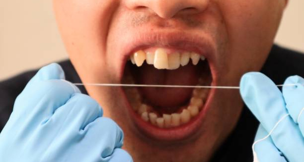
Dentist demonstrating dental flossing as part of gum disease treatment

Treatment of Gum Disease

Treatment of Gum Disease The treatment of Gum Disease focuses on stopping infection, reducing inflammation, and preventing further damage to the gums and surrounding structures. Because the treatment of Gum Disease depends on its stage and severity, options range from improved oral hygiene and professional cleaning to more advanced surgical procedures. Gum disease, also called periodontal disease, starts slowly. At first, it appears as gingivitis, which causes red or swollen gums. If treated early, this stage is usually easy to reverse. However, if left untreated, it can get worse and turn into periodontitis. At that point, the infection can damage the bone and tissue that hold your teeth in place. Fortunately, early action makes a big difference. With regular brushing, flossing, and professional cleanings, most cases can be stopped or even reversed. Over time, consistent care helps protect both your teeth and gums. As a result, you can keep your smile healthy and strong for many years to come 1. Early Stage Treatment – Gingivitis When Gum Disease is diagnosed in its early stage—gingivitis—the treatment is often simple and non-invasive. Key interventions include: a. Improved Oral Hygiene Brushing twice daily with fluoride toothpaste Flossing once a day to remove plaque between teeth Using an antibacterial mouthwash to reduce harmful bacteria Switching to a soft-bristled toothbrush or electric brush for better plaque removal Daily care can reverse gingivitis in a matter of weeks. b. Professional Dental Cleaning Known as scaling and polishing, this removes plaque and tartar from above and just below the gumline Performed by a dentist or dental hygienist during regular check-ups Helps reduce inflammation and bleeding of the gums This is often sufficient when the disease has not yet affected the bone. 2. Treatment of Periodontitis If the condition progresses to periodontitis, deeper cleaning and possibly surgical intervention are required. a. Scaling and Root Planing (Deep Cleaning) A non-surgical procedure performed under local anaesthesia Involves removing plaque and tartar from deep pockets around teeth (scaling) Smoothing root surfaces to help gums reattach (root planing) This may require several appointments and follow-up visits to monitor healing. b. Antibiotic Therapy May be applied locally as gels or fibres placed into gum pockets Oral antibiotics may be prescribed for more widespread infection Helps reduce bacterial load and inflammation when combined with mechanical cleaning Antibiotics alone are not effective without physical plaque removal. c. Laser Therapy Some dental clinics use lasers to remove infected tissue and bacteria Minimally invasive and can promote faster healing Not a replacement for standard treatments, but may be a helpful addition Laser treatment is still under investigation in some parts of the world, but shows promise. 3. Surgical Treatments for Advanced Cases When gum pockets are deep or tissue loss is severe, surgical procedures may be recommended: a. Flap Surgery (Pocket Reduction Surgery) Gums are lifted back to remove deep tartar deposits Irregular bone surfaces are smoothed to limit bacterial growth Gums are stitched back for a tighter fit around the teeth This reduces pocket depth and improves access for future cleaning. b. Bone Grafts Used when Gum Disease has destroyed underlying bone Bone from the patient, a donor, or synthetic source is used to promote new bone growth Helps stabilise teeth and supports gum tissue regeneration c. Soft Tissue Grafts Involves taking tissue (often from the palate) and attaching it to areas with receding gums Helps cover exposed roots and reduce sensitivity Improves gum appearance and strengthens the tissue d. Guided Tissue Regeneration A small mesh is placed between bone and gum tissue Encourages new bone growth while keeping gum tissue from growing into the area Often used in conjunction with flap surgery Surgical treatments are more common in advanced periodontitis and are tailored to individual cases. 4. Maintenance Therapy Following treatment, maintenance is essential to prevent recurrence: Regular dental visits every 3 to 6 months for professional cleaning Ongoing reinforcement of oral hygiene at home Close monitoring of any signs of relapse, such as bleeding or pocket depth increase Patients with a history of Gum Disease require lifelong maintenance to avoid further deterioration. 5. Lifestyle and Supportive Changes Managing risk factors is crucial: Quit smoking – smoking increases the risk of periodontitis and reduces treatment success Manage diabetes – high blood sugar levels increase infection risk Balanced diet – rich in vitamin C, calcium, and anti-inflammatory nutrients Stress management – stress can impact immune function and oral hygiene habits Combining lifestyle support with dental treatment ensures the best outcomes. Conclusion | Treatment of Gum Disease The treatment of gum disease begins with early detection. Once identified, the next step is to remove plaque and bacteria from the teeth and gums. First, daily brushing and flossing play a key role. Then, dentists may recommend professional deep cleanings to reach below the gumline. In some cases, antibiotics are used to fight infection. If the disease is more advanced, surgery may be needed to repair damage. Throughout treatment, a personalised care plan helps guide each step. As a result, patients receive the care that fits their specific needs. Over time, with regular follow-ups and good home care, the gums can begin to heal. Most importantly, this consistent approach can stop the disease from getting worse. In the long run, strong daily habits—combined with dental support—make a big difference. Not only can treatment protect your teeth, but it can also improve your overall health. Therefore, taking gum disease seriously leads to better outcomes for both your mouth and your body. [Next: Complications of Gum Disease →]

Treatment of Gum Disease Read More »

Scroll to Top