Mental Matters

Health Conditions

Find information on various health conditions affecting people of all ages. This category covers causes, symptoms, diagnosis, and management options for physical illnesses and chronic diseases. Get insights to help you understand different health challenges and make informed decisions about your wellbeing.

Symptoms of Edwards’ Syndrome

Symptoms of Edwards’ Syndrome

Symptoms of Edwards’ Syndrome The symptoms of Edwards’ syndrome are wide-ranging and often visible before or shortly after birth. The condition affects growth, development, and organ function. Babies with Edwards’ syndrome usually show multiple physical abnormalities, as well as severe intellectual and developmental delays. These signs help doctors suspect and confirm the diagnosis early. During pregnancy, ultrasound scans may show: Low birth weight Excess amniotic fluid (polyhydramnios) A small placenta Reduced foetal movement Congenital heart defects Once the baby is born, common physical features include: A small, misshapen head (microcephaly) A prominent back part of the head (occiput) Low-set ears A small jaw and mouth Clenched fists with overlapping fingers Rocker-bottom feet Short breastbone Narrow eyelid openings Many babies also have structural problems in the heart, kidneys, lungs, and digestive system. Heart defects are particularly common and include holes between heart chambers (ventricular or atrial septal defects) or valve abnormalities. These can lead to severe breathing and circulation problems. Feeding difficulties are also a major symptom. Many babies struggle to suck or swallow effectively, leading to poor weight gain and increased risk of aspiration or infections. Breathing may be irregular due to underdeveloped lungs or nerve dysfunction. Developmentally, most children with Edwards’ syndrome do not reach typical milestones. They may not sit, crawl, or speak. Some may respond to sound or touch, but progress is extremely limited. Seizures and neurological impairments are also common. Symptoms of Edwards’ Syndrome In South Africa, detection of symptoms may be delayed if the baby is born in a setting without trained staff or access to specialised care. Community healthcare workers play an essential role in identifying signs early and referring families for support. While the symptoms of Edwards’ syndrome are often severe, the degree can vary—especially in mosaic or partial trisomy 18. Some children may live longer than expected and show small signs of progress with the right medical and emotional care. Recognising the symptoms of Edwards’ syndrome allows for early intervention, accurate diagnosis, and compassionate care. Families facing this diagnosis deserve access to honest, supportive information that helps them make the best decisions for their unique circumstances. [Next: Diagnosis of Edwards’ Syndrome→]

Symptoms of Edwards’ Syndrome Read More »

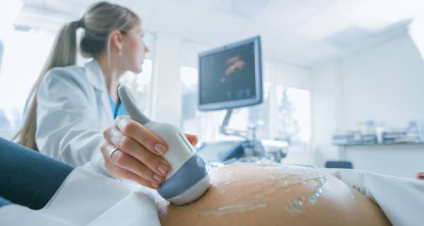
Pregnant woman undergoing ultrasound screening to detect chromosomal abnormalities like Edwards’ Syndrome.

Diagnosis of Edwards’ Syndrome

Diagnosis of Edwards’ Syndrome The diagnosis of Edwards’ syndrome can happen during pregnancy or shortly after birth. Since Edwards’ syndrome leads to a wide range of physical abnormalities, doctors may suspect the condition early, especially when routine pregnancy scans show signs of abnormal growth or development. Early diagnosis gives families more time to understand the condition and prepare for possible outcomes. In pregnancy, screening tests are often the first step. These include: Ultrasound scans, which may detect physical features such as low foetal weight, heart defects, or clenched hands Maternal blood tests, which check for abnormal levels of proteins and hormones that may signal a chromosomal condition Non-invasive prenatal testing (NIPT), which analyses foetal DNA in the mother’s blood to detect extra chromosomes These screenings cannot confirm the condition but can indicate a higher risk. If results show a high chance of Edwards’ syndrome, doctors may recommend diagnostic tests such as: Chorionic villus sampling (CVS) – done around 10–13 weeks, where a small piece of placental tissue is tested Amniocentesis – done around 15–20 weeks, where a sample of amniotic fluid is tested for chromosomal abnormalities Both tests carry a small risk of miscarriage but provide a definitive diagnosis by checking the baby’s chromosomes directly. After birth, a baby showing typical signs—such as clenched fists, small jaw, or heart problems—may be tested using a karyotype analysis, which examines the number and structure of chromosomes. This test confirms whether full, mosaic, or partial trisomy 18 is present. Diagnosis of Edwards’ Syndrome In South Africa, access to these prenatal tests may vary widely depending on whether a woman uses public or private healthcare. While private clinics in urban areas often offer NIPT and early ultrasounds, many rural facilities may only detect the condition after birth. This highlights the importance of strengthening prenatal services across the country. Once a diagnosis is confirmed, genetic counselling is usually offered to help parents understand the condition, possible outcomes, and future reproductive choices. Counsellors provide information in an empathetic way, helping families process complex emotions during a difficult time. The diagnosis of Edwards’ syndrome gives families a clearer understanding of their child’s health and life expectancy. It also allows healthcare professionals to plan the most appropriate care and support. [Next: Treatment of Edwards’ Syndrome →]

Diagnosis of Edwards’ Syndrome Read More »

Newborn with Edwards’ Syndrome receiving medical care in a neonatal unit.

Treatment of Edwards’ Syndrome

Treatment of Edwards’ Syndrome The treatment of Edwards’ syndrome focuses on easing symptoms and improving the quality of life. Since Edwards’ syndrome has no cure, treatment is based on the baby’s specific needs, the severity of the condition, and the family’s preferences. Medical teams aim to provide either supportive or palliative care, depending on the baby’s condition. At birth, babies with Edwards’ syndrome often have serious health challenges. Treatment in the neonatal intensive care unit (NICU) may be required for: Breathing difficulties, supported by oxygen or mechanical ventilation Feeding problems, managed with feeding tubes or special bottles Heart defects, which may require medication or surgery in rare cases Infections, treated with antibiotics Some babies may not need all these interventions, especially in cases of mosaic or partial trisomy 18, where symptoms are milder. In these situations, treatment plans may involve developmental support, physical therapy, and regular medical monitoring. In many cases, families and doctors focus on palliative care. This approach prioritises comfort, dignity, and love—supporting the baby with gentle care while avoiding aggressive medical treatments that may cause distress or have limited benefit. Palliative care includes pain management, warm clothing, skin-to-skin contact, and emotional support for the family. Medical teams work closely with parents to create a care plan that respects their wishes and beliefs. In South Africa, palliative care services are growing, though access remains uneven. Some families in public hospitals may not receive the same level of specialised care as those in private facilities. Advocacy is helping improve access to compassionate, community-based support in more regions. For babies who survive beyond infancy, long-term treatment may involve: Ongoing heart monitoring Nutritional support Management of seizures or reflux Speech and physiotherapy Assistive devices for movement and communication Treatment of Edwards’ Syndrome Support for the family is vital. Parents may need counselling, respite care, or financial assistance. In South Africa, several non-profit organisations help families living with trisomy conditions, offering guidance, equipment, and emotional support. The treatment of Edwards’ syndrome is highly individualised. Some babies may need intensive support early on, while others may surprise doctors with longer-than-expected survival. Every child is different, and every family deserves support in making informed, compassionate decisions. [Next: Complications of Edwards’ Syndrome →]

Treatment of Edwards’ Syndrome Read More »

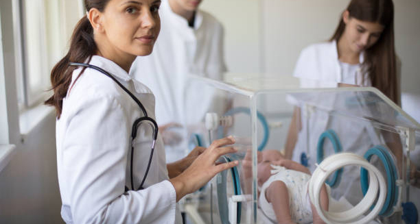
Neonatal doctors attending to a newborn in intensive care for complications of Edwards’ Syndrome.

Complications of Edwards’ Syndrome

Complications of Edwards’ Syndrome The complications of Edwards’ syndrome are often severe and affect multiple organs and systems. Because Edwards’ syndrome involves an extra chromosome in every cell (in full trisomy 18), it leads to widespread developmental challenges that begin before birth and continue throughout life. The most serious complications include: Congenital heart defects – present in over 90% of affected babies, these include holes in the heart or valve abnormalities. These can cause breathlessness, poor circulation, and failure to thrive. Breathing difficulties – due to weak muscles, irregular brain signals, or structural lung abnormalities. Some babies require constant oxygen or assisted breathing. Feeding problems – poor sucking reflexes, low energy, and underdeveloped digestive systems make feeding difficult. Malnutrition and dehydration are common. Neurological issues – including seizures, intellectual disability, and abnormal brain structure. Kidney and urinary problems, which can affect body chemistry and growth. Care From Birth Many babies are born very small and have weak immune systems, making them vulnerable to infections such as pneumonia and urinary tract infections. Repeated hospital stays are common, and some babies may need long-term medical care from birth. In those who survive past the newborn stage, delayed development is expected. Children may not walk, talk, or feed independently. Many cannot sit up or hold their head without assistance. Some can recognise familiar voices or respond to music or touch, but overall development remains severely limited. Emotional complications also affect families. Parents face uncertainty, difficult decisions, and grief—whether after a miscarriage, stillbirth, or the early death of a newborn. Mental health support for parents, siblings, and extended families is essential for healing and adjustment. In South Africa, complications are often made worse by limited access to paediatric specialists, ventilators, or early intervention services. Rural hospitals may not have NICU facilities, leaving families with fewer treatment options. Public health education and improved transport for critical cases can help reduce preventable losses. Complications of Edwards’ Syndrome Healthcare professionals must also be trained to recognise signs of Edwards’ syndrome and offer appropriate care options that respect cultural values and family preferences. Although the condition is life-limiting, not all children are the same. A small number of children with mosaic or partial trisomy 18 survive into their teens. Their lives may be complex, but they bring meaning and joy to their families. These rare cases remind us of the importance of individualised care and ongoing research. Understanding the complications of Edwards’ syndrome helps parents, doctors, and communities provide informed, respectful, and supportive care—ensuring every child, no matter their condition, is treated with dignity. [Next: Back to Overview →]

Complications of Edwards’ Syndrome Read More »

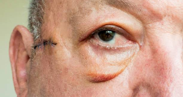
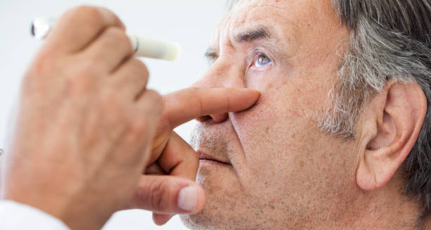
Close-up of an elderly man's eye showing signs of ectropion with visible eyelid turning outward.

Ectropion

Ectropion Ectropion is a condition in which the lower eyelid turns outward, away from the eye. This outward turning exposes the inner surface of the eyelid, which normally rests against the eyeball. Ectropion can cause discomfort, dryness, and irritation because the exposed surface is not properly protected or lubricated by tears. This condition most often affects older adults, especially those over 60, as the muscles and tissues supporting the eyelid naturally weaken with age. However, ectropion can also result from facial nerve damage, trauma, scarring, or certain skin conditions. It may affect one or both eyes, depending on the underlying cause. When the eyelid is not in contact with the eye, it disrupts the normal tear drainage process. Tears may not spread evenly, leading to dryness. Additionally, tears may overflow continuously down the cheek, a condition known as epiphora. This can cause social discomfort and increase the risk of eye infections or damage to the cornea. Ectropion In South Africa and other parts of the world, access to eye care varies. Older adults in rural or under-resourced communities may not receive early diagnosis or treatment, which can lead to worsening symptoms and complications. Public awareness campaigns around age-related eye conditions like ectropion can help improve outcomes. Fortunately, ectropion is treatable. In mild cases, artificial tears and ointments may provide relief. For more severe cases or those involving scarring or nerve damage, surgical correction is usually recommended to restore the eyelid to its normal position. Understanding ectropion and its symptoms can lead to earlier diagnosis, better eye health, and improved quality of life—especially for ageing populations. [Next: Causes of Ectropion →]

Ectropion Read More »

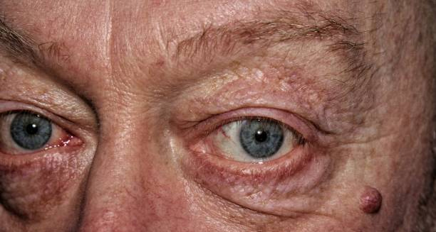
Close-up of a man's eyes showing sagging lower eyelids, a common sign of ectropion causes.

Causes of Ectropion

Causes of Ectropion The causes of ectropion are varied, but the condition most commonly results from age-related changes in the tissues and muscles around the eye. As we age, the connective tissues supporting the eyelids lose elasticity and tone, causing the lower eyelid to sag and turn outward. This form is known as involutional ectropion and is the most frequent type seen in clinical practice. Another significant cause is facial nerve paralysis, particularly of the seventh cranial nerve, also known as the facial nerve. When this nerve is damaged—due to Bell’s palsy, stroke, or head trauma—it affects the muscles that hold the eyelid in place. As a result, the eyelid can droop outward, leading to paralytic ectropion. Scarring from previous surgeries, burns, trauma, or chronic eye infections can pull the eyelid outward. This is known as cicatricial ectropion. Inflammation or damage to the skin and tissues around the eye alters the normal structure, preventing the eyelid from sitting properly against the eye. Chronic allergic reactions or infections can also cause the eyelid to swell and lose its normal positioning over time. In rare cases, tumours near the eye can push the eyelid outwards, creating mechanical ectropion. Certain congenital conditions—present from birth—may also lead to ectropion. Although rare, babies born with eyelid deformities may experience symptoms early in life, requiring specialist care and sometimes surgery. Causes of Ectropion In South Africa, trauma and burns are significant contributors to ectropion, particularly in rural or low-income areas where open cooking fires and untreated skin infections are more common. Lack of access to prompt treatment can result in scar formation, increasing the risk of cicatricial ectropion. Long-term use of certain eye medications can also contribute to tissue changes around the eyelid. Prolonged irritation may weaken the eyelid’s structure and lead to eventual outward turning. Regardless of the cause, early recognition and treatment are key. Left untreated, ectropion can lead to chronic discomfort, tearing, and even permanent damage to the eye surface. Understanding the causes of ectropion allows for prevention in some cases and supports faster intervention in others. [Next: Symptoms of Ectropion →]

Causes of Ectropion Read More »

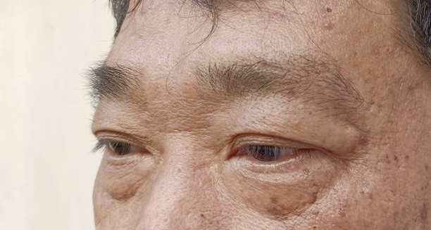
Man with visible eye irritation and sagging lower eyelids, a common symptom of ectropion.

Symptoms of Ectropion

Symptoms of Ectropion The symptoms of ectropion primarily result from the eyelid no longer sitting flush against the eye. Without this natural barrier, the eye becomes vulnerable to dryness, irritation, and infection. Recognising the symptoms of ectropion early allows for effective treatment before complications arise. One of the first signs is excessive tearing, also known as epiphora. Though this may seem counterintuitive, the exposed inner eyelid disrupts the normal tear drainage system. As tears no longer flow properly into the tear ducts, they spill over the lower eyelid and down the cheek. Other common symptoms include: Redness and irritation of the eye Dryness and grittiness, often described as a feeling of sand in the eye Light sensitivity or discomfort in bright environments Chronic conjunctivitis, or frequent eye infections Blurred vision, particularly when the cornea becomes irritated or damaged The exposed eye surface is more likely to become inflamed or infected. Without the eyelid to spread tears evenly, the eye loses its natural lubrication and becomes vulnerable to debris and bacteria. This dryness can lead to keratitis, an inflammation of the cornea that may cause pain, scarring, and visual impairment. In some people, the eyelid itself may become red, thickened, or swollen. The inner surface of the eyelid can dry out and become irritated from constant exposure to air and environmental elements. The symptoms of ectropion may worsen gradually. In age-related cases, people often ignore the early signs, assuming tearing or irritation is simply due to “old age.” However, if left untreated, the problem can progress to a more serious condition that threatens vision. Symptoms of Ectropion In South Africa, people living in areas with limited access to eye care may suffer from long-term symptoms without knowing the cause. Poor access to clean water, environmental pollutants, and untreated infections can worsen symptoms, especially among the elderly. Sometimes, symptoms are intermittent. For example, the eyelid may only turn outwards when the face is relaxed or when the person is tired. However, even temporary symptoms should not be ignored, as they can indicate underlying weakness in the eyelid structures. Understanding the symptoms of ectropion helps encourage early medical attention. With proper treatment—whether lubricating eye drops or surgery—most patients experience significant relief and a return to normal eyelid function. [Next: Diagnosis of Ectropion→]

Symptoms of Ectropion Read More »

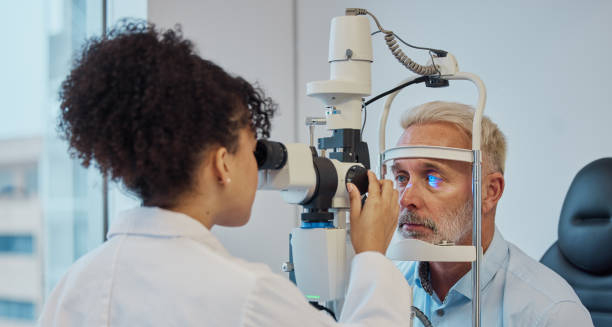
Ophthalmologist examining an older man's eye for signs of ectropion using a slit lamp.

Diagnosis of Ectropion

Diagnosis of Ectropion The diagnosis of ectropion is usually straightforward and can be made during a physical examination by an eye care professional. Because ectropion presents visible outward turning of the lower eyelid. Doctors can often confirm the condition by simply inspecting the eye and eyelid. The doctor may gently pull on the lower eyelid to observe its position and elasticity. In some cases, they may ask the patient to blink or look in different directions to see how the eyelid behaves during movement. A slit lamp examination—using a microscope with a light—may also be done to inspect the cornea and inner eyelid more closely for signs of irritation, dryness, or damage. It’s important to determine the underlying cause of the ectropion. If trauma, scarring, or a previous surgical procedure is suspected. The doctor will take a full medical history and examine the skin and tissues surrounding the eye. If facial nerve paralysis is present. They may assess muscle strength and facial movement to evaluate the severity of the nerve damage. In cases of suspected cicatricial ectropion—where scar tissue is pulling the eyelid outward. Doctors may also examine the tightness of the skin around the cheeks and temple. If a tumour or growth is suspected, imaging studies such as MRI or CT scans may be ordered. Diagnosis of Ectropion For children or newborns with congenital ectropion, further evaluation by a paediatric ophthalmologist may be needed to assess for related conditions. In South Africa, ectropion is often diagnosed by general practitioners or optometrists, especially in rural areas where access to ophthalmologists is limited. Community clinics may refer patients with persistent symptoms or complications to public hospitals for specialist care. Awareness campaigns on age-related eye conditions can help ensure earlier detection, particularly in the elderly. Early diagnosis of ectropion is essential to prevent complications such as corneal ulcers or vision loss. Once identified, doctors can begin appropriate treatment to relieve symptoms and protect the eye. [Next: Treatment of Ectropion →]

Diagnosis of Ectropion Read More »

Treatment of Ectropion

Treatment of Ectropion

Treatment of Ectropion The treatment of ectropion depends on the cause and severity of the condition. In mild or temporary cases, non-surgical options may offer relief. However, most long-term cases of ectropion—especially those caused by ageing or scarring—require surgical correction to restore the eyelid to its normal position. Non-surgical treatments include: Lubricating eye drops and ointments to prevent dryness Tear substitutes to reduce irritation and support corneal health Antibiotic eye drops if infection is present Moisture shields or eye patches, particularly during sleep These measures do not correct the position of the eyelid but provide temporary relief from symptoms. Doctors often use them while patients wait for surgery or when patients cannot undergo surgical procedures due to other health issues. Surgical treatment is the most effective and permanent solution. For involutional ectropion (age-related), the surgeon tightens the muscles and tendons of the eyelid in a procedure known as lateral tarsal strip surgery. This helps the eyelid sit closer to the eye and function normally. In cases of cicatricial ectropion, where scar tissue pulls the eyelid outward, surgeons may perform skin grafting to lengthen the eyelid and release the tension. If facial nerve paralysis causes the ectropion, surgeons may perform additional support or lifting procedures to stabilise the lower lid. Surgery is usually performed under local anaesthetic and is often a day procedure. Recovery is quick, and most patients experience immediate improvement in eye comfort and tear drainage. Treatment of Ectropion In South Africa, surgical treatment for ectropion is available at major public hospitals and private eye clinics. However, access in rural areas may be limited, and patients are sometimes placed on waiting lists. Early referral and follow-up are crucial for managing symptoms and preventing long-term damage. Patient education is also important. Avoiding eye rubbing, using protective eyewear in dusty environments, and managing skin conditions can reduce the risk of recurrence after surgery. The treatment of ectropion restores eyelid function, protects the eye, and improves quality of life. With modern techniques and good aftercare, most cases can be corrected successfully and without complications. [Next: Complications of Ectropion →]

Treatment of Ectropion Read More »

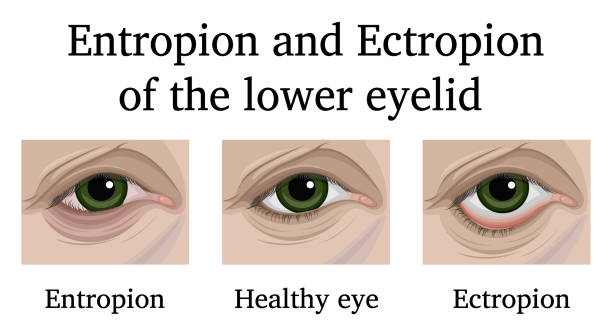
Medical illustration showing a healthy eye compared to eyes with entropion and ectropion of the lower eyelid.

Complications of Ectropion

Complications of Ectropion The complications of ectropion mainly arise when the condition is left untreated. Since ectropion causes the lower eyelid to pull away from the eye, it leaves the eye surface exposed and unprotected. This can lead to ongoing discomfort, recurrent infections, and long-term damage to vision if not managed properly. The most common complication is dry eye syndrome. Without proper contact between the eyelid and the eye, the tear film cannot spread evenly. This leads to a dry, gritty sensation, redness, and a burning feeling in the eye. Over time, chronic dryness can damage the corneal surface. Another major issue is chronic tearing (epiphora). Although it may seem like the eye is producing too many tears, the problem lies in tear drainage. Because the lower eyelid is turned outward, tears cannot enter the tear ducts and instead spill over onto the face. Corneal damage is a more serious complication. If the cornea becomes dry or scratched, it may lead to keratitis (inflammation), corneal ulcers, or even vision loss. This is especially dangerous for elderly patients or those with weakened immune systems, who may not detect the signs of infection early. Recurrent conjunctivitis (inflammation of the eye lining) is also common. Without adequate eyelid protection, the eye becomes more susceptible to bacterial or viral infections. Patients may experience frequent redness, discharge, and discomfort. In some cases, cosmetic deformity can develop. The constant exposure of the inner eyelid and tearing may cause skin irritation, darkening, or scarring. Social discomfort may follow, particularly for people who feel self-conscious about their appearance. People with paralytic ectropion face additional complications if facial nerve function is not addressed. The inability to blink properly can lead to exposure keratopathy, where the cornea becomes chronically exposed and inflamed. Complications of Ectropion In South Africa, complications from ectropion are more likely in regions with poor access to eye care. Delayed diagnosis and lack of treatment resources contribute to preventable vision problems. In many cases, simple interventions could have avoided serious outcomes. To prevent complications, early intervention is key. Lubrication, proper eye hygiene, and surgical repair when needed can restore normal eyelid function and protect vision. Regular eye check-ups—especially for older adults—can detect ectropion before it causes lasting damage. Addressing the complications of ectropion requires public awareness, timely referral to specialists, and access to affordable treatment. With proper care, most people can avoid long-term effects and maintain clear, comfortable vision. [Next: Back to Overview →]

Complications of Ectropion Read More »

Scroll to Top