Mental Matters

Health Conditions

Find information on various health conditions affecting people of all ages. This category covers causes, symptoms, diagnosis, and management options for physical illnesses and chronic diseases. Get insights to help you understand different health challenges and make informed decisions about your wellbeing.

Two women discussing health while enjoying a salad and green smoothies, representing lifestyle and dietary management in treating Crohn’s disease.

Treatment of Crohn’s Disease

Treatment of Crohn’s Disease Treatment of Crohn’s disease aims to reduce inflammation, relieve symptoms, and maintain long-term remission. As Crohn’s disease is a chronic condition with no cure, the focus is on managing flare-ups, preventing complications, and improving quality of life. Treatment is often personalised and may involve medication, lifestyle changes, and, in some cases, surgery. Medications Aminosalicylates (5-ASAs): Used in mild cases to reduce inflammation, though less commonly than in ulcerative colitis Corticosteroids (e.g. prednisolone): Effective for short-term flare-ups but not suitable for long-term use due to side effects Immunomodulators (e.g. azathioprine, methotrexate): Help suppress immune response and maintain remission Biologic therapies: Target specific parts of the immune system; includes: Anti-TNF agents (e.g. infliximab, adalimumab) Anti-integrin drugs (e.g. vedolizumab) IL-12/23 inhibitors (e.g. ustekinumab) Antibiotics: Occasionally used to treat infections or abscesses in the bowel Lifestyle and Dietary Adjustments Low-fibre diet during flare-ups to reduce bowel irritation Nutritional support, including supplements for iron, B12, and calcium Stress management through exercise, mindfulness, or counselling Smoking cessation, which is critical for symptom control and relapse prevention Surgical Treatment | Diagnosis of Crohn’s Disease Up to 70% of people with Crohn’s disease eventually require surgery to: Remove damaged bowel segments Treat complications like strictures, fistulas, or abscesses Create a stoma in severe cases (temporary or permanent) Surgery is not curative, as the disease can return elsewhere in the digestive tract. However, it often significantly improves symptoms and quality of life. Monitoring and Long-Term Care Regular follow-ups to monitor inflammation Colonoscopies every 1–3 years to screen for colorectal cancer in long-standing disease Blood tests to monitor medication side effects and nutritional status In conclusion, treatment of Crohn’s disease combines medication, lifestyle change, and sometimes surgery to control inflammation and maintain remission. Early and consistent management leads to better outcomes and fewer complications. [Next: Complications and Recovery from Crohn’s Disease →]
Treatment of Crohn’s Disease Read More »

Complications and Recovery from Crohn’s Disease

Complications and Recovery from Crohn’s Disease

Complications and Recovery from Crohn’s Disease Complications and recovery from Crohn’s disease vary widely depending on disease severity, response to treatment, and the presence of coexisting health issues. While many people with Crohn’s disease achieve good symptom control, others may face ongoing challenges requiring surgical or advanced medical intervention. Common Complications Strictures: Narrowing of the bowel due to chronic inflammation and scarring, potentially leading to obstruction Fistulas: Abnormal connections between the bowel and other organs, such as the bladder, skin, or other bowel segments Abscesses: Infected pockets of pus within the abdominal cavity or rectum Malnutrition: Due to reduced absorption of nutrients, especially in the small intestine Bone loss (osteopenia or osteoporosis): Caused by steroid use and calcium malabsorption Perianal disease: Includes fissures, fistulas, or painful skin inflammation near the anus Emotional and Mental Health Living with a chronic illness can impact mental well-being. Anxiety, depression, and stress are common, especially during flare-ups. Support from mental health professionals, support groups, and IBD charities can be invaluable. Recovery After Flare-Ups Recovery time varies: mild cases may resolve in weeks, while severe flares may last months Medication adjustments and rest are typically required Nutritional support may help restore energy and nutrient balance Post-Surgery Recovery | Complications and Recovery from Crohn’s Disease Hospital stays range from a few days to over a week depending on the procedure Full recovery can take several weeks, and dietary changes may be necessary Follow-up appointments are essential to monitor for recurrence or new complications Preventing Long-Term Damage Early treatment to control inflammation helps prevent complications Ongoing monitoring via imaging and blood tests ensures that treatment remains effective Vaccinations and regular screenings protect against infections and medication side effects In summary, complications and recovery from Crohn’s disease depend on individual response and timely management. With proper care, many people live full, productive lives, even while managing this lifelong condition. [Next: Back to Overview →]

Complications and Recovery from Crohn’s Disease Read More »

Creutzfeldt-Jakob Disease

Creutzfeldt-Jakob Disease

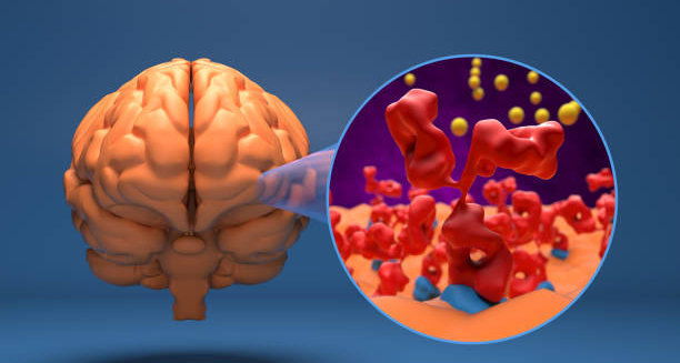
Creutzfeldt-Jakob Disease – Overview Creutzfeldt-Jakob disease is a rare, rapidly progressive, and fatal neurodegenerative condition that affects the brain. Caused by abnormal proteins called prions, CJD leads to brain tissue damage, resulting in severe mental deterioration, physical disability, and ultimately death. It is one of several conditions classified as prion diseases or transmissible spongiform encephalopathies (TSEs). CJD is most often sporadic, meaning it occurs without any identifiable cause or risk factor. However, in some cases, it may be inherited through genetic mutations, acquired via contaminated medical procedures, or linked to the consumption of infected meat products, as in variant CJD (vCJD). The disease causes brain cells to die, creating tiny holes in the brain tissue and giving it a sponge-like appearance. Creutzfeldt-Jakob Disease Symptoms of CJD typically begin with memory loss, personality changes, or impaired judgement. As the disease progresses, individuals experience muscle stiffness, difficulty speaking or swallowing, involuntary movements, and eventually lose the ability to walk or communicate. The condition worsens rapidly, with most patients dying within a year of symptom onset. Although extremely rare—affecting roughly one to two people per million annually—CJD is devastating. There is no cure or effective treatment, and diagnosis can be difficult during life. However, supportive care and accurate diagnosis are important for managing symptoms and providing comfort. In summary, CJD is a fatal brain disorder caused by misfolded proteins. While rare, its rapid progression and severe symptoms make it a significant neurological emergency that requires immediate attention. [Next: Causes of Creutzfeldt-Jakob Disease →]

Creutzfeldt-Jakob Disease Read More »

Causes of Creutzfeldt-Jakob Disease

Causes of Creutzfeldt-Jakob Disease

Causes of Creutzfeldt-Jakob Disease Causes of Creutzfeldt-Jakob disease revolve around the accumulation of prions—misfolded proteins that cause normal brain proteins to fold abnormally. While prions are not viruses or bacteria, they are infectious in the sense that they induce abnormal protein folding, leading to brain cell death. Understanding the causes of Creutzfeldt-Jakob disease is vital for identifying at-risk individuals and preventing transmission in healthcare settings. 1. Sporadic CJD This is the most common form, accounting for approximately 85–90% of cases. It occurs without any clear reason, usually in people over the age of 60. Doctors do not fully understand the exact mechanism behind sporadic CJD. Researchers believe it involves spontaneous changes in prion proteins in the brain. 2. Genetic CJD (Familial) Inherited mutations in the PRNP gene, which codes for the prion protein, are responsible for 5–15% of CJD cases. Inherited forms can pass from one generation to the next and typically develop earlier than sporadic CJD. 3. Acquired CJD | Causes of Creutzfeldt-Jakob Disease This extremely rare form is contracted through exposure to prions from an external source: 4. Prion Transmission Mechanism Prions are incredibly resistant to standard sterilisation methods. Once introduced into the body, they propagate by inducing the misfolding of normal proteins, triggering a chain reaction that leads to widespread brain damage. 5. Not Contagious CJD is not spread through casual contact, touch, or air. Only specific medical or surgical exposures carry a transmission risk, which is now strictly controlled with enhanced sterilisation protocols. The causes of Creutzfeldt-Jakob disease are unique in that they involve no bacteria, viruses, or parasites—just a corrupted protein that triggers a cascade of brain destruction. Early recognition of risk factors, especially in surgical and genetic contexts, is key to prevention and surveillance. [Next: Symptoms of Creutzfeldt-Jakob Disease →]

Causes of Creutzfeldt-Jakob Disease Read More »

Symptoms of Creutzfeldt-Jakob Disease

Symptoms of Creutzfeldt-Jakob Disease

Symptoms of Creutzfeldt-Jakob Disease Symptoms of Creutzfeldt-Jakob disease typically begin subtly and progress rapidly over weeks or months. The hallmark of Creutzfeldt-Jakob disease is a swift and severe decline in mental and physical function, with death usually occurring within 6 to 12 months after symptoms appear. Recognising these symptoms early is critical for diagnosis and supportive care. 1. Early Cognitive Changes The first signs often include memory loss, impaired judgement, confusion, or difficulty concentrating. These may mimic Alzheimer’s disease but progress far more rapidly. Personality changes, depression, and anxiety may also be early features. 2. Motor Dysfunction As the disease advances, muscle stiffness (rigidity), jerky movements (myoclonus), coordination problems (ataxia), and speech difficulties emerge. Patients often struggle to walk, use their hands, or perform basic tasks due to deteriorating muscle control. 3. Visual Disturbances Blurred vision, double vision, and even partial blindness can occur due to involvement of the visual cortex. Some patients also report hallucinations or visual illusions. 4. Behavioural and Psychiatric Symptoms In some types of CJD, especially variant CJD, mood swings, paranoia, hallucinations, or social withdrawal may precede neurological symptoms. These can lead to misdiagnosis as a psychiatric illness in the early stages. 5. Seizures and Muscle Spasms Myoclonus, characterised by sudden, involuntary muscle jerks, is common. Seizures may also develop in some cases, especially in later stages. 6. Dementia | Symptoms of Creutzfeldt-Jakob Disease Rapid-onset dementia is a defining feature. Patients lose the ability to think clearly, remember, make decisions, or interact socially. Speech becomes incoherent, and eventually, verbal communication ceases altogether. 7. Coma and Death In the final stages, individuals become immobile, unresponsive, and eventually fall into a coma. Death typically results from infections, such as pneumonia, or from complications related to immobility and organ failure. The symptoms of Creutzfeldt-Jakob disease progress quickly and devastatingly, often leading to full dependency within weeks. Early identification allows families to prepare for palliative care and emotional support, even though a cure is currently unavailable. [Next: Diagnosis of Creutzfeldt-Jakob Disease →]

Symptoms of Creutzfeldt-Jakob Disease Read More »

Diagnosis of Creutzfeldt-Jakob Disease

Diagnosis of Creutzfeldt-Jakob Disease

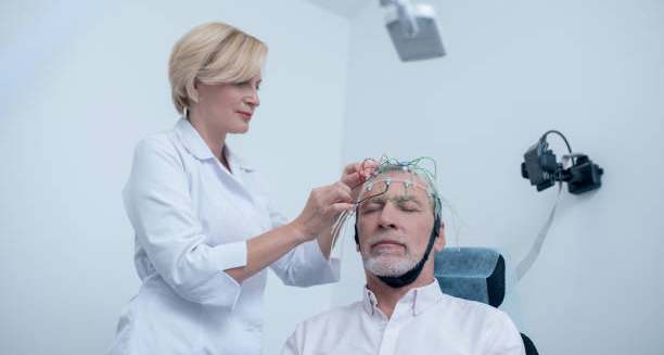
Diagnosis of Creutzfeldt-Jakob Disease Diagnosis of Creutzfeldt-Jakob disease is challenging due to its rarity and symptom overlap with other neurological disorders. However, a combination of clinical evaluation, brain imaging, EEG, and laboratory testing can strongly suggest Creutzfeldt-Jakob disease, even before post-mortem confirmation. 1. Medical History and Neurological Exam Doctors first review the patient’s medical history and conduct a detailed neurological assessment. Rapidly progressing dementia, myoclonus, and coordination issues raise suspicion for CJD, especially if the decline occurs over weeks rather than years. 2. Electroencephalogram (EEG) An EEG records electrical activity in the brain. In classic (sporadic) CJD, characteristic periodic sharp wave complexes may be seen, which help distinguish it from other forms of dementia. 3. MRI Scan Magnetic resonance imaging of the brain often shows distinctive changes in the basal ganglia, thalamus, or cortex. The “pulvinar sign” on MRI is particularly suggestive of variant CJD. 4. Cerebrospinal Fluid (CSF) Tests A lumbar puncture (spinal tap) allows analysis of cerebrospinal fluid. Elevated levels of certain proteins, such as 14-3-3, tau protein, and RT-QuIC positivity, support the diagnosis of CJD. 5. Genetic Testing | Diagnosis of Creutzfeldt-Jakob Disease In suspected familial cases, genetic testing of the PRNP gene can identify mutations associated with inherited forms of CJD. 6. Brain Biopsy (Rare) In uncertain cases, a brain biopsy may be considered. However, due to risks and difficulty in obtaining viable tissue, it is rarely performed. A definitive diagnosis is usually confirmed after death by autopsy and neuropathological examination. 7. Rule Out Other Conditions Doctors must exclude other causes of rapid dementia, such as Alzheimer’s, stroke, encephalitis, or metabolic disorders. Imaging and lab tests help in this differential diagnosis. Although confirmation often comes post-mortem, an accurate diagnosis of Creutzfeldt-Jakob disease during life enables supportive care, palliative planning, and proper infection control precautions in healthcare settings. [Next: Treatment of Creutzfeldt-Jakob Disease →]

Diagnosis of Creutzfeldt-Jakob Disease Read More »

Treatment of Creutzfeldt-Jakob Disease

Treatment of Creutzfeldt-Jakob Disease

Treatment of Creutzfeldt-Jakob Disease Treatment of Creutzfeldt-Jakob disease focuses on supportive and palliative care, as there is currently no cure or disease-modifying therapy available. Because Creutzfeldt-Jakob disease progresses rapidly, the primary goals are comfort, symptom control, and maintaining dignity throughout the disease process. 1. Symptom Management Doctors use medications to alleviate distressing symptoms: 2. Nutritional Support As patients lose the ability to eat and swallow safely. A feeding tube can provide comfort and nutrition, but the decision depends on individual preferences. 3. Mobility and Safety | Treatment of Creutzfeldt-Jakob Disease Physiotherapists and occupational therapists may provide support during the early stages to reduce fall risk and encourage gentle movement. With proper nursing care, patients avoid bedsores and infections linked to immobility. 4. Psychological and Emotional Support Both patients and families require emotional support. Counselling, support groups, and hospice services are important resources to help families prepare for end-of-life decisions and cope with grief. 5. Palliative and End-of-Life Care As the disease progresses, most individuals require round-the-clock care. Hospice services focus on comfort, dignity, and emotional well-being. Clinicians and families should address artificial nutrition, hydration, and resuscitation decisions early in care planning. 6. Infection Control Although CJD is not contagious in everyday contact, special precautions are taken in hospitals during surgical procedures or post-mortem examination due to the durability of prions on instruments. Research is ongoing, exploring treatments that block prion propagation, but no therapies have proven effective to date. Ultimately, the treatment of Creutzfeldt-Jakob disease is centred on compassionate care and supporting both patient and family through a deeply challenging and rapid disease trajectory. [Next: Complications and Recovery from Creutzfeldt-Jakob Disease →]

Treatment of Creutzfeldt-Jakob Disease Read More »

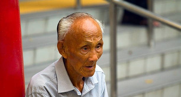
Elderly man sitting with a distant, solemn expression, symbolising cognitive and neurological decline associated with Creutzfeldt-Jakob disease.

Complications and Recovery from Creutzfeldt-Jakob Disease

Complications and Recovery from Creutzfeldt-Jakob Disease Complications and recovery from Creutzfeldt-Jakob disease must be understood in the context of a terminal illness. While Creutzfeldt-Jakob disease cannot be cured or reversed, supportive measures aim to reduce suffering and manage complications that arise during its rapid course. 1. Loss of Independence Patients quickly lose the ability to walk, talk, eat, and care for themselves. Most become bedbound within a few months, requiring full-time assistance for even the simplest tasks. 2. Communication Difficulties As cognitive and language abilities deteriorate, communication becomes limited. Speech therapists may offer early support, but in late stages, families rely on non-verbal cues and emotional presence. 3. Infections | Complications and Recovery from Creutzfeldt-Jakob Disease Immobilised patients are at high risk of chest infections, bedsores, and urinary tract infections. Pneumonia is a common cause of death. Good hygiene and positioning help reduce risk. 4. Seizures and Neurological Crises Frequent seizures, muscle rigidity, and startle responses may cause distress. These are controlled with medications but often worsen as the disease progresses. 5. Nutritional and Hydration Issues Difficulty swallowing and appetite loss lead to malnutrition and dehydration. Families must make difficult decisions about artificial feeding and end-of-life preferences. 6. Psychological and Social Impact CJD is traumatic for families, as loved ones often change dramatically within weeks. Counselling and hospice involvement are crucial for emotional support and grief management. Recovery Outlook Recovery from CJD is not possible. The disease progresses inevitably toward coma and death. However, with early diagnosis and excellent palliative care, many patients experience a peaceful and dignified end-of-life phase. In conclusion, complications and recovery from Creutzfeldt-Jakob disease must focus on maximising comfort, providing emotional support, and ensuring that patients pass with dignity. The rare and devastating nature of this disease underscores the importance of early recognition and compassionate care. [Next: Back to Overview →]

Complications and Recovery from Creutzfeldt-Jakob Disease Read More »

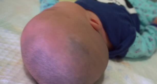
Close-up of an infant’s head from behind, showing abnormal skull shape suggestive of craniosynostosis.

Craniosynostosis

Craniosynostosis – Overview Craniosynostosis is a birth defect in which one or more of the sutures in a baby’s skull close too early. Before the brain has finished growing. This premature fusion affects the shape of the head and may increase pressure inside the skull. Craniosynostosis can vary in severity. Depending on how many sutures are involved and whether it occurs as part of a genetic syndrome or in isolation. The human skull is made up of several bones joined by flexible sutures. Furthermore, allowing the skull to expand as the brain grows. In craniosynostosis, these sutures fuse too early, forcing the brain to grow in the direction of the remaining open sutures. This results in an abnormally shaped head, and in severe cases, it can lead to developmental delays, visual impairment, and neurological issues. Craniosynostosis Craniosynostosis can be non-syndromic, which means it occurs without other abnormalities, or syndromic, where it is associated with genetic conditions such as Apert, Crouzon, or Pfeiffer syndrome. Non-syndromic cases are more common and typically involve just one suture. Early detection and treatment are crucial. In many cases, surgery is required to correct the shape of the skull and allow the brain to grow normally. When treated promptly, the long-term outlook is often positive. In summary, craniosynostosis is a condition involving premature skull suture fusion that affects head shape and brain development. With early diagnosis and appropriate intervention, most children go on to lead healthy lives. Importance of Early Intervention Early diagnosis of craniosynostosis is key to achieving the best possible outcomes. Pediatricians and specialists often detect abnormal head shapes during routine check-ups, prompting imaging tests to confirm the diagnosis. When addressed promptly — usually within the first year of life — surgical treatment can relieve pressure on the brain, correct deformities, and support normal development. Delays in treatment may increase the risk of complications, highlighting the importance of early intervention. Supporting Families and Long-Term Outlook Raising a child with craniosynostosis can be emotionally challenging for families, especially in syndromic cases requiring multiple surgeries or ongoing care. Support groups, genetic counselling, and coordinated medical care can ease the journey. Fortunately, with appropriate medical and surgical management, many children go on to reach developmental milestones and lead healthy, active lives. Advances in surgical techniques and multidisciplinary care continue to improve both cosmetic and functional outcomes. [Next: Causes of Craniosynostosis →]

Craniosynostosis Read More »

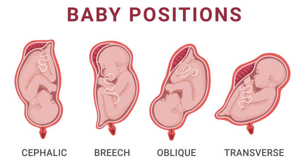
Illustration of various fetal positions in the womb, including cephalic, breech, oblique, and transverse, which may influence cranial development.

Causes of Craniosynostosis

Causes of Craniosynostosis Causes of craniosynostosis can be genetic, environmental, or a combination of both. While many cases arise without a known reason, understanding the underlying mechanisms can help parents and healthcare providers identify the condition early. Craniosynostosis occurs when one or more cranial sutures close too soon, disrupting normal skull and brain growth. Genetic Factors | Causes of Craniosynostosis Approximately 15–30% of cases are associated with known genetic syndromes, such as: Apert syndrome Crouzon syndrome Pfeiffer syndrome Muenke syndrome These syndromic forms of craniosynostosis often involve multiple sutures and come with other physical abnormalities, such as limb deformities or facial asymmetry. Mutations in genes such as FGFR2, FGFR3, and TWIST1 have been identified as common culprits. Non-Syndromic Causes In most cases, craniosynostosis occurs in isolation, without other medical conditions or inherited traits. These non-syndromic cases may result from: Spontaneous gene mutations Intrauterine constraints, such as limited space in the womb Abnormal foetal positioning Multiple pregnancies, which increase pressure on the baby’s head Environmental Factors While not as well-established, certain environmental factors may contribute to craniosynostosis, including: Maternal smoking Advanced paternal age Certain fertility treatments Use of specific medications during pregnancy More research is needed to fully understand how these factors interact with genetic predispositions. Prematurity and Metabolic Disorders Some premature infants are at greater risk due to altered bone development. Additionally, metabolic disorders affecting calcium or phosphate levels can influence early suture fusion. In conclusion, causes of craniosynostosis vary widely, ranging from genetic mutations to external pressures and unknown factors. Recognising risk factors helps guide screening, especially in newborns with abnormal head shapes or family history of related conditions. [Next: Symptoms of Craniosynostosis →]

Causes of Craniosynostosis Read More »

Scroll to Top