Mental Matters

Health Conditions

Find information on various health conditions affecting people of all ages. This category covers causes, symptoms, diagnosis, and management options for physical illnesses and chronic diseases. Get insights to help you understand different health challenges and make informed decisions about your wellbeing.

DNA mutation representing the genetic cause of Glutaric Aciduria Type 1

Causes of Glutaric Aciduria Type 1

Causes of Glutaric Aciduria Type 1 The causes of glutaric aciduria type 1 lie in inherited genetic mutations that affect how the body processes certain amino acids. Glutaric aciduria type 1 is caused by mutations in the GCDH gene, which result in a deficiency or complete lack of the enzyme glutaryl-CoA dehydrogenase. This enzyme is essential for the breakdown of lysine, hydroxylysine, and tryptophan—amino acids that are part of our regular diet and necessary for growth and development. When this enzyme is absent or not functioning correctly, toxic by-products build up in the body, particularly in the brain. The accumulation of these substances, especially glutaric acid and 3-hydroxyglutaric acid, causes damage to brain tissue and impairs motor control. Understanding the causes of glutaric aciduria type 1 is crucial for both treatment and family planning. Genetic Inheritance Pattern Glutaric aciduria type 1 is inherited in an autosomal recessive manner. This means: A child must inherit two copies of the defective gene, one from each parent, to develop the disorder If both parents are carriers (having one normal and one defective copy), there is: A 25% chance the child will have the condition A 50% chance the child will be a carrier A 25% chance the child will inherit two normal genes Carrier parents typically show no symptoms and are unaware they carry the faulty gene until a child is affected. Mutations in the GCDH Gene The GCDH gene provides instructions for producing the enzyme glutaryl-CoA dehydrogenase. This enzyme is active in mitochondria—the energy-producing centres of cells—and is involved in: Breaking down lysine, hydroxylysine, and tryptophan Preventing the build-up of glutaric acid, which is toxic in high amounts Over 200 different mutations in the GCDH gene have been identified. The severity of glutaric aciduria type 1 may depend on the nature of the mutation: Null mutations (producing no enzyme at all) tend to result in more severe outcomes Missense mutations (producing partially functioning enzymes) may result in milder disease Genetic testing allows families to identify the specific mutation involved, which can help with prognosis and inform future pregnancies. Population and Ethnic Factors Certain populations have a higher incidence of glutaric aciduria type 1 due to the founder effect, where a mutation becomes more common in a closed population over generations: Amish and Mennonite communities in the United States Specific regions in Sweden and Turkey North African, Middle Eastern, and South Asian populations with high rates of consanguinity In these groups, routine carrier screening may be offered as part of antenatal or pre-marital counselling programmes. Environmental and Dietary Triggers While the genetic mutation is the underlying cause, environmental stressors often act as triggers for symptoms in children who have not yet been diagnosed or treated. These include: Infections, particularly viral illnesses with fever Vaccinations, if not managed appropriately in undiagnosed children Fasting or poor feeding, especially during infancy High protein intake, which increases the burden of amino acid breakdown These triggers can precipitate a metabolic crisis, characterised by rapid neurological decline, often occurring between 6 and 24 months of age. Role of Newborn Screening In regions where newborn screening is available, glutaric aciduria type 1 can be detected before symptoms develop. Early detection allows: Dietary restrictions to be introduced Emergency regimens during illness to be prepared Neurological outcomes to be significantly improved Parents of affected children are also counselled to seek early care during fevers or infections to prevent metabolic decompensation. Importance of Genetic Counselling When a child is diagnosed with glutaric aciduria type 1, the entire family may benefit from genetic counselling. This involves: Understanding the pattern of inheritance Offering carrier testing to siblings or extended family members Discussing options for prenatal diagnosis in future pregnancies Planning for preimplantation genetic diagnosis (PGD) if desired In populations with higher prevalence, preconception screening programmes may help reduce the incidence of affected births. Summary of Causes Cause Category Description Genetic mutation Mutation in the GCDH gene affects enzyme production Autosomal recessive Requires two defective copies to cause the disease Environmental triggers Illness, fasting, or high-protein diets may trigger symptoms High-risk populations Founder effect increases prevalence in certain groups Inheritance risk 25% if both parents are carriers Conclusion | Causes of Glutaric Aciduria Type 1 The causes of glutaric aciduria type 1 are rooted in genetic mutations that disrupt amino acid metabolism. While the condition itself is rare, its impact can be profound if left untreated. Early diagnosis through newborn screening, coupled with awareness of the causes of glutaric aciduria type 1, allows for preventive strategies that can dramatically improve outcomes and quality of life. [Next: Symptoms of Glutaric Aciduria Type 1→]

Causes of Glutaric Aciduria Type 1 Read More »

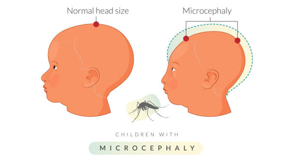
Illustration comparing normal head size to microcephaly in children

Symptoms of Glutaric Aciduria Type 1

Symptoms of Glutaric Aciduria Type 1 The symptoms of glutaric aciduria type 1 can vary widely from person to person, making early detection both crucial and challenging. In most affected children, the symptoms of glutaric aciduria type 1 become apparent during infancy or early childhood, often triggered by common illnesses such as fevers or respiratory infections. Without early diagnosis and management, the disease can cause severe, irreversible neurological damage. Glutaric aciduria type 1 is a metabolic disorder that disrupts the breakdown of certain amino acids, leading to the accumulation of toxic substances in the brain. The parts of the brain most affected are the basal ganglia, which help control movement. As a result, many of the hallmark symptoms involve motor function and coordination, but the condition can also impact growth, cognition, and behaviour. Early Signs in Infancy Some infants may show signs of glutaric aciduria type 1 from birth, while others appear healthy until a “triggering event” causes a sudden deterioration. Early signs include: Macrocephaly (an abnormally large head): Often one of the earliest clues Poor feeding or vomiting during infancy Hypotonia (reduced muscle tone), making the infant appear floppy Mild developmental delays, particularly in motor milestones like sitting or crawling Increased irritability or lethargy, especially during minor illnesses Macrocephaly is especially important to note, as it may precede neurological decline even in otherwise asymptomatic infants. Symptoms Triggered by Metabolic Crises A metabolic crisis is often the event that reveals the condition. These crises typically occur during times of physiological stress, such as infections, immunisations, fasting, or trauma. During a metabolic crisis, symptoms can appear suddenly and escalate rapidly: Loss of motor skills: Previously acquired abilities like sitting, walking, or grasping may regress Dystonia: Involuntary muscle contractions leading to twisted, rigid postures Chorea or athetosis: Jerky or writhing movements Seizures: Common in acute phases, often difficult to control Coma: In severe cases, untreated crises can lead to unconsciousness A single crisis can result in lasting brain damage, with most of the injury occurring in the basal ganglia. This can lead to permanent motor impairment. Long-Term Neurological Manifestations Children who experience a crisis, or those with delayed diagnosis, may develop chronic symptoms such as: Spasticity or stiffness of the limbs Dystonic cerebral palsy, which can impair walking and fine motor skills Poor balance and coordination Swallowing difficulties (dysphagia) and risk of aspiration Speech delays or difficulty speaking clearly In some children, these symptoms are progressive, particularly if the disorder is not managed carefully through diet and medical therapy. Cognitive and Behavioural Features While many individuals with glutaric aciduria type 1 have normal intelligence, cognitive and behavioural concerns can still arise: Learning difficulties, particularly with tasks requiring motor planning or attention Memory issues, especially in those with significant neurological injury Hyperactivity or impulsivity, sometimes misdiagnosed as ADHD Emotional distress, stemming from communication challenges or physical limitations Some children may require special educational support or behavioural therapy to help them reach their full potential. Adult Presentation Though rare, some individuals are not diagnosed until adolescence or adulthood. In these cases, symptoms may be milder or atypical: Mild tremors Subtle balance issues Fatigue or poor exercise tolerance Occasional speech changes or stiffness Adult-onset cases tend to be less severe but still benefit from metabolic management and nutritional intervention. Importance of Early Recognition Prompt recognition of the symptoms of glutaric aciduria type 1 is essential to prevent irreversible damage. Early symptoms such as macrocephaly, feeding difficulties, and developmental delays should prompt metabolic testing. In families with known GCDH mutations, even subtle changes warrant medical review. Neonatal screening has made early detection more achievable, dramatically reducing the number of children who suffer metabolic crises. Monitoring Symptoms Over Time Children with glutaric aciduria type 1 require lifelong monitoring. Healthcare teams track: Growth and head circumference Motor and cognitive development Nutritional status Frequency and severity of illnesses Adherence to dietary restrictions and emergency plans With careful oversight, many children remain stable and avoid further neurological decline. Conclusion | Symptoms of Glutaric Aciduria Type 1 The symptoms of glutaric aciduria type 1 range from early subtle signs like macrocephaly to sudden, life-altering neurological crises. Recognising the early warning signs and intervening before a crisis occurs is key to protecting brain function. Once diagnosed, ongoing attention to the symptoms of glutaric aciduria type 1 helps families and clinicians manage the condition and maintain a good quality of life. [Next: Diagnosis of Glutaric Aciduria Type 1 →]

Symptoms of Glutaric Aciduria Type 1 Read More »

Doctor examining newborn baby with a stethoscope

Diagnosis of Glutaric Aciduria Type 1

Diagnosis of Glutaric Aciduria Type 1 The diagnosis of glutaric aciduria type 1 is essential for preventing serious neurological complications and enabling early treatment. In many countries, the diagnosis of glutaric aciduria type 1 is now possible through routine newborn screening, which has significantly improved outcomes by identifying affected infants before symptoms develop. A deficiency in the enzyme glutaryl-CoA dehydrogenase causes glutaric aciduria type 1, leading to the accumulation of toxic compounds in the body. If undetected, these substances can damage the brain, particularly the basal ganglia, leading to permanent motor disability. Early and accurate diagnosis ensures timely interventions that can prevent irreversible injury. Newborn Screening In countries with comprehensive newborn screening programmes, diagnosis usually begins in the first days of life: Dried blood spot testing (heel prick test) uses tandem mass spectrometry to detect elevated levels of glutaric acid derivatives Screening typically identifies markers such as glutaric acid, 3-hydroxyglutaric acid, and C5DC-carnitine Positive results trigger confirmatory testing and immediate referral to a metabolic specialist Early detection allows treatment to begin before a metabolic crisis occurs, offering the best chance at normal development. Biochemical Testing In addition to screening, diagnostic biochemical tests confirm the presence of the condition: 1. Urine Organic Acid Analysis Performed using gas chromatography–mass spectrometry (GC-MS) Detects elevated glutaric acid, 3-hydroxyglutaric acid, and glutaconic acid 2. Plasma Acylcarnitine Profile Elevated glutarylcarnitine (C5DC) is a hallmark of the condition Supports the urine analysis findings These tests are non-invasive and form the cornerstone of diagnosis in symptomatic individuals and newborns flagged during screening. Enzyme Assay Where available, direct measurement of glutaryl-CoA dehydrogenase activity in: Fibroblasts (from a skin biopsy) White blood cells (leukocytes) Clinicians use this functional assay to confirm the deficiency and possibly predict disease severity, but genetic testing is now more common and less invasive. Genetic Testing Molecular genetic analysis of the GCDH gene: Confirms the presence of pathogenic mutations Can detect carriers within the family Supports prenatal diagnosis in future pregnancies Identifies genotype–phenotype correlations that may influence management Researchers have reported over 200 mutations, with some causing milder or more severe clinical outcomes Neuroimaging For children presenting with symptoms before diagnosis, MRI of the brain can be useful: Shows characteristic changes in the basal ganglia May reveal widening of the Sylvian fissures (open operculum) Helps differentiate glutaric aciduria type 1 from other neurological conditions Doctors typically do not require an MRI if screening confirms the diagnosis and the patient shows no symptoms. Differential Diagnosis Symptoms such as macrocephaly, dystonia, and regression may also occur in other conditions. Therefore, it is important to rule out: Other organic acidurias, such as methylmalonic or propionic aciduria Mitochondrial disorders Perinatal brain injury Cerebral palsy Doctors ensure accurate diagnosis so they can quickly implement targeted dietary and emergency treatment plans. Family Testing and Genetic Counselling Doctors recommend family screening as soon as they confirm a diagnosis: Doctors can test parents and siblings to determine their carrier status Genetic counselling provides information on recurrence risks Genetic counselors can offer prenatal testing or preimplantation genetic diagnosis (PGD) for future pregnancies Counselling also helps families understand the implications of the diagnosis and the importance of treatment adherence. Follow-Up After Diagnosis Following diagnosis: A personalised management plan is developed Regular monitoring of growth, development, and metabolic markers is initiated Emergency protocols are prepared for illness or fasting Nutritional therapy is started immediately Ongoing education for parents, caregivers, and schools ensures the child’s needs are met in all environments. Conclusion | Diagnosis of Glutaric Aciduria Type 1 The diagnosis of glutaric aciduria type 1 is most effective when made early through newborn screening. Furthermore, followed by confirmatory biochemical and genetic testing. Timely and accurate diagnosis of glutaric aciduria type 1 enables intervention before the onset of serious neurological symptoms, improving outcomes and quality of life. With proper diagnosis, families can be empowered to manage the condition confidently and proactively. [Next: Treatment of Glutaric Aciduria Type 1→]

Diagnosis of Glutaric Aciduria Type 1 Read More »

Brain model and reflex hammer used in neurological assessments

Treatment of Glutaric Aciduria Type 1

Treatment of Glutaric Aciduria Type 1 The treatment of glutaric aciduria type 1 focuses on preventing metabolic crises and minimising the accumulation of toxic metabolites that can damage the brain. Because the condition affects how the body breaks down certain amino acids, the treatment of glutaric aciduria type 1 centres around dietary management, emergency protocols, and long-term neurological support. When implemented early—ideally before symptoms appear—this approach can significantly reduce or prevent neurological complications. Glutaric aciduria type 1 is caused by a deficiency of the enzyme glutaryl-CoA dehydrogenase, which is necessary for breaking down lysine, hydroxylysine, and tryptophan. Without proper treatment, toxic compounds like glutaric acid build up and cause damage, especially in the basal ganglia of the brain. However, with early diagnosis and a dedicated management plan, many children can develop normally and lead fulfilling lives. Dietary Management The cornerstone of treatment is a protein-restricted diet that limits the intake of lysine and tryptophan, the amino acids that lead to toxic metabolite production. 1. Amino Acid-Modified Medical Formula Special formulas provide all essential nutrients while limiting lysine and tryptophan These are typically prescribed and adjusted by a metabolic dietitian Often used from infancy through early childhood, and sometimes lifelong 2. Natural Protein Restriction Natural foods high in protein—especially meat, eggs, cheese, and legumes—are restricted The exact amount of allowed protein depends on age, growth rate, and plasma amino acid levels Protein needs are closely monitored to ensure growth and development continue 3. L-Carnitine Supplementation Many patients are prescribed L-carnitine, which helps remove toxic compounds from the body It plays a role in energy production and may reduce the buildup of harmful metabolites Regular blood tests help adjust the dose and monitor for deficiency Diet must be supervised regularly by a specialist team to ensure it remains balanced, effective, and appropriate for the child’s growth and health status. Emergency Illness Protocols Metabolic crises often occur during illness, injury, surgery, or fasting. Therefore, all individuals with glutaric aciduria type 1 must have an emergency treatment plan: Immediate hospitalisation if fever, vomiting, or poor feeding occurs Intravenous glucose to prevent the body from breaking down protein for energy Avoidance of fasting, even for short periods Frequent meals and snacks during illness, with emergency feeds as needed Use of an emergency letter or care plan to inform clinicians unfamiliar with the condition These interventions can prevent acute encephalopathy, the primary cause of neurological damage in untreated children. Monitoring and Follow-Up Successful treatment requires ongoing evaluation by a multidisciplinary team. Routine follow-up includes: Growth measurements and nutritional assessment Plasma amino acid levels to adjust dietary plans Monitoring of carnitine levels and supplementation response Regular developmental assessments and neurological evaluations MRI scans, if needed, to monitor brain structure Careful long-term management improves quality of life and helps detect and address any emerging complications early. Neurological and Supportive Therapies Even with optimal management, some individuals develop neurological symptoms and benefit from additional therapies: Physiotherapy and occupational therapy to improve mobility and coordination Speech and language therapy, particularly if dysarthria or communication delays are present Assistive devices, such as walkers or wheelchairs, if movement is limited Special education support for those with learning difficulties or motor impairments Early and consistent therapeutic intervention can dramatically improve independence and participation in school and daily life. Family Education and Emergency Planning Educating families is vital to effective care. This includes: Training parents and caregivers on emergency feeding plans and when to seek hospital care Ensuring that schools, nurseries, and extended family understand the child’s needs Providing written emergency protocols and alert bracelets Encouraging families to carry a “metabolic emergency pack” with supplies and instructions Empowered families are better prepared to act quickly during critical moments and reduce the risk of serious outcomes. Transition to Adult Care As children grow, treatment evolves. In adolescence and adulthood: The strictest dietary restrictions may be relaxed, depending on clinical history and risk Emergency protocols remain important, especially during surgery or illness Psychological support may be needed during the transition to adult healthcare services Coordination between paediatric and adult metabolic teams ensures continuity of care and reduces the chance of lapses in treatment adherence. Conclusion | Treatment of Glutaric Aciduria Type 1 The treatment of glutaric aciduria type 1 requires a comprehensive, lifelong approach that includes dietary restriction, emergency planning, and close monitoring. With early diagnosis and careful adherence, the treatment of glutaric aciduria type 1 can significantly reduce the risk of neurological complications and enable affected individuals to live healthy, productive lives. Early intervention saves brain function—making awareness, family education, and proactive care essential. [Next: Complications of Glutaric Aciduria Type 1 →]

Treatment of Glutaric Aciduria Type 1 Read More »

Elderly woman holding leg in pain while seated on a couch

Complications of Glutaric Aciduria Type 1

Complications of Glutaric Aciduria Type 1 The complications of glutaric aciduria type 1 can be severe, especially if the condition is undiagnosed or inadequately managed. Most of the serious outcomes are neurological and often irreversible, but with early diagnosis and proper treatment, many of the complications of glutaric aciduria type 1 can be avoided entirely. Glutaric aciduria type 1 is caused by a defect in the enzyme responsible for breaking down certain amino acids, resulting in the build-up of toxic compounds. These substances damage the basal ganglia, the part of the brain responsible for movement control. Preventing or limiting this damage is the primary goal of lifelong management. If missed or mismanaged, however, complications can profoundly affect physical, cognitive, and social functioning. 1. Dystonic Cerebral Palsy One of the most common and debilitating complications is dystonic cerebral palsy, a movement disorder that results from damage to the basal ganglia: Muscle stiffness and twisting movements (dystonia) Involuntary contractions that interfere with mobility Postural abnormalities and joint contractures Pain or discomfort during movement Dystonia typically develops after a metabolic crisis and can be permanent. Physical therapy and medications such as muscle relaxants or botulinum toxin may be required to manage symptoms. 2. Developmental Delays Children who experience metabolic crises may suffer: Delays in walking, speaking, or self-care skills Fine motor difficulties, such as writing or buttoning clothing Intellectual disability, in more severe cases Developmental progress varies widely, but early intervention with therapy and special education often improves outcomes. 3. Seizures Epileptic seizures may occur during or after a metabolic crisis. Seizures can be: Focal or generalised Frequent or rare Responsive or resistant to medication Seizure activity may further impair learning, behaviour, and motor coordination. Long-term neurological follow-up is essential. 4. Swallowing Difficulties and Nutrition Issues Damage to motor control can result in: Dysphagia (difficulty swallowing), increasing the risk of aspiration Feeding problems, which may lead to poor growth The need for feeding tubes in severe cases Nutritional deficits can affect immune function, growth, and overall energy levels. Speech and feeding therapists can offer valuable support. 5. Orthopaedic Problems Over time, muscle imbalance from dystonia and spasticity can lead to: Joint deformities Scoliosis Hip dislocation Orthopaedic management may include physiotherapy, braces, or surgical interventions in more severe cases. 6. Behavioural and Emotional Difficulties Children and adolescents with chronic health conditions may experience: Frustration or depression, particularly if mobility or communication is affected Anxiety, especially about illness or social participation Behavioural issues, such as aggression or withdrawal Support from psychologists, social workers, and peer groups can improve emotional resilience and quality of life. 7. Increased Risk During Illness Even well-managed individuals are vulnerable to complications during: Fever, vomiting, or fasting, which can trigger metabolic decompensation Surgery or trauma, which increases energy demand and stress Delays in emergency treatment, which may lead to permanent damage Every illness must be treated promptly with emergency glucose therapy and metabolic protocols. 8. Reduced Independence in Adulthood Some individuals, especially those with neurological injury, may require: Assisted living or long-term care Support with daily activities, such as dressing or bathing Vocational rehabilitation or specialised education programmes However, many affected individuals can live semi-independently or independently if properly supported and treated from an early age. Preventing Complications While these complications are serious, most are preventable. Prevention includes: Early diagnosis, ideally through newborn screening Strict dietary and emergency management Prompt hospital care during illness Regular monitoring and supportive therapies Family education is key—knowing how to manage illness, recognise danger signs, and communicate with healthcare professionals can change outcomes dramatically. Conclusion | Complications of Glutaric Aciduria Type 1 The complications of glutaric aciduria type 1 can affect physical mobility, cognitive development, emotional wellbeing, and overall independence. But with early detection, proactive management, and lifelong support, many of these complications can be reduced or completely avoided. Understanding and preventing the complications of glutaric aciduria type 1 remains central to improving quality of life and long-term prognosis. [Next: Back to Overview →]

Complications of Glutaric Aciduria Type 1 Read More »

Doctor examining young boy’s ear with an otoscope

Glue Ear

Glue Ear Glue ear is a common condition in children where the middle ear becomes filled with a thick, sticky fluid instead of air. This fluid dampens the movement of the eardrum and the small bones in the ear, leading to temporary hearing loss. Although glue ear is most prevalent in young children, it can also affect adults, especially after infections or upper respiratory issues. The condition is also known as otitis media with effusion (OME) and can occur in one or both ears. It is not usually painful, unlike acute ear infections, which involve inflammation and fever. Despite being painless, glue ear can significantly affect a child’s ability to hear, speak, and learn if it persists for long periods. What Happens Inside the Ear? The ear is divided into three sections: the outer ear, middle ear, and inner ear. The middle ear, located behind the eardrum, normally contains air, which helps conduct sound efficiently to the inner ear. A small tube called the Eustachian tube connects the middle ear to the back of the nose, allowing air to flow in and fluid to drain out. In glue ear: The Eustachian tube becomes blocked or doesn’t function properly Air in the middle ear is replaced with sticky fluid Sound vibrations are muffled by the fluid, reducing hearing clarity The condition may develop slowly over weeks or months Children are more susceptible because their Eustachian tubes are shorter and more horizontal, making drainage less effective. Who Is Affected? Glue ear can affect people of any age but is most common in: Children aged 1 to 6 years Boys more than girls (slightly higher incidence) Children who attend day care or nursery Children with allergies, frequent colds, or family history of ear problems Those with cleft palate, Down’s syndrome, or other craniofacial abnormalities It is estimated that up to 80% of children will experience at least one episode of glue ear before the age of 10. Symptoms of Glue Ear Glue ear often goes unnoticed by parents or caregivers because it doesn’t typically cause pain. However, signs to watch for include: Muffled hearing or selective listening Delayed speech or language development Poor attention or concentration Frustration, behavioural changes, or social withdrawal Balance issues in younger children Talking loudly or turning up the volume on electronic devices In school-aged children, teachers may be the first to notice that a child is not responding appropriately in class. Diagnosis Glue ear is diagnosed through a combination of clinical history and specialised ear examinations: 1. Otoscopy A small lighted scope is used to examine the eardrum The eardrum may appear dull, retracted, or show fluid behind it 2. Tympanometry Measures how well the eardrum moves in response to pressure changes A flat or abnormal curve suggests fluid in the middle ear 3. Hearing Tests Audiometry or age-appropriate hearing assessments evaluate the degree of hearing loss Conductive hearing loss is usually mild to moderate in glue ear Routine hearing checks are especially important for children with speech delays or learning difficulties. Duration and Course of the Condition Glue ear is often self-limiting: Most cases resolve within 6 to 10 weeks If it persists for longer than three months, it may be considered chronic Recurrent episodes are common, particularly during winter or allergy seasons Some children experience glue ear repeatedly, especially during their early school years, but often outgrow the condition as their Eustachian tubes mature. Impact on Development Persistent glue ear can affect: Speech and language development Social interaction Educational performance Children may miss key sounds and words during a critical period of language acquisition, leading to longer-term communication challenges. When to Seek Medical Advice Parents should consult a healthcare provider if: Hearing problems last longer than a few weeks Speech development appears delayed There are concerns about learning or behaviour The child is experiencing frequent ear infections alongside glue ear Early assessment can prevent unnecessary complications and ensure timely intervention if needed. Conclusion | Glue Ear Glue ear is a widespread, often temporary condition that can significantly impact a child’s hearing and development if not identified early. Although glue ear usually resolves without treatment, persistent or recurrent cases require careful monitoring and, in some cases, medical or surgical intervention. Recognising the signs and seeking early advice can ensure children continue to thrive in their communication and learning milestones. [Next: Causes of Glue Ear →]

Glue Ear Read More »

Toddler touching ear with discomfort, showing signs of glue ear

Symptoms of Glue Ear

Symptoms of Glue Ear The symptoms of glue ear can be difficult to detect, particularly in young children who may not be able to describe what they’re experiencing. Many parents and teachers only realise there’s a problem when they observe behavioural changes or delays in communication. Because the symptoms of glue ear often develop gradually and are not painful, they may go unnoticed for weeks or even months. Glue ear (otitis media with effusion) affects the middle ear, where a thick, sticky fluid accumulates behind the eardrum. This fluid prevents sound vibrations from reaching the inner ear effectively, resulting in a mild to moderate hearing loss. Identifying the symptoms early is key to minimising its impact on language development, learning, and social interaction—especially in younger children. Key Indicators in Infants and Toddlers Children under three are the most affected by glue ear, but they are also the least likely to be able to describe what they’re feeling. Parents and caregivers should look for the following signs: Delayed speech or unclear words compared to children of the same age Lack of response when spoken to from behind or in noisy environments Startling only when they see someone, not when they hear them Turning the TV volume up unusually high Frequent tugging or rubbing of the ears, although this is less common than with infections Clumsiness or poor balance, since the middle ear also helps with equilibrium Many of these signs may also be seen in children with general hearing loss, so it’s important to have a proper diagnosis from a healthcare provider. Behavioural Clues in Older Children School-aged children may display a range of subtle behaviours that point to undiagnosed glue ear: Daydreaming or appearing inattentive in class Difficulty following instructions, especially in group settings Speaking loudly or repeating questions unnecessarily Frustration or emotional outbursts due to communication difficulties Mispronunciation of words or avoiding speaking entirely in group settings These behaviours can sometimes be misinterpreted as learning difficulties, ADHD, or oppositional behaviour, when in fact they stem from an undiagnosed hearing impairment. Physical Symptoms Although glue ear itself does not usually cause pain, some physical symptoms can still appear: Muffled hearing, described by older children as having “blocked ears” or “underwater” hearing Tinnitus, or a ringing/buzzing in the ears A feeling of pressure or fullness in the affected ear Mild dizziness or balance problems during movement or play In some cases, a recent cold or upper respiratory infection may have preceded the development of these symptoms, especially during the winter months. Duration and Patterns The symptoms of glue ear can fluctuate: Some children experience them intermittently, especially after viral illnesses Others have persistent issues lasting for several weeks or even months Hearing loss is often bilateral, affecting both ears, although it may be more noticeable in one Parents may find that symptoms are worse during allergy season or when their child has a stuffy nose. Long-Term Impacts If glue ear persists without treatment: Language and speech development can be significantly delayed Children may fall behind in reading, comprehension, and literacy skills Social withdrawal or low confidence may develop Children may struggle with peer interaction or be mislabelled as disruptive The long-term consequences of ignoring the symptoms of glue ear can extend well beyond childhood. That’s why early detection and management are essential. When to Seek Medical Advice You should consult a doctor or audiologist if: Your child has any persistent hearing loss or unclear speech The symptoms have lasted longer than four to six weeks There is evidence of speech delay or learning difficulties Teachers or caregivers have raised concerns about listening or behaviour The child has repeated episodes during the year Referral to an ENT (ear, nose, and throat) specialist may be recommended if symptoms are severe, chronic, or recurrent. Conclusion | Symptoms of Glue Ear The symptoms of glue ear can range from subtle hearing loss to more noticeable speech and behavioural changes. Recognising the early signs allows parents and teachers to seek medical evaluation before the condition affects the child’s development. With close observation and early intervention, the impact of the symptoms of glue ear can be minimised, ensuring better communication and learning outcomes. [Next: Diagnosis of Glue Ear →]

Symptoms of Glue Ear Read More »

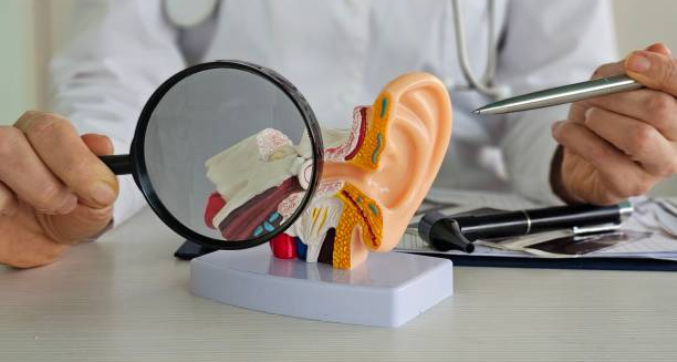
Doctor examining anatomical ear model with magnifying glass to illustrate causes of glue ear

Causes of Glue Ear

Causes of Glue Ear The causes of glue ear primarily involve malfunction or blockage of the Eustachian tube, the channel that connects the middle ear to the back of the nose. When this tube does not work properly, air cannot enter the middle ear, and fluid builds up instead. These mechanical and physiological disruptions are at the heart of most cases of glue ear in both children and adults. While the exact trigger can vary from person to person, several underlying causes contribute to Eustachian tube dysfunction and fluid accumulation. Understanding the causes of glue ear helps guide treatment decisions and prevent recurrent episodes. 1. Eustachian Tube Dysfunction The Eustachian tube plays a key role in balancing air pressure and draining fluid from the middle ear. In children, the tube is shorter, narrower, and more horizontal, making it more likely to become blocked. Common reasons for Eustachian tube dysfunction include: Immaturity of the tube in young children Swelling due to allergies or infections Nasal congestion from colds or flu Adenoid enlargement pressing on or blocking the tube Cleft palate or structural abnormalities interfering with normal function When the tube cannot open properly, negative pressure develops in the middle ear, drawing fluid into the space. 2. Respiratory Infections Frequent upper respiratory tract infections are one of the leading causes of glue ear. These infections inflame the mucous membranes lining the Eustachian tube and middle ear, resulting in: Increased mucus production Swelling and narrowing of the drainage passage Temporary blockage of normal air exchange Children in nursery or school settings are more exposed to colds, which increases their risk of developing glue ear. 3. Allergies Allergic rhinitis (hay fever) and other airborne allergies can lead to chronic inflammation of the nasal passages and Eustachian tubes. This can cause: Persistent fluid build-up in the middle ear Recurring or long-term episodes of glue ear Greater susceptibility to coexisting ear infections In allergic children, symptoms of glue ear may worsen during pollen seasons or when exposed to dust mites, pet dander, or mould. 4. Environmental and Lifestyle Factors Certain environmental exposures can raise the risk of developing glue ear, including: Passive smoking – increases inflammation in the airways and middle ear Bottle feeding in a lying-down position – can allow milk to enter the Eustachian tube Air pollution – irritates the respiratory tract and contributes to inflammation Poor ventilation and damp homes – increase exposure to allergens and infections Lifestyle changes, such as avoiding second-hand smoke and using upright feeding techniques, can help prevent some cases of glue ear. 5. Enlarged Adenoids Adenoids are lymphatic tissue located behind the nasal cavity. In young children, enlarged adenoids can: Physically block the Eustachian tubes Harbour bacteria that increase infection risk Cause chronic nasal obstruction and snoring Surgical removal of the adenoids (adenoidectomy) is sometimes recommended for children with recurrent or persistent glue ear. 6. Recurrent Ear Infections While glue ear itself is not an infection, recurrent ear infections can increase the risk of fluid build-up in the middle ear. Following an infection, the residual inflammation and fluid can persist even after the acute symptoms subside. Children with frequent ear infections may experience cycles of: Infection → fluid retention → temporary improvement → re-infection Breaking this cycle may require antibiotics, grommets, or addressing predisposing factors like allergies or enlarged adenoids. 7. Genetic and Anatomical Factors Some children inherit structural characteristics that make them more prone to glue ear. These may include: Narrow Eustachian tubes Familial tendency toward allergies or sinus issues Craniofacial syndromes such as Down’s syndrome Children with congenital anomalies affecting the skull or palate often require earlier and more proactive ENT (ear, nose, and throat) management. Summary of Key Causes Cause Role in Glue Ear Eustachian tube dysfunction Prevents air flow and fluid drainage Respiratory infections Inflammation and mucus production Allergies Chronic swelling of nasal and ear passages Passive smoking Increases airway inflammation Enlarged adenoids Block drainage and trap bacteria Recurrent ear infections Repeated inflammation and fluid retention Anatomical/genetic predisposition Increased susceptibility due to structure or family history Conclusion | Causes of Glue Ear The causes of glue ear are varied but often involve blockages or inflammation that prevent proper drainage of the middle ear. By identifying and addressing these underlying factors, healthcare professionals can reduce the risk of chronic episodes and help children maintain normal hearing and development. Whether caused by infection, allergy, or anatomy, early recognition of the causes of glue ear supports better outcomes and informed treatment planning. [Next: Symptoms of Glue Ear→]

Causes of Glue Ear Read More »

Doctor examining young boy's ear for signs of glue ear during a clinical assessment

Diagnosis of Glue Ear

Diagnosis of Glue Ear The diagnosis of glue ear relies on a careful combination of history, clinical examination, and specialised hearing tests. Because many children with this condition appear otherwise healthy, the diagnosis of glue ear is often delayed until a parent, caregiver, or teacher notices something is wrong—typically with hearing, speech, or behaviour. Glue ear (otitis media with effusion) causes a build-up of thick fluid behind the eardrum, reducing its ability to vibrate and conduct sound. Although painless, the condition can significantly affect a child’s ability to hear, especially in noisy settings like classrooms. Early and accurate diagnosis is critical to ensuring timely treatment and preventing long-term developmental issues. First Steps in Clinical Evaluation The diagnosis typically begins with: A detailed medical history, including frequency of colds, allergies, or previous ear infections Observation of any speech delays, listening difficulties, or behavioural concerns Parental and teacher reports regarding academic performance or social interaction A physical examination to check for nasal congestion, mouth breathing, or enlarged tonsils/adenoids These details help determine the likelihood that middle ear fluid is present and affecting hearing. Otoscopy Otoscopy is the first visual tool used by a GP or ENT specialist: A small handheld light is used to view the eardrum (tympanic membrane) In glue ear, the eardrum may appear dull, retracted, fluid-filled, or grey/yellow in colour Air bubbles or fluid levels may also be visible in the middle ear While this method provides immediate clues, it may not confirm the diagnosis on its own. Tympanometry Tympanometry is one of the most important tests for confirming glue ear: It measures how the eardrum responds to changes in air pressure In a healthy ear, the eardrum moves freely, creating a peak in the pressure reading In glue ear, movement is restricted, producing a flat or abnormal trace This test is painless and quick, and it’s suitable for children as young as six months. Audiometry Hearing tests are crucial in determining the degree and type of hearing loss: Pure-tone audiometry is used for children over 3 years The child responds to tones through headphones, allowing the audiologist to map hearing ability across different frequencies In glue ear, the results usually show conductive hearing loss, where sound transmission through the middle ear is impaired For younger children, visual reinforcement audiometry or play audiometry is used to assess hearing in a fun and engaging way. Observation Period Not all cases of glue ear require immediate treatment: If hearing loss is mild and recent, doctors may suggest a “watch and wait” approach for 6–12 weeks In many cases, the fluid clears spontaneously without medical intervention Follow-up appointments and repeat hearing tests help track progress However, prolonged or severe cases often need further evaluation. Additional Investigations If glue ear is persistent or recurrent, additional assessments may include: Speech and language evaluation, especially if the child is underperforming Assessment of the adenoids and tonsils, using nasal endoscopy or X-rays Allergy testing, if seasonal patterns or chronic nasal symptoms are present Children with craniofacial anomalies or developmental conditions may require more comprehensive evaluation and care coordination. When to Refer to a Specialist Referral to an ENT specialist is usually indicated when: Glue ear lasts longer than 3 months Hearing loss exceeds 20–30 decibels There is significant speech delay or academic struggle Episodes recur multiple times a year There is suspicion of anatomical issues (e.g. cleft palate, large adenoids) The specialist may recommend interventions such as grommet insertion or adenoidectomy depending on the severity and recurrence of symptoms. Diagnosis in Adults While less common, glue ear can affect adults, especially following: Air travel or scuba diving Sinus infections or nasal congestion Sudden hearing loss after a cold Diagnosis in adults follows a similar process but may require imaging if symptoms persist or are associated with dizziness or other complications. Conclusion | Diagnosis of Glue Ear The diagnosis of glue ear is based on careful clinical observation, otoscopy, tympanometry, and hearing assessments. Because the condition can affect hearing without causing pain, it is often overlooked. However, an early and accurate diagnosis of glue ear allows for appropriate treatment, minimising the impact on speech, learning, and quality of life. Regular follow-up ensures that temporary hearing loss does not become a long-term barrier to development. [Next: Treatment of Glue Ear→]

Diagnosis of Glue Ear Read More »

Doctor using otoscope to examine a child's ear for glue ear treatment options

Treatment of Glue Ear

Treatment of Glue Ear The treatment of glue ear depends on how long the condition has persisted, the severity of the hearing loss, and whether it is affecting speech or learning development. While the treatment of glue ear is often unnecessary in mild, short-term cases, persistent or recurrent episodes may require medical or surgical intervention to prevent long-term effects. Glue ear, also known as otitis media with effusion, causes a thick, sticky fluid to accumulate in the middle ear, reducing the ear’s ability to conduct sound. This may result in temporary hearing loss, which can affect a child’s ability to learn, speak clearly, and socialise confidently. Treatment decisions are based on a combination of clinical observation, hearing tests, and the child’s developmental needs. Watchful Waiting In most children, glue ear resolves on its own within a few weeks. Doctors may recommend a “watchful waiting” approach, especially if: The condition is recent (less than 3 months) The hearing loss is mild The child is otherwise developing normally During this observation period, parents are advised to: Monitor their child’s hearing, speech, and responsiveness Avoid passive smoking or other environmental triggers Encourage regular nose blowing to keep Eustachian tubes clear Return for follow-up assessments after 6–12 weeks Many children improve spontaneously as their Eustachian tubes mature or when seasonal allergies subside. Medical Treatments Although there is no medication that directly clears the fluid of glue ear, supportive treatments can relieve associated symptoms: 1. Decongestants and Antihistamines Often used if nasal congestion or allergies are contributing to Eustachian tube blockage May offer short-term relief but are not recommended for long-term use in young children 2. Nasal Steroid Sprays Reduce inflammation in the nasal passages and Eustachian tubes Useful in children with allergic rhinitis or enlarged adenoids Can be prescribed for 6–12 weeks under medical supervision 3. Antibiotics Not usually effective for glue ear unless there is a concurrent infection May be used in specific cases, particularly when glue ear follows acute otitis media It’s important to note that over-reliance on medication may delay more appropriate interventions if the condition is chronic. Grommet Insertion (Tympanostomy Tubes) Surgical grommet insertion is the most effective and widely used treatment for chronic glue ear. A grommet is a tiny ventilation tube placed in the eardrum to: Allow air into the middle ear Drain the sticky fluid Restore hearing almost immediately Indications for grommet insertion include: Glue ear lasting more than 3 months Hearing loss greater than 20–30 decibels Associated speech delay, learning difficulties, or behavioural problems Frequent recurrences affecting quality of life The procedure is performed under general anaesthetic and usually takes less than 15 minutes. Most children go home the same day. Grommets typically remain in place for 6–12 months before falling out naturally. Hearing usually returns to normal during this period. In some cases, a second procedure may be needed if glue ear returns. Adenoidectomy If enlarged adenoids are contributing to Eustachian tube blockage or recurrent infections, adenoidectomy (surgical removal of the adenoids) may be recommended alongside grommet insertion. This is especially helpful for children who: Snore or breathe through the mouth Have chronic nasal obstruction Experience frequent upper respiratory infections Removing the adenoids can improve middle ear ventilation and reduce the likelihood of recurrence. Hearing Aids In cases where surgery is not an option—such as in children with certain health conditions—temporary hearing aids may be offered to support communication while monitoring the ears for natural recovery. Bone-conduction hearing aids may also be used for children with structural issues that prevent effective grommet placement. Managing Glue Ear in Schools and Homes Even while waiting for treatment or monitoring the condition: Minimise background noise when speaking to the child Encourage face-to-face communication Use clear speech and visual cues Inform teachers and support staff about the child’s needs Speech therapy may be considered if there are concerns about language delays or articulation problems. Treatment in Adults Adults with glue ear often require different treatment approaches: Nasal sprays, decongestants, or allergy management Valsalva manoeuvre (gently blowing while pinching the nose) to equalise ear pressure Myringotomy or grommet insertion if fluid persists beyond 3 months Persistent cases in adults may signal underlying sinus issues or tumours and should be thoroughly investigated. Conclusion | Treatment of Glue Ear The treatment of glue ear varies depending on how the condition presents and whether it resolves on its own or causes complications. While observation is often sufficient, persistent cases may require grommets or other interventions. By tailoring the treatment of glue ear to the individual child or adult, long-term impacts on hearing, speech, and learning can be effectively minimised. [Next: Complications of Glue Ear →]

Treatment of Glue Ear Read More »

Scroll to Top