Mental Matters

Health Conditions

Find information on various health conditions affecting people of all ages. This category covers causes, symptoms, diagnosis, and management options for physical illnesses and chronic diseases. Get insights to help you understand different health challenges and make informed decisions about your wellbeing.

Child covering ears while watching TV, indicating potential hearing issues from glue ear

Complications of Glue Ear

Complications of Glue Ear The complications of glue ear can vary depending on the duration and severity of the condition. While many cases resolve without lasting effects, persistent glue ear can interfere with hearing, speech development, behaviour, and quality of life. Recognising the potential complications of glue ear helps ensure early diagnosis, appropriate treatment, and long-term success in communication and learning. Glue ear occurs when fluid accumulates in the middle ear, reducing the ear’s ability to conduct sound efficiently. This fluid build-up may last weeks or months and often fluctuates. Although painless, the condition can still lead to a range of health and developmental issues if left untreated. 1. Hearing Loss Mild to moderate conductive hearing loss is the most common complication of glue ear. This can affect both ears and: Leads to difficulty hearing soft or high-pitched sounds Makes it harder to understand speech in noisy environments May fluctuate from day to day, causing confusion for both child and caregivers Long-standing hearing loss may go unnoticed, especially in young children who cannot communicate their experience. 2. Delayed Speech and Language Development Children rely heavily on hearing to develop speech and language. When glue ear interferes with this process: Vocabulary acquisition is slowed Sentence formation may be delayed or incorrect Speech may be muffled, unclear, or difficult to understand Phonetic errors (mispronunciations) can become habitual Speech delays are most concerning in children under age 5, as this is a critical period for language development. These delays may continue even after hearing is restored. 3. Academic and Behavioural Difficulties Undiagnosed glue ear can affect a child’s school performance: Trouble following instructions or concentrating Perceived as inattentive, disobedient, or struggling academically Risk of being misdiagnosed with learning disabilities or behavioural disorders such as ADHD Poor hearing in a classroom setting leads to frustration and social withdrawal, especially when children are unable to keep up with their peers. 4. Balance Problems The middle ear plays a role in balance regulation. When fluid builds up: Children may appear clumsy or unsteady Toddlers may be delayed in walking or climbing Older children may experience dizziness, particularly during fast movement or exercise These issues can affect a child’s confidence in play and physical activity. 5. Ear Infections Glue ear increases the risk of developing acute ear infections (otitis media): The trapped fluid can become infected by bacteria Sudden pain, fever, and pus discharge may occur Repeated infections may cause scarring of the eardrum or perforation Children with glue ear are often more prone to ear infections in the winter months or following colds. 6. Tympanic Membrane Damage In some cases, the persistent presence of fluid and recurrent infections can lead to: Retraction pockets in the eardrum Thinning or perforation of the tympanic membrane Cholesteatoma, a rare growth of skin cells behind the eardrum These complications require ENT referral and possibly surgery to restore hearing and prevent further damage. 7. Delayed Social and Emotional Development Children with untreated glue ear may become: Frustrated or isolated due to communication difficulties Less engaged in conversation or group activities Less confident in expressing their thoughts Emotionally distressed, which may be misinterpreted as poor behaviour Early treatment and support are essential to avoid long-term social consequences. 8. Impact on Family Life Glue ear affects not only the child but also their caregivers: Parents may struggle to understand why their child is unresponsive or frustrated Repeated medical visits and uncertainty may cause stress and anxiety Costs related to hearing aids, speech therapy, or private medical care can be burdensome Family support, good communication with teachers, and early intervention can all help to reduce this impact. Summary of Common Complications Complication Description Hearing loss Reduced ability to hear, often mild to moderate Speech and language delays Slowed vocabulary growth, unclear speech Learning and behavioural issues Misdiagnosis or frustration in school Balance problems Clumsiness, dizziness, delayed mobility Ear infections Painful episodes that can scar the ear Eardrum damage Perforation or cholesteatoma in rare cases Social and emotional effects Isolation, low confidence, emotional distress Conclusion | Complications of Glue Ear The complications of glue ear are wide-ranging and can affect every aspect of a child’s development—from hearing and language to behaviour and emotional wellbeing. Fortunately, most complications are preventable with early identification and appropriate care. Understanding the potential complications of glue ear empowers parents, teachers, and healthcare professionals to act promptly, ensuring the best possible outcome for each child. [Next: Back to Overview →]


Complications of Glue Ear Read More »

Cartoon kidney with plaster and sad face symbolising glomerulonephritis

Glomerulonephritis

Glomerulonephritis Glomerulonephritis is a group of kidney conditions characterised by inflammation of the glomeruli, the tiny filtering units within the kidneys. These glomeruli remove waste, excess fluid, and toxins from the blood, producing urine. When damaged, the filtration process is disrupted, which can lead to protein and blood leaking into the urine. If glomerulonephritis is left untreated, it can progress to chronic kidney disease or even kidney failure. The condition can develop suddenly (acute) or gradually over time (chronic), and its severity varies significantly depending on the cause and how quickly it is identified. Prompt diagnosis and appropriate treatment are crucial to preserving kidney function and preventing long-term complications. Understanding the Glomeruli The kidneys contain approximately one million glomeruli each. These tiny capillary networks play a vital role in filtering blood, retaining essential proteins, and excreting waste. When glomeruli become inflamed or scarred, their ability to filter properly is impaired, which can lead to: Haematuria (blood in the urine) Proteinuria (excess protein in the urine) Reduced glomerular filtration rate (GFR) Fluid retention, causing swelling in various parts of the body In severe cases, the build-up of toxins and fluid imbalances can lead to high blood pressure, fatigue, and systemic complications. Types of Glomerulonephritis Glomerulonephritis is not a single disease but a spectrum of related conditions. It is typically classified into: 1. Acute Glomerulonephritis Develops rapidly over days or weeks Often follows an infection, such as strep throat or skin infections Symptoms include dark urine, puffiness around the eyes, and reduced urine output Can resolve spontaneously or progress to more serious kidney damage 2. Chronic Glomerulonephritis Develops gradually over months or years May be asymptomatic in the early stages Can lead to progressive loss of kidney function and end-stage kidney disease 3. Primary Glomerulonephritis The kidney is the main organ affected Includes diseases like minimal change disease, focal segmental glomerulosclerosis (FSGS), and IgA nephropathy 4. Secondary Glomerulonephritis Occurs as part of a systemic condition, such as: Lupus Diabetes Vasculitis Hepatitis B or C HIV Who Is at Risk? Glomerulonephritis can affect people of all ages, but certain groups face a higher risk: Children and young adults: More likely to develop acute forms after infections People with autoimmune diseases: Higher risk of chronic or secondary forms Individuals with a family history of kidney disorders People with diabetes, lupus, or viral infections Some forms are more prevalent in specific populations—for instance, FSGS is more common in people of African descent. Common Symptoms Symptoms vary depending on the type and severity but may include: Foamy or bubbly urine (due to protein loss) Blood in the urine, which may appear pink, red, or cola-coloured High blood pressure Swelling, particularly in the face, hands, feet, and abdomen Fatigue and weakness Decreased urination Unexplained weight gain from fluid retention In mild cases, symptoms may be subtle or entirely absent, making routine screening important for those at risk. Diagnosis Early and accurate diagnosis is essential for managing glomerulonephritis. A full workup typically includes: Urinalysis to detect blood, protein, and other abnormalities Blood tests to measure kidney function (e.g., creatinine, urea, GFR) Serological tests to identify autoimmune or infectious causes Imaging (such as ultrasound) to assess kidney size and structure Kidney biopsy for definitive diagnosis in unclear or severe cases A biopsy helps determine the specific type and extent of glomerular damage, guiding treatment decisions. Complications Untreated or poorly managed glomerulonephritis can lead to: Chronic kidney disease (CKD) End-stage renal disease (ESRD) requiring dialysis or transplant Nephrotic syndrome (high protein loss, low albumin, swelling) High blood pressure Electrolyte imbalances, such as high potassium Anaemia The prognosis depends on early detection, the underlying cause, and the individual’s response to treatment. Living with Glomerulonephritis People diagnosed with glomerulonephritis should work closely with a nephrologist. Management often includes: Regular monitoring of kidney function Blood pressure control Dietary modifications, such as reduced salt and protein intake Avoiding nephrotoxic medications, including NSAIDs Adherence to prescribed medication regimens With appropriate care, many patients can maintain stable kidney function and live normal lives. Conclusion of Glomerulonephritis Glomerulonephritis is a serious but manageable condition when caught early. By understanding the signs and seeking timely medical advice, individuals can protect their kidneys and prevent irreversible damage. With the right treatment and lifestyle changes, long-term outcomes are often positive. [Next: Causes of Glomerulonephritis→]

Glomerulonephritis Read More »

Illustration showing causes of glomerulonephritis with damaged kidneys

Causes of Glomerulonephritis

Causes of Glomerulonephritis Glomerulonephritis has many causes, including infections, autoimmune diseases, and inherited conditions. It develops when the glomeruli—the kidney’s filtering units—become inflamed, which damages kidney function. This inflammation may result from an immune response, direct injury to kidney tissue, or a broader systemic disease. Understanding the underlying cause is essential for selecting the most effective treatment to prevent long-term kidney damage. In many cases, the root cause determines whether the condition is temporary and treatable or chronic and progressive. Immune-Mediated Causes A significant number of glomerulonephritis cases are immune-mediated, meaning the body’s immune system makes the mistake of attacking its own tissues or reacts abnormally to infection. 1. Post-Infectious Glomerulonephritis Often follows streptococcal infections, such as strep throat or impetigo More common in children and adolescents Develops 1 to 3 weeks after infection Triggered by immune complex deposition in the glomeruli Other infections that can cause this include: Hepatitis B and C HIV Malaria Infective endocarditis Most cases resolve with supportive care, although some may progress to chronic disease. 2. IgA Nephropathy (Berger’s Disease) One of the most common causes of glomerulonephritis worldwide Caused by deposits of IgA antibodies in the glomeruli Leads to recurring blood in the urine, often after upper respiratory infections Can progress slowly over decades 3. Lupus Nephritis Occurs in people with systemic lupus erythaematosus (SLE) Autoantibodies attack the glomeruli Can lead to proteinuria, haematuria, and kidney failure Severity ranges from mild to rapidly progressive 4. ANCA-Associated Vasculitis This group includes granulomatosis with polyangiitis (GPA) and microscopic polyangiitis (MPA). Because this can worsen fast, treatment needs to start right away with strong medicines that calm down the immune system (immunosuppressive therapy). These conditions happen when the immune system mistakenly attacks small blood vessels in the body. As a result, they can quickly damage the kidneys, leading to a serious condition called rapidly progressive glomerulonephritis (RPGN). Genetic and Hereditary Causes Some forms of glomerulonephritis are inherited, often manifesting in childhood or adolescence. 1. Alport Syndrome Caused by cells that mutate too rapidly in the COL4A5 gene, affecting collagen in kidney cell-membranes Leads to progressive kidney failure Often associated with hearing loss and eye abnormalities 2. Thin Basement Membrane Disease Causes microscopic haematuria Usually benign but may progress to kidney issues in some families 3. Familial FSGS (Focal Segmental Glomerulosclerosis) Genetic defects in podocyte function May resist steroid treatment, causing issues. Can progress to ESRD Secondary to Systemic Diseases Glomerulonephritis may develop from issues caused by other illnesses: 1. Diabetes Mellitus Can lead to diabetic nephropathy, a leading cause of kidney failure Though not traditionally categorised as glomerulonephritis, similar pathology occurs 2. Goodpasture Syndrome Autoimmune disorder where antibodies attack both lungs and kidneys Causes bleeding in the lungs and rapidly progressive kidney failure Rare but extremely dangerous if untreated 3. Amyloidosis Abnormal protein deposits in organs, including kidneys Disrupt glomerular function Associated with chronic inflammation or blood disorders Environmental and Lifestyle-Related Causes Certain environmental factors may trigger glomerulonephritis or exacerbate existing inflammation. These include: Prolonged use of NSAIDs (e.g. ibuprofen) Exposure to heavy metals or toxins Use of recreational drugs, such as heroin Poorly controlled high blood pressure Though not direct causes in most cases, these factors can worsen the kidney damage and cause issues for existing glomerular disease. Idiopathic Glomerulonephritis In some cases, no clear cause is identified. These are termed idiopathic glomerulonephritis. They may still be managed effectively with: Immunosuppressive medications Lifestyle modifications Close monitoring to track kidney function Idiopathic conditions include minimal change disease and idiopathic membranous nephropathy. Summary of Main Causes Cause Type Examples Post-infectious Strep throat, hepatitis B/C, HIV Autoimmune Lupus, ANCA vasculitis, Goodpasture’s Genetic Alport syndrome, familial FSGS Systemic illness Diabetes, amyloidosis Environmental/lifestyle NSAIDs, toxins, drugs Idiopathic No identifiable cause Conclusion | Causes of Glomerulonephritis The causes of glomerulonephritis are diverse, ranging from infections and autoimmune disorders to inherited conditions. Identifying the specific cause is vital to guide treatment and assess the risk of progression. With early diagnosis and appropriate care, many people with glomerulonephritis can maintain healthy kidney function and avoid serious complications. [Next: Symptoms of Glomerulonephritis→]

Causes of Glomerulonephritis Read More »

Urine colour scale indicating dehydration and possible glomerulonephritis symptoms

Symptoms of Glomerulonephritis

Symptoms of Glomerulonephritis The symptoms of glomerulonephritis can vary significantly depending on the type, cause, and progression of the disease. Some people experience sudden and severe symptoms, while others live for years without noticing anything abnormal. In many cases, glomerulonephritis is only diagnosed during routine blood or urine tests. This is why awareness of early warning signs is so important, especially for those with known risk factors. The disease may present acutely—with a rapid onset of noticeable symptoms—or chronically, with subtle signs that appear gradually. In both forms, the underlying issue is inflammation of the glomeruli, the tiny filters in the kidneys responsible for removing waste and excess fluid from the blood. Early Signs and Subtle Symptoms In the early stages, symptoms may be non-specific or mild, making diagnosis difficult without targeted testing. Common early indicators include: Foamy or bubbly urine – a sign of proteinuria (protein in the urine) Dark, tea-coloured, or red urine – may indicate haematuria (blood in the urine) Puffiness around the eyes, especially in the morning Mild swelling in the ankles, feet, or hands These signs are often overlooked or mistaken for temporary water retention or urinary tract infections. Symptoms of Acute Glomerulonephritis Acute glomerulonephritis often appears suddenly and can be more dramatic in its presentation. This form is more common in children and young adults and may follow an infection like strep throat. Symptoms may include: Swelling (oedema) in the face, particularly around the eyes, as well as in the legs and feet Reduced urine output, even with normal fluid intake High blood pressure, sometimes severe Headaches and blurred vision, due to elevated blood pressure Shortness of breath, especially if fluid builds up in the lungs Fatigue and lethargy, caused by retained waste products in the blood (uraemia) In many cases, acute glomerulonephritis resolves on its own with supportive care. However, in severe cases, it may progress rapidly and require hospitalisation. Symptoms of Chronic Glomerulonephritis Chronic glomerulonephritis develops slowly over time, often without noticeable symptoms in the early stages. Many people are unaware they have the condition until significant kidney damage has already occurred. Symptoms may include: Persistent proteinuria or haematuria, detected through routine urine tests Gradual swelling, particularly in the lower limbs Long-standing high blood pressure that does not respond well to treatment Unexplained weight gain, due to fluid retention Nausea and loss of appetite Itching and dry skin, especially in later stages Muscle cramps or twitching, caused by electrolyte imbalances Difficulty concentrating or mental confusion in advanced cases These symptoms may worsen as kidney function declines, eventually leading to signs of chronic kidney disease or renal failure. Nephrotic Syndrome as a Symptom Complex Some types of glomerulonephritis, such as minimal change disease and membranous nephropathy, can lead to nephrotic syndrome, a distinct clinical presentation characterised by: Heavy proteinuria (more than 3.5 grams per day) Low blood albumin levels (hypoalbuminaemia) Severe oedema, particularly in the face, abdomen, and legs High cholesterol levels This syndrome is often associated with significant fatigue, increased risk of infections, and a tendency for blood clots. Red Flag Symptoms That Require Urgent Attention While glomerulonephritis is often manageable, certain symptoms should prompt immediate medical attention: Sudden swelling and rapid weight gain Severe high blood pressure or headaches Visible blood in the urine Marked decrease in urination Difficulty breathing or chest discomfort These signs could indicate acute kidney injury, pulmonary oedema, or hypertensive emergencies—all of which need urgent intervention. Paediatric Symptoms In children, symptoms may be similar to adults but may also include: Irritability or changes in behaviour Bedwetting or reduced daytime urination Loss of appetite or slower growth Because young children cannot always articulate their symptoms, parental vigilance and routine paediatric check-ups play a crucial role in early detection. How Symptoms Relate to Disease Progression Stage of Glomerulonephritis Typical Symptoms Early Fatigue, foamy urine, subtle swelling Moderate Hypertension, visible haematuria, oedema Advanced Nausea, mental confusion, shortness of breath End-stage renal disease Little or no urine, severe swelling, systemic symptoms Conclusion | Symptoms of Glomerulonephritis The symptoms of glomerulonephritis can be mild and easy to miss or severe and life-altering. Understanding these symptoms, especially when they occur gradually, allows for earlier diagnosis and more effective treatment. For those with known risk factors, routine screening can detect problems before irreversible damage occurs, offering the best chance at long-term kidney health. [Next: Diagnosis of Glomerulonephritis →]

Symptoms of Glomerulonephritis Read More »

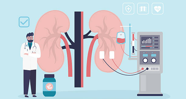
Illustration showing kidney diagnosis and dialysis setup for glomerulonephritis

Diagnosis of Glomerulonephritis

Diagnosis of Glomerulonephritis The diagnosis of glomerulonephritis is based on a combination of clinical history, physical examination, laboratory tests, and sometimes a kidney biopsy. Because glomerulonephritis can present subtly or mimic other conditions, accurate diagnosis is essential to prevent mismanagement and to tailor treatment effectively. Early diagnosis offers the best chance of preserving kidney function. With advances in diagnostic technology, doctors can now detect even small changes in kidney structure and function long before symptoms appear. Medical History and Clinical Examination The diagnostic process begins with a comprehensive medical history, where the doctor explores: Recent infections (e.g., strep throat, skin infections) Autoimmune diseases (e.g., lupus, vasculitis) Family history of kidney disease Medications, including NSAIDs or antibiotics Exposure to toxins, illicit drugs, or heavy metals A physical exam checks for: Oedema in the legs, face, or abdomen Elevated blood pressure Signs of fluid overload, such as lung crackles Skin rashes or joint inflammation (suggestive of systemic autoimmune disease) Urinalysis: The Cornerstone of Diagnosis Urine tests are often the first clue in identifying glomerulonephritis. Important findings include: Proteinuria – high levels of protein in the urine Haematuria – microscopic or visible blood Casts – tube-shaped particles made of proteins or cells, indicating kidney inflammation White or red blood cells – signalling infection or inflammation Dipstick urinalysis is useful for screening, while 24-hour urine collection quantifies protein loss. Blood Tests Several blood tests support the diagnosis and help identify underlying causes: 1. Kidney Function Tests Creatinine and urea: Elevated levels suggest impaired filtration Estimated glomerular filtration rate (eGFR): Indicates overall kidney function 2. Electrolyte and Metabolic Panels Assess sodium, potassium, calcium, phosphate Check acid-base balance 3. Complete Blood Count (CBC) May show anaemia (from chronic kidney disease) Elevated white cells in infection or inflammation 4. Autoimmune Markers ANA (antinuclear antibodies) for lupus ANCA for vasculitis Anti-GBM antibodies for Goodpasture syndrome Complement levels (C3, C4) – often low in post-infectious or lupus nephritis 5. Viral Serologies Tests for hepatitis B, hepatitis C, and HIV Essential in cases of secondary glomerulonephritis Imaging Studies While not diagnostic on their own, imaging helps assess kidney structure and rule out other issues. 1. Renal Ultrasound Non-invasive and widely used Detects kidney size, cysts, or obstruction Helps assess chronicity (e.g. shrunken kidneys in longstanding disease) 2. CT or MRI Rarely required but useful if complications are suspected Kidney Biopsy: The Definitive Test When the cause of glomerulonephritis is uncertain or the disease appears severe, a kidney biopsy may be necessary. This involves: Using a needle to take a small sample of kidney tissue Examining it under a microscope for signs of inflammation, scarring, or immune deposits A biopsy can: Confirm the specific subtype (e.g. IgA nephropathy, membranous nephropathy) Guide treatment decisions (e.g. steroid use, immunosuppressants) Help determine prognosis Biopsies are typically safe and performed under local anaesthesia with ultrasound guidance. Differential Diagnosis Several other conditions can mimic glomerulonephritis. A careful diagnostic process distinguishes it from: Urinary tract infections Interstitial nephritis Nephrotic syndrome of other causes Polycystic kidney disease Diabetic nephropathy Proper differentiation ensures the correct therapy and prevents further damage. Diagnostic Summary Table Test Purpose Urinalysis Detects blood, protein, and casts Blood tests Assess kidney function and inflammation Autoimmune markers Identify systemic causes Imaging Visualise kidney structure Kidney biopsy Definitive diagnosis and disease classification When to Refer to a Specialist Patients with signs of glomerulonephritis should be referred to a nephrologist if they: Have significant proteinuria or haematuria Show signs of rapid kidney deterioration Have resistant hypertension Need a biopsy or immunosuppressive therapy Early referral improves outcomes by allowing for targeted treatment before irreversible damage occurs. Conclusion | Diagnosis of Glomerulonephritis The diagnosis of glomerulonephritis involves a step-by-step evaluation using urine and blood tests, imaging, and sometimes biopsy. Accurate diagnosis is critical not only to confirm the condition but also to determine the underlying cause and appropriate treatment. With early detection and ongoing monitoring, patients can avoid complications and preserve their kidney health long term. [Next: Treatment of Glomerulonephritis→]

Diagnosis of Glomerulonephritis Read More »

3D illustration of human organs with capsules and tablets used for glomerulonephritis treatment

Treatment of Glomerulonephritis

Treatment of Glomerulonephritis The treatment of glomerulonephritis depends on the type, underlying cause, and severity of kidney involvement. In many cases, especially with mild or early disease, the treatment of glomerulonephritis focuses on managing symptoms, controlling blood pressure, and reducing inflammation to preserve kidney function. When an autoimmune disorder, infection, or genetic factor causes the condition, doctors must also treat the underlying cause to prevent progression. Tailoring treatment to the specific type of glomerulonephritis is essential. What works for one form may be inappropriate—or even harmful—for another. The goal is always the same: to protect the glomeruli from further damage and slow or prevent the onset of chronic kidney disease or renal failure. General Principles of Management Regardless of the underlying cause, doctors use several treatment approaches in most cases, including: 1. Blood Pressure Control High blood pressure accelerates glomerular damage, so effective management is critical. Doctors usually prescribe: ACE inhibitors or angiotensin receptor blockers (ARBs), which reduce both blood pressure and protein loss in the urine Lifestyle changes like low-sodium diets, weight loss, and regular exercise Doctors may use these medications to reduce proteinuria and protect kidney health, even in patients with normal blood pressure. 2. Dietary Adjustments Special dietary recommendations support kidney function and reduce strain on the glomeruli: Low-protein diet may be advised in some cases to reduce urea build-up Low-sodium diet to help control blood pressure and reduce oedema Restricted potassium or phosphorus intake in more advanced disease Adequate fluid intake to maintain hydration without overloading the kidneys Dieticians often work alongside nephrologists to personalise nutrition plans. 3. Diuretics If fluid retention is causing significant swelling, diuretics (such as furosemide) may be used to help the body eliminate excess fluid and reduce oedema. Treating the Underlying Cause The cornerstone of effective therapy is addressing the trigger behind the inflammation. 1. Infection-Related Glomerulonephritis Often self-limiting, particularly in children Antibiotics may be used if an active infection is still present Supportive care (rest, blood pressure control) is often sufficient Immunosuppressive drugs are generally avoided unless complications arise 2. Autoimmune or Immune-Mediated Types For conditions like lupus nephritis, IgA nephropathy, or ANCA-associated vasculitis, immunosuppressive therapy may be required: Corticosteroids (e.g. prednisone) to reduce inflammation Cyclophosphamide, azathioprine, or mycophenolate mofetil as steroid-sparing agents Rituximab for certain forms of vasculitis or resistant disease Close monitoring of immune markers, kidney function, and side effects is essential Immunosuppressive drugs can increase infection risk and require careful management. 3. Nephrotic Syndrome For nephrotic-range proteinuria, treatment focuses on: Controlling oedema with diuretics and salt restriction Reducing protein loss using ACE inhibitors or ARBs Lowering cholesterol, often with statins Corticosteroids or immunosuppressants depending on the underlying cause (e.g. minimal change disease, membranous nephropathy) Plasmapheresis and Advanced Therapies In rapidly progressive or life-threatening cases, such as Goodpasture syndrome or severe ANCA vasculitis, more aggressive measures may be used: Plasmapheresis (plasma exchange) to remove harmful antibodies Combined with high-dose steroids and immunosuppressants Used in hospital under specialist care These treatments are reserved for severe cases and require close monitoring in a renal or intensive care setting. Management of Chronic Glomerulonephritis For chronic forms, the emphasis is on slowing progression and preventing complications: Regular monitoring of kidney function, urine protein levels, and blood pressure Early detection of declining kidney function allows for preemptive planning Managing comorbidities like diabetes, anaemia, and cardiovascular disease In advanced stages, patients may need: Erythropoietin-stimulating agents to treat anaemia Phosphate binders or vitamin D supplements Dialysis or consideration for kidney transplant if kidney failure develops Patient Education and Lifestyle Empowering patients is a key part of managing glomerulonephritis: Understanding medication regimens Learning to monitor blood pressure at home Attending all scheduled blood and urine tests Avoiding non-prescribed NSAIDs or nephrotoxic drugs Vaccination against influenza, hepatitis B, and pneumococcus Education helps reduce hospitalisation rates and supports long-term kidney health. Conclusion | Treatment of Glomerulonephritis The treatment of glomerulonephritis is complex but often highly effective when tailored to the specific cause. Whether it involves immune suppression, infection control, or supportive care, the treatment of glomerulonephritis can slow progression, preserve function, and improve quality of life. Early diagnosis, consistent follow-up, and proactive patient involvement are the keys to successful outcomes. [Next: Complications of Glomerulonephritis →]

Treatment of Glomerulonephritis Read More »

Person holding lower back in pain, indicating kidney-related complications of glomerulonephritis

Complications of Glomerulonephritis

Complications of Glomerulonephritis The complications of glomerulonephritis can be serious, particularly when diagnosis is delayed or treatment is inadequate. While many cases resolve with minimal long-term damage, others lead to chronic kidney disease, systemic complications, or even kidney failure. Identifying the complications of glomerulonephritis early can prevent life-altering consequences and improve long-term outcomes. These complications arise as a result of ongoing inflammation, immune dysfunction, or scarring in the kidneys. The severity and likelihood of complications depend on the type of glomerulonephritis, the underlying cause, and how promptly it is managed. 1. Chronic Kidney Disease (CKD) One of the most common long-term outcomes of glomerulonephritis is chronic kidney disease. Repeated inflammation or ongoing damage to the glomeruli can lead to: Gradual decline in kidney function Increased serum creatinine and reduced GFR Accumulation of waste products in the blood CKD develops in stages and may remain stable for years if well managed. However, without appropriate treatment, it may eventually progress to kidney failure. 2. End-Stage Renal Disease (ESRD) If glomerular damage continues unchecked, end-stage renal disease can occur. This is the most severe complication and requires: Dialysis, either haemodialysis or peritoneal dialysis, to perform the kidney’s filtering role Kidney transplantation, when suitable ESRD significantly affects quality of life and life expectancy. It is more common in chronic or severe cases of glomerulonephritis, particularly those resistant to treatment. 3. Nephrotic Syndrome Several forms of glomerulonephritis can lead to nephrotic syndrome, a condition marked by: Severe proteinuria Low blood albumin levels Generalised oedema High cholesterol and blood lipid levels Nephrotic syndrome increases the risk of: Blood clots (venous thromboembolism) Infections, due to loss of immunoglobulins Malnutrition, if protein loss is significant Early and aggressive treatment can reverse nephrotic syndrome in many cases. 4. Hypertension (High Blood Pressure) Inflamed or scarred kidneys struggle to regulate fluid and salt, leading to high blood pressure, which in turn accelerates glomerular damage. Complications of uncontrolled hypertension include: Heart disease Stroke Vision loss Further kidney damage Managing blood pressure is essential to prevent worsening of glomerulonephritis. 5. Electrolyte and Metabolic Imbalances Advanced glomerular disease can disrupt electrolyte levels and cause: Hyperkalaemia (high potassium), increasing the risk of cardiac arrhythmias Metabolic acidosis, leading to fatigue and bone loss Calcium and phosphate imbalances, which contribute to bone disease These imbalances require close monitoring and may need specific medications or dietary adjustments. 6. Anaemia Reduced kidney function impairs the production of erythropoietin, a hormone required for red blood cell production. This can lead to: Chronic anaemia Fatigue, weakness, and reduced concentration Anaemia of chronic disease is common in later stages and often requires iron supplementation or erythropoiesis-stimulating agents. 7. Cardiovascular Disease Glomerulonephritis increases the risk of heart disease through several mechanisms: Hypertension High cholesterol (especially in nephrotic syndrome) Inflammation and oxidative stress Anaemia, which increases cardiac strain Cardiovascular events are a leading cause of death in people with chronic kidney disease, making prevention vital. 8. Psychological and Quality-of-Life Impacts Living with chronic illness can result in: Depression or anxiety Loss of independence Work or school disruptions Financial strain due to medical costs Early referral to counselling services, social workers, and patient support groups can help mitigate these challenges. Summary of Common Complications Complication Impact Chronic kidney disease Gradual loss of kidney function Kidney failure (ESRD) Requires dialysis or transplant Nephrotic syndrome Severe protein loss and immune risk Hypertension Heart disease, stroke, kidney damage Electrolyte imbalances Bone, heart, and muscle complications Anaemia Fatigue, reduced productivity Cardiovascular disease Increased mortality risk Mental health challenges Depression, anxiety, social impact Conclusion | Complications of Glomerulonephritis The complications of glomerulonephritis can range from manageable metabolic issues to life-threatening kidney failure. However, with early detection, appropriate treatment, and regular follow-up, many of these complications can be avoided or delayed. Addressing the complications of glomerulonephritis proactively not only protects kidney health but also improves overall quality of life and long-term outcomes. [Next: Back to Overview →]

Complications of Glomerulonephritis Read More »

Glaucoma

Glaucoma

Glaucoma Glaucoma is a group of eye conditions that damage the optic nerve, often due to abnormally high pressure inside the eye. Left untreated, glaucoma can lead to irreversible vision loss or even total blindness. It is one of the leading causes of preventable blindness worldwide, particularly among people over the age of 60. However, early diagnosis and proper treatment can preserve sight and significantly slow progression. The disease is typically painless in its early stages, which makes it particularly dangerous. Many people do not realise they have glaucoma until permanent damage has already occurred. Because of this, regular eye tests—especially after the age of 40—are vital to detect the condition before symptoms become apparent. What Is Glaucoma? Glaucoma refers to a family of eye diseases that share a common characteristic: progressive damage to the optic nerve, which connects the eye to the brain and is responsible for transmitting visual signals. Damage to the nerve impairs peripheral vision initially and can eventually affect central vision. In many cases, the damage is associated with elevated intraocular pressure (IOP), which results from a build-up of aqueous humour—the fluid inside the front part of the eye. When this fluid does not drain properly, pressure increases and damages the nerve fibres in the optic nerve. However, not all glaucoma patients have high eye pressure. In normal-tension glaucoma, optic nerve damage occurs despite IOP being within the normal range, likely due to poor blood flow or other risk factors. Types of Glaucoma There are several different forms of this condition, each with its own causes and characteristics: 1. Primary Open-Angle Glaucoma The most common type worldwide Caused by slow clogging of the eye’s drainage canals Develops gradually and often without symptoms Peripheral vision is affected first 2. Angle-Closure Glaucoma (Acute Glaucoma) Less common but more dangerous Occurs when the iris bulges forward and blocks the drainage angle Sudden increase in IOP leads to eye pain, redness, headache, and blurred vision A medical emergency that requires immediate treatment 3. Normal-Tension Glaucoma Optic nerve damage occurs despite normal IOP levels Risk factors include poor circulation, migraines, and vascular conditions 4. Congenital Glaucoma Present at birth due to developmental defects in the eye Symptoms include clouded cornea, enlarged eyes, and excessive tearing Requires surgery and lifelong monitoring 5. Secondary Glaucoma Caused by an underlying condition such as eye injury, inflammation, steroids, or diabetes Can mimic open-angle or angle-closure types Risk Factors Certain factors increase the risk of developing this condition: Age over 60 Family history of this condition African, Asian, or Hispanic ancestry High eye pressure (IOP) Thin corneas Diabetes or hypertension Prolonged use of corticosteroids Eye injuries or surgery history Because many of these risk factors are silent, routine eye screening is the most effective way to catch glaucoma early. Symptoms In its early stages, this conditionoften has no noticeable symptoms. This is why it is often called the “silent thief of sight.” In Primary Open-Angle Glaucoma: Vision loss begins at the edges of visual field May go unnoticed until significant peripheral vision is lost Gradually progresses to central vision if untreated In Acute Angle-Closure Glaucoma: Sudden onset of severe eye pain Redness, haloes around lights, and blurred vision Nausea and vomiting Requires immediate medical intervention to prevent permanent blindness How Glaucoma Is Diagnosed Routine eye exams include several tests to detect this condition, such as: Tonometry: Measures eye pressure Ophthalmoscopy: Examines the optic nerve Visual field testing: Detects peripheral vision loss Pachymetry: Measures corneal thickness Gonioscopy: Checks the drainage angle Most people are diagnosed during routine check-ups, underscoring the importance of regular eye care, especially in those with risk factors. Prevention and Screening While glaucoma cannot always be prevented, its progression can be significantly delayed. Prevention strategies include: Regular eye exams: Every 2 years for those over 40; annually if at risk Managing diabetes and blood pressure Protecting eyes from injury Avoiding prolonged steroid use unless medically necessary Early detection remains the most powerful tool in preserving vision. The Impact of Untreated Glaucoma If glaucoma is not diagnosed or managed, it can lead to: Permanent vision loss Blindness Reduced independence and quality of life Vision lost to glaucoma cannot be restored, which is why timely diagnosis and ongoing treatment are essential. Living with Glaucoma With proper care, most people with this condition retain good vision throughout life. Key to this is: Adherence to medication Routine follow-up with an ophthalmologist Reporting any new vision changes promptly Understanding glaucoma empowers individuals to seek appropriate care, protect their sight, and lead full, independent lives. [Next: Causes of Glaucoma →]

Glaucoma Read More »

Causes of Glaucoma

Causes of Glaucoma

Causes of Glaucoma The causes of glaucoma vary depending on the specific type of the condition. At its core, glaucoma is linked to damage of the optic nerve, often—but not always—caused by elevated intraocular pressure. Understanding what leads to increased pressure, as well as what can damage the optic nerve in normal-tension cases, is key to recognising risks and preventing sight loss. Although many factors contribute to the development of glaucoma, most involve the build-up of fluid in the eye, poor drainage, or damage from compromised blood supply. Genetics and systemic health conditions also play significant roles. Eye Fluid Dynamics The eye continuously produces a fluid called aqueous humour, which: Nourishes the eye tissues Maintains proper pressure inside the eye Drains through the trabecular meshwork at the drainage angle between the iris and cornea When this drainage system becomes blocked or inefficient, fluid builds up, leading to: Increased intraocular pressure (IOP) Compression of the optic nerve fibres Gradual death of nerve cells responsible for vision This process is central to primary open-angle glaucoma, the most common form of the disease. Causes of Different Types of Glaucoma 1. Primary Open-Angle Glaucoma This is usually caused by: Age-related changes in the eye’s drainage system Resistance to fluid outflow, even though the angle is open Genetic mutations affecting pressure regulation While eye pressure increases slowly, it eventually reaches a level that damages the optic nerve. Some people with high eye pressure never develop nerve damage, while others with normal pressure can develop glaucoma. 2. Angle-Closure Glaucoma This occurs when: The iris bulges forward, narrowing or blocking the drainage angle Fluid builds up rapidly, causing a sudden spike in IOP The eye cannot drain fluid quickly enough, creating acute symptoms Common triggers include: Pupil dilation in low light Certain medications Hyperopia (farsightedness), which can alter eye anatomy This type is more common in women, older adults, and people of Asian descent. 3. Normal-Tension Glaucoma In these cases, optic nerve damage occurs despite normal eye pressure. Possible causes include: Poor blood flow to the optic nerve Vascular conditions like low blood pressure or Raynaud’s disease Genetic factors Structural vulnerability of the optic nerve This form is more common in people of Japanese ancestry and those with a family history of glaucoma. 4. Congenital Glaucoma This rare form occurs in infants and young children. Causes include: Malformation of the drainage system during development Genetic defects affecting eye growth Inherited mutations passed from parents Early surgical treatment is often required to preserve vision. 5. Secondary Glaucoma Secondary glaucoma results from another condition that interferes with fluid drainage or increases pressure. Causes include: Eye trauma or surgery Inflammatory eye diseases (e.g. uveitis) Steroid medications Diabetes, which can cause neovascular glaucoma Tumours or lens dislocation Managing the underlying condition is essential for treating this form of glaucoma. Genetic and Family Factors Genetics play a significant role in all types of glaucoma. Those with a family history—especially first-degree relatives—are at four to nine times greater risk of developing the disease. Specific genes linked to glaucoma include: MYOC – associated with juvenile open-angle glaucoma OPTN and TBK1 – linked to normal-tension glaucoma CYP1B1 – associated with congenital glaucoma Because of the genetic link, screening and early testing are advised for relatives of diagnosed patients. Systemic Conditions That Increase Risk Certain systemic illnesses and lifestyle factors can contribute to glaucoma: Diabetes mellitus Hypertension or hypotension Obstructive sleep apnoea Migraine Use of steroids (oral, inhaled, or topical) Smoking and poor cardiovascular health These conditions may either raise eye pressure or reduce blood supply to the optic nerve. Environmental and Behavioural Factors While less clearly established, other potential contributors include: Chronic eye strain Low oxygen levels at high altitudes Excessive caffeine intake, which may raise IOP temporarily Prolonged stress, affecting vascular tone Though not direct causes, these factors may compound risk in already susceptible individuals. Summary of Main Causes Glaucoma Type Primary Cause Open-Angle Slow fluid drainage, age, genetics Angle-Closure Iris position, anatomy, sudden blockage Normal-Tension Poor blood flow, nerve vulnerability Congenital Developmental defects, genetic mutation Secondary Trauma, inflammation, medications Conclusion | Causes of Glaucoma The causes of glaucoma are diverse and often interrelated. Elevated eye pressure, poor fluid drainage, genetic predisposition, and vascular problems all contribute to the development of this sight-threatening condition. Identifying risk factors early and understanding the underlying mechanisms allows for better prevention, early diagnosis, and timely treatment. [Next: Symptoms of Glaucoma→]

Causes of Glaucoma Read More »

Symptoms of Glaucoma

Symptoms of Glaucoma

Symptoms of Glaucoma The symptoms of glaucoma depend on the type and stage of the disease. In its early stages, glaucoma is often silent and symptomless, which is why it is commonly referred to as the “silent thief of sight.” Many people do not realise they have the condition until irreversible damage to their vision has already occurred. Recognising the different symptom patterns between the various types of glaucoma is crucial for timely diagnosis and prevention of vision loss. In most cases, symptoms progress gradually, affecting peripheral vision first. However, certain types—such as acute angle-closure glaucoma—can cause rapid, painful symptoms that require emergency intervention. Understanding what to look out for, even in the absence of discomfort, empowers individuals to protect their eyesight through regular screenings and prompt action. Symptoms of Primary Open-Angle Glaucoma This is the most common form of glaucoma globally, accounting for approximately 70–90% of cases. It progresses slowly and often goes unnoticed for years. Typical symptoms include: Gradual loss of peripheral (side) vision Tunnel vision in advanced stages Difficulty adjusting to low light or glare Blurred vision in the late stages Because central vision remains intact until the disease is advanced, many people do not realise their peripheral vision is deteriorating. By the time vision loss is noticed, substantial and irreversible optic nerve damage may have already occurred. Symptoms of Acute Angle-Closure Glaucoma This type presents suddenly and requires urgent treatment to prevent permanent vision loss. It is less common but far more dramatic in presentation. Common signs and symptoms include: Severe eye pain, often in one eye Redness of the eye Blurry vision or haloes around lights Sudden visual loss Headache, especially on the affected side Nausea and vomiting This occurs when the drainage angle in the eye becomes abruptly blocked, causing a rapid rise in intraocular pressure. If not treated within hours, it can lead to permanent blindness. Anyone experiencing these symptoms should seek emergency medical attention. Symptoms of Normal-Tension Glaucoma Despite normal intraocular pressure, optic nerve damage still occurs in this form of the disease. The symptoms mirror those of open-angle glaucoma but may appear in people without the traditional risk of high eye pressure. These symptoms include: Gradual peripheral vision loss Difficulty seeing in dim light Occasional blurred or distorted vision Because pressure readings may be normal, diagnosis can be delayed unless the optic nerve is thoroughly assessed during routine eye exams. Symptoms of Secondary Glaucoma Secondary glaucoma arises from other medical conditions, eye injuries, or medications. Symptoms vary depending on the underlying cause but may include: Blurred vision Eye pain or redness Increased sensitivity to light Visual disturbances related to inflammation, trauma, or cataract complications Sometimes the symptoms of the primary condition—such as uveitis or steroid-induced IOP elevation—overshadow the signs of glaucoma, making early detection more challenging. Symptoms in Congenital Glaucoma This rare form affects newborns and infants. The signs may include: Cloudy cornea Excessive tearing (epiphora) Enlarged eyes (buphthalmos) Light sensitivity (photophobia) Failure to thrive due to discomfort Parents often notice that their infant has unusually large, shiny eyes. Prompt referral to a paediatric ophthalmologist is essential to prevent lifelong visual impairment. Vision Loss Pattern in Glaucoma Glaucoma typically affects peripheral vision first. As the disease progresses: Vision loss extends towards the centre Blind spots develop in the visual field Tunnel vision becomes more pronounced In the final stages, central vision is lost, potentially leading to complete blindness Because this pattern of vision loss is painless and slow, people may adapt unconsciously—turning their heads more often or relying on the unaffected eye—without realising the extent of the damage. Psychological and Functional Symptoms As vision deteriorates, people with glaucoma may experience: Difficulty reading or driving Reduced mobility, especially in unfamiliar environments Increased risk of falls or collisions Anxiety, frustration, or depression due to declining independence While vision cannot be restored, timely treatment can preserve what remains and slow the progression of symptoms. Summary of Symptoms by Type Type of Glaucoma Key Symptoms Open-angle Slow peripheral vision loss, tunnel vision Angle-closure Sudden pain, haloes, blurred vision, nausea Normal-tension Vision loss with normal IOP, often unnoticed Secondary Depends on underlying condition (pain, redness) Congenital Cloudy eyes, tearing, light sensitivity in infants Conclusion | Symptoms of Glaucoma The symptoms of glaucoma range from subtle and progressive to sudden and severe, depending on the type. Because early stages are often asymptomatic, regular eye examinations are essential—especially for those at increased risk. Early detection offers the best chance of preserving vision and maintaining quality of life. [Next: Diagnosis of Glaucoma →]

Symptoms of Glaucoma Read More »

Scroll to Top