Mental Matters

Author name: admin

Toddler touching ear with discomfort, showing signs of glue ear

Symptoms of Glue Ear

Symptoms of Glue Ear The symptoms of glue ear can be difficult to detect, particularly in young children who may not be able to describe what they’re experiencing. Many parents and teachers only realise there’s a problem when they observe behavioural changes or delays in communication. Because the symptoms of glue ear often develop gradually and are not painful, they may go unnoticed for weeks or even months. Glue ear (otitis media with effusion) affects the middle ear, where a thick, sticky fluid accumulates behind the eardrum. This fluid prevents sound vibrations from reaching the inner ear effectively, resulting in a mild to moderate hearing loss. Identifying the symptoms early is key to minimising its impact on language development, learning, and social interaction—especially in younger children. Key Indicators in Infants and Toddlers Children under three are the most affected by glue ear, but they are also the least likely to be able to describe what they’re feeling. Parents and caregivers should look for the following signs: Delayed speech or unclear words compared to children of the same age Lack of response when spoken to from behind or in noisy environments Startling only when they see someone, not when they hear them Turning the TV volume up unusually high Frequent tugging or rubbing of the ears, although this is less common than with infections Clumsiness or poor balance, since the middle ear also helps with equilibrium Many of these signs may also be seen in children with general hearing loss, so it’s important to have a proper diagnosis from a healthcare provider. Behavioural Clues in Older Children School-aged children may display a range of subtle behaviours that point to undiagnosed glue ear: Daydreaming or appearing inattentive in class Difficulty following instructions, especially in group settings Speaking loudly or repeating questions unnecessarily Frustration or emotional outbursts due to communication difficulties Mispronunciation of words or avoiding speaking entirely in group settings These behaviours can sometimes be misinterpreted as learning difficulties, ADHD, or oppositional behaviour, when in fact they stem from an undiagnosed hearing impairment. Physical Symptoms Although glue ear itself does not usually cause pain, some physical symptoms can still appear: Muffled hearing, described by older children as having “blocked ears” or “underwater” hearing Tinnitus, or a ringing/buzzing in the ears A feeling of pressure or fullness in the affected ear Mild dizziness or balance problems during movement or play In some cases, a recent cold or upper respiratory infection may have preceded the development of these symptoms, especially during the winter months. Duration and Patterns The symptoms of glue ear can fluctuate: Some children experience them intermittently, especially after viral illnesses Others have persistent issues lasting for several weeks or even months Hearing loss is often bilateral, affecting both ears, although it may be more noticeable in one Parents may find that symptoms are worse during allergy season or when their child has a stuffy nose. Long-Term Impacts If glue ear persists without treatment: Language and speech development can be significantly delayed Children may fall behind in reading, comprehension, and literacy skills Social withdrawal or low confidence may develop Children may struggle with peer interaction or be mislabelled as disruptive The long-term consequences of ignoring the symptoms of glue ear can extend well beyond childhood. That’s why early detection and management are essential. When to Seek Medical Advice You should consult a doctor or audiologist if: Your child has any persistent hearing loss or unclear speech The symptoms have lasted longer than four to six weeks There is evidence of speech delay or learning difficulties Teachers or caregivers have raised concerns about listening or behaviour The child has repeated episodes during the year Referral to an ENT (ear, nose, and throat) specialist may be recommended if symptoms are severe, chronic, or recurrent. Conclusion | Symptoms of Glue Ear The symptoms of glue ear can range from subtle hearing loss to more noticeable speech and behavioural changes. Recognising the early signs allows parents and teachers to seek medical evaluation before the condition affects the child’s development. With close observation and early intervention, the impact of the symptoms of glue ear can be minimised, ensuring better communication and learning outcomes. [Next: Diagnosis of Glue Ear →]

Symptoms of Glue Ear Read More »

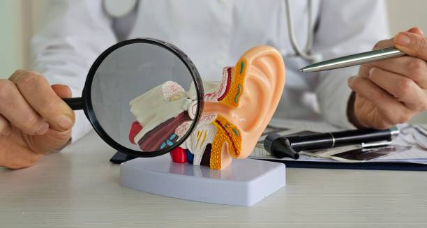
Doctor examining anatomical ear model with magnifying glass to illustrate causes of glue ear

Causes of Glue Ear

Causes of Glue Ear The causes of glue ear primarily involve malfunction or blockage of the Eustachian tube, the channel that connects the middle ear to the back of the nose. When this tube does not work properly, air cannot enter the middle ear, and fluid builds up instead. These mechanical and physiological disruptions are at the heart of most cases of glue ear in both children and adults. While the exact trigger can vary from person to person, several underlying causes contribute to Eustachian tube dysfunction and fluid accumulation. Understanding the causes of glue ear helps guide treatment decisions and prevent recurrent episodes. 1. Eustachian Tube Dysfunction The Eustachian tube plays a key role in balancing air pressure and draining fluid from the middle ear. In children, the tube is shorter, narrower, and more horizontal, making it more likely to become blocked. Common reasons for Eustachian tube dysfunction include: Immaturity of the tube in young children Swelling due to allergies or infections Nasal congestion from colds or flu Adenoid enlargement pressing on or blocking the tube Cleft palate or structural abnormalities interfering with normal function When the tube cannot open properly, negative pressure develops in the middle ear, drawing fluid into the space. 2. Respiratory Infections Frequent upper respiratory tract infections are one of the leading causes of glue ear. These infections inflame the mucous membranes lining the Eustachian tube and middle ear, resulting in: Increased mucus production Swelling and narrowing of the drainage passage Temporary blockage of normal air exchange Children in nursery or school settings are more exposed to colds, which increases their risk of developing glue ear. 3. Allergies Allergic rhinitis (hay fever) and other airborne allergies can lead to chronic inflammation of the nasal passages and Eustachian tubes. This can cause: Persistent fluid build-up in the middle ear Recurring or long-term episodes of glue ear Greater susceptibility to coexisting ear infections In allergic children, symptoms of glue ear may worsen during pollen seasons or when exposed to dust mites, pet dander, or mould. 4. Environmental and Lifestyle Factors Certain environmental exposures can raise the risk of developing glue ear, including: Passive smoking – increases inflammation in the airways and middle ear Bottle feeding in a lying-down position – can allow milk to enter the Eustachian tube Air pollution – irritates the respiratory tract and contributes to inflammation Poor ventilation and damp homes – increase exposure to allergens and infections Lifestyle changes, such as avoiding second-hand smoke and using upright feeding techniques, can help prevent some cases of glue ear. 5. Enlarged Adenoids Adenoids are lymphatic tissue located behind the nasal cavity. In young children, enlarged adenoids can: Physically block the Eustachian tubes Harbour bacteria that increase infection risk Cause chronic nasal obstruction and snoring Surgical removal of the adenoids (adenoidectomy) is sometimes recommended for children with recurrent or persistent glue ear. 6. Recurrent Ear Infections While glue ear itself is not an infection, recurrent ear infections can increase the risk of fluid build-up in the middle ear. Following an infection, the residual inflammation and fluid can persist even after the acute symptoms subside. Children with frequent ear infections may experience cycles of: Infection → fluid retention → temporary improvement → re-infection Breaking this cycle may require antibiotics, grommets, or addressing predisposing factors like allergies or enlarged adenoids. 7. Genetic and Anatomical Factors Some children inherit structural characteristics that make them more prone to glue ear. These may include: Narrow Eustachian tubes Familial tendency toward allergies or sinus issues Craniofacial syndromes such as Down’s syndrome Children with congenital anomalies affecting the skull or palate often require earlier and more proactive ENT (ear, nose, and throat) management. Summary of Key Causes Cause Role in Glue Ear Eustachian tube dysfunction Prevents air flow and fluid drainage Respiratory infections Inflammation and mucus production Allergies Chronic swelling of nasal and ear passages Passive smoking Increases airway inflammation Enlarged adenoids Block drainage and trap bacteria Recurrent ear infections Repeated inflammation and fluid retention Anatomical/genetic predisposition Increased susceptibility due to structure or family history Conclusion | Causes of Glue Ear The causes of glue ear are varied but often involve blockages or inflammation that prevent proper drainage of the middle ear. By identifying and addressing these underlying factors, healthcare professionals can reduce the risk of chronic episodes and help children maintain normal hearing and development. Whether caused by infection, allergy, or anatomy, early recognition of the causes of glue ear supports better outcomes and informed treatment planning. [Next: Symptoms of Glue Ear→]

Causes of Glue Ear Read More »

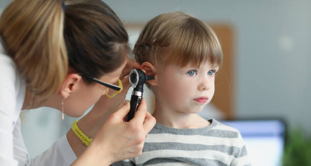
Doctor examining young boy's ear for signs of glue ear during a clinical assessment

Diagnosis of Glue Ear

Diagnosis of Glue Ear The diagnosis of glue ear relies on a careful combination of history, clinical examination, and specialised hearing tests. Because many children with this condition appear otherwise healthy, the diagnosis of glue ear is often delayed until a parent, caregiver, or teacher notices something is wrong—typically with hearing, speech, or behaviour. Glue ear (otitis media with effusion) causes a build-up of thick fluid behind the eardrum, reducing its ability to vibrate and conduct sound. Although painless, the condition can significantly affect a child’s ability to hear, especially in noisy settings like classrooms. Early and accurate diagnosis is critical to ensuring timely treatment and preventing long-term developmental issues. First Steps in Clinical Evaluation The diagnosis typically begins with: A detailed medical history, including frequency of colds, allergies, or previous ear infections Observation of any speech delays, listening difficulties, or behavioural concerns Parental and teacher reports regarding academic performance or social interaction A physical examination to check for nasal congestion, mouth breathing, or enlarged tonsils/adenoids These details help determine the likelihood that middle ear fluid is present and affecting hearing. Otoscopy Otoscopy is the first visual tool used by a GP or ENT specialist: A small handheld light is used to view the eardrum (tympanic membrane) In glue ear, the eardrum may appear dull, retracted, fluid-filled, or grey/yellow in colour Air bubbles or fluid levels may also be visible in the middle ear While this method provides immediate clues, it may not confirm the diagnosis on its own. Tympanometry Tympanometry is one of the most important tests for confirming glue ear: It measures how the eardrum responds to changes in air pressure In a healthy ear, the eardrum moves freely, creating a peak in the pressure reading In glue ear, movement is restricted, producing a flat or abnormal trace This test is painless and quick, and it’s suitable for children as young as six months. Audiometry Hearing tests are crucial in determining the degree and type of hearing loss: Pure-tone audiometry is used for children over 3 years The child responds to tones through headphones, allowing the audiologist to map hearing ability across different frequencies In glue ear, the results usually show conductive hearing loss, where sound transmission through the middle ear is impaired For younger children, visual reinforcement audiometry or play audiometry is used to assess hearing in a fun and engaging way. Observation Period Not all cases of glue ear require immediate treatment: If hearing loss is mild and recent, doctors may suggest a “watch and wait” approach for 6–12 weeks In many cases, the fluid clears spontaneously without medical intervention Follow-up appointments and repeat hearing tests help track progress However, prolonged or severe cases often need further evaluation. Additional Investigations If glue ear is persistent or recurrent, additional assessments may include: Speech and language evaluation, especially if the child is underperforming Assessment of the adenoids and tonsils, using nasal endoscopy or X-rays Allergy testing, if seasonal patterns or chronic nasal symptoms are present Children with craniofacial anomalies or developmental conditions may require more comprehensive evaluation and care coordination. When to Refer to a Specialist Referral to an ENT specialist is usually indicated when: Glue ear lasts longer than 3 months Hearing loss exceeds 20–30 decibels There is significant speech delay or academic struggle Episodes recur multiple times a year There is suspicion of anatomical issues (e.g. cleft palate, large adenoids) The specialist may recommend interventions such as grommet insertion or adenoidectomy depending on the severity and recurrence of symptoms. Diagnosis in Adults While less common, glue ear can affect adults, especially following: Air travel or scuba diving Sinus infections or nasal congestion Sudden hearing loss after a cold Diagnosis in adults follows a similar process but may require imaging if symptoms persist or are associated with dizziness or other complications. Conclusion | Diagnosis of Glue Ear The diagnosis of glue ear is based on careful clinical observation, otoscopy, tympanometry, and hearing assessments. Because the condition can affect hearing without causing pain, it is often overlooked. However, an early and accurate diagnosis of glue ear allows for appropriate treatment, minimising the impact on speech, learning, and quality of life. Regular follow-up ensures that temporary hearing loss does not become a long-term barrier to development. [Next: Treatment of Glue Ear→]

Diagnosis of Glue Ear Read More »

Doctor using otoscope to examine a child's ear for glue ear treatment options

Treatment of Glue Ear

Treatment of Glue Ear The treatment of glue ear depends on how long the condition has persisted, the severity of the hearing loss, and whether it is affecting speech or learning development. While the treatment of glue ear is often unnecessary in mild, short-term cases, persistent or recurrent episodes may require medical or surgical intervention to prevent long-term effects. Glue ear, also known as otitis media with effusion, causes a thick, sticky fluid to accumulate in the middle ear, reducing the ear’s ability to conduct sound. This may result in temporary hearing loss, which can affect a child’s ability to learn, speak clearly, and socialise confidently. Treatment decisions are based on a combination of clinical observation, hearing tests, and the child’s developmental needs. Watchful Waiting In most children, glue ear resolves on its own within a few weeks. Doctors may recommend a “watchful waiting” approach, especially if: The condition is recent (less than 3 months) The hearing loss is mild The child is otherwise developing normally During this observation period, parents are advised to: Monitor their child’s hearing, speech, and responsiveness Avoid passive smoking or other environmental triggers Encourage regular nose blowing to keep Eustachian tubes clear Return for follow-up assessments after 6–12 weeks Many children improve spontaneously as their Eustachian tubes mature or when seasonal allergies subside. Medical Treatments Although there is no medication that directly clears the fluid of glue ear, supportive treatments can relieve associated symptoms: 1. Decongestants and Antihistamines Often used if nasal congestion or allergies are contributing to Eustachian tube blockage May offer short-term relief but are not recommended for long-term use in young children 2. Nasal Steroid Sprays Reduce inflammation in the nasal passages and Eustachian tubes Useful in children with allergic rhinitis or enlarged adenoids Can be prescribed for 6–12 weeks under medical supervision 3. Antibiotics Not usually effective for glue ear unless there is a concurrent infection May be used in specific cases, particularly when glue ear follows acute otitis media It’s important to note that over-reliance on medication may delay more appropriate interventions if the condition is chronic. Grommet Insertion (Tympanostomy Tubes) Surgical grommet insertion is the most effective and widely used treatment for chronic glue ear. A grommet is a tiny ventilation tube placed in the eardrum to: Allow air into the middle ear Drain the sticky fluid Restore hearing almost immediately Indications for grommet insertion include: Glue ear lasting more than 3 months Hearing loss greater than 20–30 decibels Associated speech delay, learning difficulties, or behavioural problems Frequent recurrences affecting quality of life The procedure is performed under general anaesthetic and usually takes less than 15 minutes. Most children go home the same day. Grommets typically remain in place for 6–12 months before falling out naturally. Hearing usually returns to normal during this period. In some cases, a second procedure may be needed if glue ear returns. Adenoidectomy If enlarged adenoids are contributing to Eustachian tube blockage or recurrent infections, adenoidectomy (surgical removal of the adenoids) may be recommended alongside grommet insertion. This is especially helpful for children who: Snore or breathe through the mouth Have chronic nasal obstruction Experience frequent upper respiratory infections Removing the adenoids can improve middle ear ventilation and reduce the likelihood of recurrence. Hearing Aids In cases where surgery is not an option—such as in children with certain health conditions—temporary hearing aids may be offered to support communication while monitoring the ears for natural recovery. Bone-conduction hearing aids may also be used for children with structural issues that prevent effective grommet placement. Managing Glue Ear in Schools and Homes Even while waiting for treatment or monitoring the condition: Minimise background noise when speaking to the child Encourage face-to-face communication Use clear speech and visual cues Inform teachers and support staff about the child’s needs Speech therapy may be considered if there are concerns about language delays or articulation problems. Treatment in Adults Adults with glue ear often require different treatment approaches: Nasal sprays, decongestants, or allergy management Valsalva manoeuvre (gently blowing while pinching the nose) to equalise ear pressure Myringotomy or grommet insertion if fluid persists beyond 3 months Persistent cases in adults may signal underlying sinus issues or tumours and should be thoroughly investigated. Conclusion | Treatment of Glue Ear The treatment of glue ear varies depending on how the condition presents and whether it resolves on its own or causes complications. While observation is often sufficient, persistent cases may require grommets or other interventions. By tailoring the treatment of glue ear to the individual child or adult, long-term impacts on hearing, speech, and learning can be effectively minimised. [Next: Complications of Glue Ear →]

Treatment of Glue Ear Read More »

Child covering ears while watching TV, indicating potential hearing issues from glue ear

Complications of Glue Ear

Complications of Glue Ear The complications of glue ear can vary depending on the duration and severity of the condition. While many cases resolve without lasting effects, persistent glue ear can interfere with hearing, speech development, behaviour, and quality of life. Recognising the potential complications of glue ear helps ensure early diagnosis, appropriate treatment, and long-term success in communication and learning. Glue ear occurs when fluid accumulates in the middle ear, reducing the ear’s ability to conduct sound efficiently. This fluid build-up may last weeks or months and often fluctuates. Although painless, the condition can still lead to a range of health and developmental issues if left untreated. 1. Hearing Loss Mild to moderate conductive hearing loss is the most common complication of glue ear. This can affect both ears and: Leads to difficulty hearing soft or high-pitched sounds Makes it harder to understand speech in noisy environments May fluctuate from day to day, causing confusion for both child and caregivers Long-standing hearing loss may go unnoticed, especially in young children who cannot communicate their experience. 2. Delayed Speech and Language Development Children rely heavily on hearing to develop speech and language. When glue ear interferes with this process: Vocabulary acquisition is slowed Sentence formation may be delayed or incorrect Speech may be muffled, unclear, or difficult to understand Phonetic errors (mispronunciations) can become habitual Speech delays are most concerning in children under age 5, as this is a critical period for language development. These delays may continue even after hearing is restored. 3. Academic and Behavioural Difficulties Undiagnosed glue ear can affect a child’s school performance: Trouble following instructions or concentrating Perceived as inattentive, disobedient, or struggling academically Risk of being misdiagnosed with learning disabilities or behavioural disorders such as ADHD Poor hearing in a classroom setting leads to frustration and social withdrawal, especially when children are unable to keep up with their peers. 4. Balance Problems The middle ear plays a role in balance regulation. When fluid builds up: Children may appear clumsy or unsteady Toddlers may be delayed in walking or climbing Older children may experience dizziness, particularly during fast movement or exercise These issues can affect a child’s confidence in play and physical activity. 5. Ear Infections Glue ear increases the risk of developing acute ear infections (otitis media): The trapped fluid can become infected by bacteria Sudden pain, fever, and pus discharge may occur Repeated infections may cause scarring of the eardrum or perforation Children with glue ear are often more prone to ear infections in the winter months or following colds. 6. Tympanic Membrane Damage In some cases, the persistent presence of fluid and recurrent infections can lead to: Retraction pockets in the eardrum Thinning or perforation of the tympanic membrane Cholesteatoma, a rare growth of skin cells behind the eardrum These complications require ENT referral and possibly surgery to restore hearing and prevent further damage. 7. Delayed Social and Emotional Development Children with untreated glue ear may become: Frustrated or isolated due to communication difficulties Less engaged in conversation or group activities Less confident in expressing their thoughts Emotionally distressed, which may be misinterpreted as poor behaviour Early treatment and support are essential to avoid long-term social consequences. 8. Impact on Family Life Glue ear affects not only the child but also their caregivers: Parents may struggle to understand why their child is unresponsive or frustrated Repeated medical visits and uncertainty may cause stress and anxiety Costs related to hearing aids, speech therapy, or private medical care can be burdensome Family support, good communication with teachers, and early intervention can all help to reduce this impact. Summary of Common Complications Complication Description Hearing loss Reduced ability to hear, often mild to moderate Speech and language delays Slowed vocabulary growth, unclear speech Learning and behavioural issues Misdiagnosis or frustration in school Balance problems Clumsiness, dizziness, delayed mobility Ear infections Painful episodes that can scar the ear Eardrum damage Perforation or cholesteatoma in rare cases Social and emotional effects Isolation, low confidence, emotional distress Conclusion | Complications of Glue Ear The complications of glue ear are wide-ranging and can affect every aspect of a child’s development—from hearing and language to behaviour and emotional wellbeing. Fortunately, most complications are preventable with early identification and appropriate care. Understanding the potential complications of glue ear empowers parents, teachers, and healthcare professionals to act promptly, ensuring the best possible outcome for each child. [Next: Back to Overview →]

Complications of Glue Ear Read More »

Cartoon kidney with plaster and sad face symbolising glomerulonephritis

Glomerulonephritis

Glomerulonephritis Glomerulonephritis is a group of kidney conditions characterised by inflammation of the glomeruli, the tiny filtering units within the kidneys. These glomeruli remove waste, excess fluid, and toxins from the blood, producing urine. When damaged, the filtration process is disrupted, which can lead to protein and blood leaking into the urine. If glomerulonephritis is left untreated, it can progress to chronic kidney disease or even kidney failure. The condition can develop suddenly (acute) or gradually over time (chronic), and its severity varies significantly depending on the cause and how quickly it is identified. Prompt diagnosis and appropriate treatment are crucial to preserving kidney function and preventing long-term complications. Understanding the Glomeruli The kidneys contain approximately one million glomeruli each. These tiny capillary networks play a vital role in filtering blood, retaining essential proteins, and excreting waste. When glomeruli become inflamed or scarred, their ability to filter properly is impaired, which can lead to: Haematuria (blood in the urine) Proteinuria (excess protein in the urine) Reduced glomerular filtration rate (GFR) Fluid retention, causing swelling in various parts of the body In severe cases, the build-up of toxins and fluid imbalances can lead to high blood pressure, fatigue, and systemic complications. Types of Glomerulonephritis Glomerulonephritis is not a single disease but a spectrum of related conditions. It is typically classified into: 1. Acute Glomerulonephritis Develops rapidly over days or weeks Often follows an infection, such as strep throat or skin infections Symptoms include dark urine, puffiness around the eyes, and reduced urine output Can resolve spontaneously or progress to more serious kidney damage 2. Chronic Glomerulonephritis Develops gradually over months or years May be asymptomatic in the early stages Can lead to progressive loss of kidney function and end-stage kidney disease 3. Primary Glomerulonephritis The kidney is the main organ affected Includes diseases like minimal change disease, focal segmental glomerulosclerosis (FSGS), and IgA nephropathy 4. Secondary Glomerulonephritis Occurs as part of a systemic condition, such as: Lupus Diabetes Vasculitis Hepatitis B or C HIV Who Is at Risk? Glomerulonephritis can affect people of all ages, but certain groups face a higher risk: Children and young adults: More likely to develop acute forms after infections People with autoimmune diseases: Higher risk of chronic or secondary forms Individuals with a family history of kidney disorders People with diabetes, lupus, or viral infections Some forms are more prevalent in specific populations—for instance, FSGS is more common in people of African descent. Common Symptoms Symptoms vary depending on the type and severity but may include: Foamy or bubbly urine (due to protein loss) Blood in the urine, which may appear pink, red, or cola-coloured High blood pressure Swelling, particularly in the face, hands, feet, and abdomen Fatigue and weakness Decreased urination Unexplained weight gain from fluid retention In mild cases, symptoms may be subtle or entirely absent, making routine screening important for those at risk. Diagnosis Early and accurate diagnosis is essential for managing glomerulonephritis. A full workup typically includes: Urinalysis to detect blood, protein, and other abnormalities Blood tests to measure kidney function (e.g., creatinine, urea, GFR) Serological tests to identify autoimmune or infectious causes Imaging (such as ultrasound) to assess kidney size and structure Kidney biopsy for definitive diagnosis in unclear or severe cases A biopsy helps determine the specific type and extent of glomerular damage, guiding treatment decisions. Complications Untreated or poorly managed glomerulonephritis can lead to: Chronic kidney disease (CKD) End-stage renal disease (ESRD) requiring dialysis or transplant Nephrotic syndrome (high protein loss, low albumin, swelling) High blood pressure Electrolyte imbalances, such as high potassium Anaemia The prognosis depends on early detection, the underlying cause, and the individual’s response to treatment. Living with Glomerulonephritis People diagnosed with glomerulonephritis should work closely with a nephrologist. Management often includes: Regular monitoring of kidney function Blood pressure control Dietary modifications, such as reduced salt and protein intake Avoiding nephrotoxic medications, including NSAIDs Adherence to prescribed medication regimens With appropriate care, many patients can maintain stable kidney function and live normal lives. Conclusion of Glomerulonephritis Glomerulonephritis is a serious but manageable condition when caught early. By understanding the signs and seeking timely medical advice, individuals can protect their kidneys and prevent irreversible damage. With the right treatment and lifestyle changes, long-term outcomes are often positive. [Next: Causes of Glomerulonephritis→]

Glomerulonephritis Read More »

Illustration showing causes of glomerulonephritis with damaged kidneys

Causes of Glomerulonephritis

Causes of Glomerulonephritis Glomerulonephritis has many causes, including infections, autoimmune diseases, and inherited conditions. It develops when the glomeruli—the kidney’s filtering units—become inflamed, which damages kidney function. This inflammation may result from an immune response, direct injury to kidney tissue, or a broader systemic disease. Understanding the underlying cause is essential for selecting the most effective treatment to prevent long-term kidney damage. In many cases, the root cause determines whether the condition is temporary and treatable or chronic and progressive. Immune-Mediated Causes A significant number of glomerulonephritis cases are immune-mediated, meaning the body’s immune system makes the mistake of attacking its own tissues or reacts abnormally to infection. 1. Post-Infectious Glomerulonephritis Often follows streptococcal infections, such as strep throat or impetigo More common in children and adolescents Develops 1 to 3 weeks after infection Triggered by immune complex deposition in the glomeruli Other infections that can cause this include: Hepatitis B and C HIV Malaria Infective endocarditis Most cases resolve with supportive care, although some may progress to chronic disease. 2. IgA Nephropathy (Berger’s Disease) One of the most common causes of glomerulonephritis worldwide Caused by deposits of IgA antibodies in the glomeruli Leads to recurring blood in the urine, often after upper respiratory infections Can progress slowly over decades 3. Lupus Nephritis Occurs in people with systemic lupus erythaematosus (SLE) Autoantibodies attack the glomeruli Can lead to proteinuria, haematuria, and kidney failure Severity ranges from mild to rapidly progressive 4. ANCA-Associated Vasculitis This group includes granulomatosis with polyangiitis (GPA) and microscopic polyangiitis (MPA). Because this can worsen fast, treatment needs to start right away with strong medicines that calm down the immune system (immunosuppressive therapy). These conditions happen when the immune system mistakenly attacks small blood vessels in the body. As a result, they can quickly damage the kidneys, leading to a serious condition called rapidly progressive glomerulonephritis (RPGN). Genetic and Hereditary Causes Some forms of glomerulonephritis are inherited, often manifesting in childhood or adolescence. 1. Alport Syndrome Caused by cells that mutate too rapidly in the COL4A5 gene, affecting collagen in kidney cell-membranes Leads to progressive kidney failure Often associated with hearing loss and eye abnormalities 2. Thin Basement Membrane Disease Causes microscopic haematuria Usually benign but may progress to kidney issues in some families 3. Familial FSGS (Focal Segmental Glomerulosclerosis) Genetic defects in podocyte function May resist steroid treatment, causing issues. Can progress to ESRD Secondary to Systemic Diseases Glomerulonephritis may develop from issues caused by other illnesses: 1. Diabetes Mellitus Can lead to diabetic nephropathy, a leading cause of kidney failure Though not traditionally categorised as glomerulonephritis, similar pathology occurs 2. Goodpasture Syndrome Autoimmune disorder where antibodies attack both lungs and kidneys Causes bleeding in the lungs and rapidly progressive kidney failure Rare but extremely dangerous if untreated 3. Amyloidosis Abnormal protein deposits in organs, including kidneys Disrupt glomerular function Associated with chronic inflammation or blood disorders Environmental and Lifestyle-Related Causes Certain environmental factors may trigger glomerulonephritis or exacerbate existing inflammation. These include: Prolonged use of NSAIDs (e.g. ibuprofen) Exposure to heavy metals or toxins Use of recreational drugs, such as heroin Poorly controlled high blood pressure Though not direct causes in most cases, these factors can worsen the kidney damage and cause issues for existing glomerular disease. Idiopathic Glomerulonephritis In some cases, no clear cause is identified. These are termed idiopathic glomerulonephritis. They may still be managed effectively with: Immunosuppressive medications Lifestyle modifications Close monitoring to track kidney function Idiopathic conditions include minimal change disease and idiopathic membranous nephropathy. Summary of Main Causes Cause Type Examples Post-infectious Strep throat, hepatitis B/C, HIV Autoimmune Lupus, ANCA vasculitis, Goodpasture’s Genetic Alport syndrome, familial FSGS Systemic illness Diabetes, amyloidosis Environmental/lifestyle NSAIDs, toxins, drugs Idiopathic No identifiable cause Conclusion | Causes of Glomerulonephritis The causes of glomerulonephritis are diverse, ranging from infections and autoimmune disorders to inherited conditions. Identifying the specific cause is vital to guide treatment and assess the risk of progression. With early diagnosis and appropriate care, many people with glomerulonephritis can maintain healthy kidney function and avoid serious complications. [Next: Symptoms of Glomerulonephritis→]

Causes of Glomerulonephritis Read More »

Urine colour scale indicating dehydration and possible glomerulonephritis symptoms

Symptoms of Glomerulonephritis

Symptoms of Glomerulonephritis The symptoms of glomerulonephritis can vary significantly depending on the type, cause, and progression of the disease. Some people experience sudden and severe symptoms, while others live for years without noticing anything abnormal. In many cases, glomerulonephritis is only diagnosed during routine blood or urine tests. This is why awareness of early warning signs is so important, especially for those with known risk factors. The disease may present acutely—with a rapid onset of noticeable symptoms—or chronically, with subtle signs that appear gradually. In both forms, the underlying issue is inflammation of the glomeruli, the tiny filters in the kidneys responsible for removing waste and excess fluid from the blood. Early Signs and Subtle Symptoms In the early stages, symptoms may be non-specific or mild, making diagnosis difficult without targeted testing. Common early indicators include: Foamy or bubbly urine – a sign of proteinuria (protein in the urine) Dark, tea-coloured, or red urine – may indicate haematuria (blood in the urine) Puffiness around the eyes, especially in the morning Mild swelling in the ankles, feet, or hands These signs are often overlooked or mistaken for temporary water retention or urinary tract infections. Symptoms of Acute Glomerulonephritis Acute glomerulonephritis often appears suddenly and can be more dramatic in its presentation. This form is more common in children and young adults and may follow an infection like strep throat. Symptoms may include: Swelling (oedema) in the face, particularly around the eyes, as well as in the legs and feet Reduced urine output, even with normal fluid intake High blood pressure, sometimes severe Headaches and blurred vision, due to elevated blood pressure Shortness of breath, especially if fluid builds up in the lungs Fatigue and lethargy, caused by retained waste products in the blood (uraemia) In many cases, acute glomerulonephritis resolves on its own with supportive care. However, in severe cases, it may progress rapidly and require hospitalisation. Symptoms of Chronic Glomerulonephritis Chronic glomerulonephritis develops slowly over time, often without noticeable symptoms in the early stages. Many people are unaware they have the condition until significant kidney damage has already occurred. Symptoms may include: Persistent proteinuria or haematuria, detected through routine urine tests Gradual swelling, particularly in the lower limbs Long-standing high blood pressure that does not respond well to treatment Unexplained weight gain, due to fluid retention Nausea and loss of appetite Itching and dry skin, especially in later stages Muscle cramps or twitching, caused by electrolyte imbalances Difficulty concentrating or mental confusion in advanced cases These symptoms may worsen as kidney function declines, eventually leading to signs of chronic kidney disease or renal failure. Nephrotic Syndrome as a Symptom Complex Some types of glomerulonephritis, such as minimal change disease and membranous nephropathy, can lead to nephrotic syndrome, a distinct clinical presentation characterised by: Heavy proteinuria (more than 3.5 grams per day) Low blood albumin levels (hypoalbuminaemia) Severe oedema, particularly in the face, abdomen, and legs High cholesterol levels This syndrome is often associated with significant fatigue, increased risk of infections, and a tendency for blood clots. Red Flag Symptoms That Require Urgent Attention While glomerulonephritis is often manageable, certain symptoms should prompt immediate medical attention: Sudden swelling and rapid weight gain Severe high blood pressure or headaches Visible blood in the urine Marked decrease in urination Difficulty breathing or chest discomfort These signs could indicate acute kidney injury, pulmonary oedema, or hypertensive emergencies—all of which need urgent intervention. Paediatric Symptoms In children, symptoms may be similar to adults but may also include: Irritability or changes in behaviour Bedwetting or reduced daytime urination Loss of appetite or slower growth Because young children cannot always articulate their symptoms, parental vigilance and routine paediatric check-ups play a crucial role in early detection. How Symptoms Relate to Disease Progression Stage of Glomerulonephritis Typical Symptoms Early Fatigue, foamy urine, subtle swelling Moderate Hypertension, visible haematuria, oedema Advanced Nausea, mental confusion, shortness of breath End-stage renal disease Little or no urine, severe swelling, systemic symptoms Conclusion | Symptoms of Glomerulonephritis The symptoms of glomerulonephritis can be mild and easy to miss or severe and life-altering. Understanding these symptoms, especially when they occur gradually, allows for earlier diagnosis and more effective treatment. For those with known risk factors, routine screening can detect problems before irreversible damage occurs, offering the best chance at long-term kidney health. [Next: Diagnosis of Glomerulonephritis →]

Symptoms of Glomerulonephritis Read More »

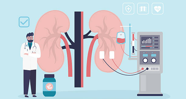
Illustration showing kidney diagnosis and dialysis setup for glomerulonephritis

Diagnosis of Glomerulonephritis

Diagnosis of Glomerulonephritis The diagnosis of glomerulonephritis is based on a combination of clinical history, physical examination, laboratory tests, and sometimes a kidney biopsy. Because glomerulonephritis can present subtly or mimic other conditions, accurate diagnosis is essential to prevent mismanagement and to tailor treatment effectively. Early diagnosis offers the best chance of preserving kidney function. With advances in diagnostic technology, doctors can now detect even small changes in kidney structure and function long before symptoms appear. Medical History and Clinical Examination The diagnostic process begins with a comprehensive medical history, where the doctor explores: Recent infections (e.g., strep throat, skin infections) Autoimmune diseases (e.g., lupus, vasculitis) Family history of kidney disease Medications, including NSAIDs or antibiotics Exposure to toxins, illicit drugs, or heavy metals A physical exam checks for: Oedema in the legs, face, or abdomen Elevated blood pressure Signs of fluid overload, such as lung crackles Skin rashes or joint inflammation (suggestive of systemic autoimmune disease) Urinalysis: The Cornerstone of Diagnosis Urine tests are often the first clue in identifying glomerulonephritis. Important findings include: Proteinuria – high levels of protein in the urine Haematuria – microscopic or visible blood Casts – tube-shaped particles made of proteins or cells, indicating kidney inflammation White or red blood cells – signalling infection or inflammation Dipstick urinalysis is useful for screening, while 24-hour urine collection quantifies protein loss. Blood Tests Several blood tests support the diagnosis and help identify underlying causes: 1. Kidney Function Tests Creatinine and urea: Elevated levels suggest impaired filtration Estimated glomerular filtration rate (eGFR): Indicates overall kidney function 2. Electrolyte and Metabolic Panels Assess sodium, potassium, calcium, phosphate Check acid-base balance 3. Complete Blood Count (CBC) May show anaemia (from chronic kidney disease) Elevated white cells in infection or inflammation 4. Autoimmune Markers ANA (antinuclear antibodies) for lupus ANCA for vasculitis Anti-GBM antibodies for Goodpasture syndrome Complement levels (C3, C4) – often low in post-infectious or lupus nephritis 5. Viral Serologies Tests for hepatitis B, hepatitis C, and HIV Essential in cases of secondary glomerulonephritis Imaging Studies While not diagnostic on their own, imaging helps assess kidney structure and rule out other issues. 1. Renal Ultrasound Non-invasive and widely used Detects kidney size, cysts, or obstruction Helps assess chronicity (e.g. shrunken kidneys in longstanding disease) 2. CT or MRI Rarely required but useful if complications are suspected Kidney Biopsy: The Definitive Test When the cause of glomerulonephritis is uncertain or the disease appears severe, a kidney biopsy may be necessary. This involves: Using a needle to take a small sample of kidney tissue Examining it under a microscope for signs of inflammation, scarring, or immune deposits A biopsy can: Confirm the specific subtype (e.g. IgA nephropathy, membranous nephropathy) Guide treatment decisions (e.g. steroid use, immunosuppressants) Help determine prognosis Biopsies are typically safe and performed under local anaesthesia with ultrasound guidance. Differential Diagnosis Several other conditions can mimic glomerulonephritis. A careful diagnostic process distinguishes it from: Urinary tract infections Interstitial nephritis Nephrotic syndrome of other causes Polycystic kidney disease Diabetic nephropathy Proper differentiation ensures the correct therapy and prevents further damage. Diagnostic Summary Table Test Purpose Urinalysis Detects blood, protein, and casts Blood tests Assess kidney function and inflammation Autoimmune markers Identify systemic causes Imaging Visualise kidney structure Kidney biopsy Definitive diagnosis and disease classification When to Refer to a Specialist Patients with signs of glomerulonephritis should be referred to a nephrologist if they: Have significant proteinuria or haematuria Show signs of rapid kidney deterioration Have resistant hypertension Need a biopsy or immunosuppressive therapy Early referral improves outcomes by allowing for targeted treatment before irreversible damage occurs. Conclusion | Diagnosis of Glomerulonephritis The diagnosis of glomerulonephritis involves a step-by-step evaluation using urine and blood tests, imaging, and sometimes biopsy. Accurate diagnosis is critical not only to confirm the condition but also to determine the underlying cause and appropriate treatment. With early detection and ongoing monitoring, patients can avoid complications and preserve their kidney health long term. [Next: Treatment of Glomerulonephritis→]

Diagnosis of Glomerulonephritis Read More »

3D illustration of human organs with capsules and tablets used for glomerulonephritis treatment

Treatment of Glomerulonephritis

Treatment of Glomerulonephritis The treatment of glomerulonephritis depends on the type, underlying cause, and severity of kidney involvement. In many cases, especially with mild or early disease, the treatment of glomerulonephritis focuses on managing symptoms, controlling blood pressure, and reducing inflammation to preserve kidney function. When an autoimmune disorder, infection, or genetic factor causes the condition, doctors must also treat the underlying cause to prevent progression. Tailoring treatment to the specific type of glomerulonephritis is essential. What works for one form may be inappropriate—or even harmful—for another. The goal is always the same: to protect the glomeruli from further damage and slow or prevent the onset of chronic kidney disease or renal failure. General Principles of Management Regardless of the underlying cause, doctors use several treatment approaches in most cases, including: 1. Blood Pressure Control High blood pressure accelerates glomerular damage, so effective management is critical. Doctors usually prescribe: ACE inhibitors or angiotensin receptor blockers (ARBs), which reduce both blood pressure and protein loss in the urine Lifestyle changes like low-sodium diets, weight loss, and regular exercise Doctors may use these medications to reduce proteinuria and protect kidney health, even in patients with normal blood pressure. 2. Dietary Adjustments Special dietary recommendations support kidney function and reduce strain on the glomeruli: Low-protein diet may be advised in some cases to reduce urea build-up Low-sodium diet to help control blood pressure and reduce oedema Restricted potassium or phosphorus intake in more advanced disease Adequate fluid intake to maintain hydration without overloading the kidneys Dieticians often work alongside nephrologists to personalise nutrition plans. 3. Diuretics If fluid retention is causing significant swelling, diuretics (such as furosemide) may be used to help the body eliminate excess fluid and reduce oedema. Treating the Underlying Cause The cornerstone of effective therapy is addressing the trigger behind the inflammation. 1. Infection-Related Glomerulonephritis Often self-limiting, particularly in children Antibiotics may be used if an active infection is still present Supportive care (rest, blood pressure control) is often sufficient Immunosuppressive drugs are generally avoided unless complications arise 2. Autoimmune or Immune-Mediated Types For conditions like lupus nephritis, IgA nephropathy, or ANCA-associated vasculitis, immunosuppressive therapy may be required: Corticosteroids (e.g. prednisone) to reduce inflammation Cyclophosphamide, azathioprine, or mycophenolate mofetil as steroid-sparing agents Rituximab for certain forms of vasculitis or resistant disease Close monitoring of immune markers, kidney function, and side effects is essential Immunosuppressive drugs can increase infection risk and require careful management. 3. Nephrotic Syndrome For nephrotic-range proteinuria, treatment focuses on: Controlling oedema with diuretics and salt restriction Reducing protein loss using ACE inhibitors or ARBs Lowering cholesterol, often with statins Corticosteroids or immunosuppressants depending on the underlying cause (e.g. minimal change disease, membranous nephropathy) Plasmapheresis and Advanced Therapies In rapidly progressive or life-threatening cases, such as Goodpasture syndrome or severe ANCA vasculitis, more aggressive measures may be used: Plasmapheresis (plasma exchange) to remove harmful antibodies Combined with high-dose steroids and immunosuppressants Used in hospital under specialist care These treatments are reserved for severe cases and require close monitoring in a renal or intensive care setting. Management of Chronic Glomerulonephritis For chronic forms, the emphasis is on slowing progression and preventing complications: Regular monitoring of kidney function, urine protein levels, and blood pressure Early detection of declining kidney function allows for preemptive planning Managing comorbidities like diabetes, anaemia, and cardiovascular disease In advanced stages, patients may need: Erythropoietin-stimulating agents to treat anaemia Phosphate binders or vitamin D supplements Dialysis or consideration for kidney transplant if kidney failure develops Patient Education and Lifestyle Empowering patients is a key part of managing glomerulonephritis: Understanding medication regimens Learning to monitor blood pressure at home Attending all scheduled blood and urine tests Avoiding non-prescribed NSAIDs or nephrotoxic drugs Vaccination against influenza, hepatitis B, and pneumococcus Education helps reduce hospitalisation rates and supports long-term kidney health. Conclusion | Treatment of Glomerulonephritis The treatment of glomerulonephritis is complex but often highly effective when tailored to the specific cause. Whether it involves immune suppression, infection control, or supportive care, the treatment of glomerulonephritis can slow progression, preserve function, and improve quality of life. Early diagnosis, consistent follow-up, and proactive patient involvement are the keys to successful outcomes. [Next: Complications of Glomerulonephritis →]

Treatment of Glomerulonephritis Read More »

Scroll to Top