Mental Matters

Author name: admin

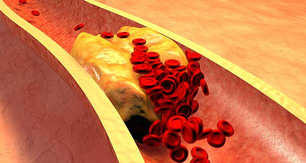
Illustration of blocked artery showing blood cells and plaque buildup, symbolising causes of Giant Cell Arteritis

Causes of Giant Cell Arteritis

Causes of Giant Cell Arteritis The causes of giant cell arteritis are still being researched. However, current evidence suggests that a combination of genetic, environmental, and immunological factors is involved. Doctors consider giant cell arteritis an autoimmune condition in which the immune system mistakenly attacks artery walls, causing inflammation and narrowing of the vessels. Understanding the underlying mechanisms helps in identifying at-risk individuals and developing better treatments. The condition is most common in older adults, especially women over 50, but doctors still do not fully understand what triggers the disease. Immune System Dysfunction The leading theory is that giant cell arteritis arises from immune dysregulation, where immune cells misidentify healthy tissue—in this case, artery walls—as harmful. This sets off a chronic inflammatory response. Key immune cells involved include: CD4+ T lymphocytes Macrophages Giant cells are large immune cells formed when multiple macrophages fuse together These cells infiltrate the tunica media and adventitia (layers of the arterial wall), forming granulomas and releasing inflammatory cytokines. The inflammation leads to: Thickening of the artery wall Narrowing or obstruction of the blood vessel Potential tissue damage due to reduced blood flow This explains the classic symptoms of ischaemia, such as headache, jaw pain, and vision disturbances. Genetic Predisposition There is a strong genetic component to giant cell arteritis. Studies have found that people with certain variations in the HLA (human leukocyte antigen) system, particularly HLA-DRB1*04, are more likely to develop the condition. While having these genes does not guarantee disease, it increases susceptibility, particularly when combined with environmental or age-related factors. Family history may also influence the risk, although doctors do not consider GCA a hereditary disease in the traditional sense. Ageing and Vascular Changes Ageing is the strongest risk factor for developing giant cell arteritis. The vast majority of cases occur in people over 50, and risk increases steadily with age. Several theories explain this age-related risk: Ageing causes degeneration of vascular structures, making them more vulnerable to inflammation The immune system becomes more prone to autoimmune activation Arterial elasticity declines, contributing to vascular stress and damage The intersection of these factors creates a favourable environment for GCA to develop. Environmental Triggers Doctors suspect that certain environmental factors may trigger the initial immune response in individuals who are predisposed. These may include: 1. Infections Viral or bacterial infections have long been hypothesised as potential triggers. Though no single pathogen has been definitively identified, several candidates include: Parvovirus B19 Varicella zoster virus (VZV) Chlamydia pneumoniae These pathogens may provoke an immune response that cross-reacts with arterial tissues, leading to autoimmunity. Some studies have found fragments of viral DNA in the arteries of patients with GCA, although evidence remains inconclusive. 2. Seasonal Variations Epidemiological data suggest a seasonal pattern to disease onset, with more diagnoses occurring during colder months. This may point to a link with seasonal infections or reduced sunlight exposure, although the exact relationship is unclear. 3. Geographic Trends Giant cell arteritis is more common in certain regions, particularly: Northern Europe and Scandinavia North America Parts of Australia and New Zealand This geographic distribution supports both genetic and environmental influences, including climate, infection exposure, and possibly vitamin D levels. Coexisting Conditions Giant cell arteritis often occurs in conjunction with polymyalgia rheumatica (PMR), an inflammatory condition that causes stiffness and pain in the shoulders and hips. Up to 50% of patients with GCA also have symptoms of PMR. This association suggests shared inflammatory mechanisms, possibly involving overlapping genetic susceptibility and immune pathways. Gender Differences Giant cell arteritis affects women more often than men, with a female-to-male ratio of approximately 2:1 to 3:1. The reasons for this are not fully understood but may involve: Hormonal influences on immune regulation Differences in vascular structure or immune gene expression Postmenopausal women are particularly at risk, further implicating hormonal changes as a contributing factor. Conclusion While the exact causes of giant cell arteritis remain under investigation, current research points to a multifactorial process involving: Immune system malfunction Genetic predisposition Age-related vascular changes Possible environmental triggers Causes of Giant Cell Arteritis Identifying and understanding these factors is crucial for early detection and better management of this potentially sight-threatening disease. [Next: Symptoms of Giant Cell Arteritis→]

Causes of Giant Cell Arteritis Read More »

Woman with grey hair touching her temples in pain, showing symptoms of Giant Cell Arteritis

Symptoms of Giant Cell Arteritis

Symptoms of Giant Cell Arteritis The symptoms of giant cell arteritis can be wide-ranging and often develop gradually, though in some cases, they appear suddenly. Giant cell arteritis is an inflammatory condition that primarily affects the large and medium arteries of the head and neck, especially the temporal arteries. Recognising the early signs of this condition is essential, as delayed diagnosis can result in serious complications, including permanent vision loss or stroke. Because the symptoms can mimic other conditions—such as tension headaches, dental problems, or sinus infections—early identification can be challenging. However, there are hallmark symptoms that should prompt immediate medical evaluation, particularly in people over the age of 50. Classic Symptoms Giant cell arteritis has a set of “classic” symptoms that often occur together or in close succession: 1. New-Onset Headache The most common symptom, affecting around two-thirds of patients Typically located at the temples, but may affect other parts of the head Often throbbing or aching, sometimes worse at night or with touch Unlike migraines or cluster headaches, GCA headaches are persistent and unresponsive to typical over-the-counter medication Patients frequently describe the pain as different from any headache they’ve had before, which should raise clinical suspicion. 2. Scalp Tenderness Pain or discomfort when brushing hair or wearing a hat The scalp may feel sore or bruised without visible signs Sensitive to light pressure, even from lying on a pillow This symptom is particularly associated with inflammation of the superficial temporal arteries. 3. Jaw Claudication Pain or cramping in the jaw when chewing, talking, or yawning Caused by reduced blood flow to the muscles of mastication Typically improves with rest Jaw claudication is highly suggestive of GCA and is rare in other conditions, making it a valuable diagnostic clue. 4. Visual Disturbances Vision symptoms can be subtle or sudden: Blurred or double vision Partial or complete loss of vision in one eye Transient blackouts of vision (amaurosis fugax) These signs often precede irreversible blindness. Prompt treatment can protect the second eye and prevent stroke. 5. Systemic Symptoms Inflammation in GCA often causes generalised symptoms such as: Low-grade fever Unexplained fatigue Night sweats Loss of appetite Weight loss These symptoms overlap with infections and malignancies, so they must be evaluated in context. Polymyalgia Rheumatica (PMR) About 40–50% of individuals with giant cell arteritis also develop polymyalgia rheumatica, a related inflammatory condition that causes: Pain and stiffness in the shoulders and hips Difficulty raising the arms or getting out of bed Symptoms worse in the morning and improving throughout the day PMR can precede, accompany, or follow the onset of GCA and may respond dramatically to corticosteroid treatment. Less Common Symptoms While the classic features are well recognised, other symptoms may also be present: Throat pain or discomfort when swallowing Earache or tinnitus Dizziness or balance issues Tongue pain, numbness, or rarely, tongue necrosis Chest pain or shortness of breath (if large arteries like the aorta are involved) These signs suggest extra-cranial arterial involvement and may indicate systemic vasculitis. Atypical Presentations In some individuals, especially those with diabetes or pre-existing cardiovascular disease, symptoms may be subtle or atypical. Headaches may be minimal, and the first sign could be vision loss or stroke. For this reason, doctors must maintain a high index of suspicion in older patients presenting with: Unexplained inflammatory markers (e.g., raised ESR or CRP) Vague symptoms like fatigue or malaise Visual complaints, even if transient Symptom Progression Without treatment, symptoms of GCA usually worsen over time, with increasing inflammation and vascular damage. Once blood flow is significantly reduced, complications can set in quickly. Vision loss from anterior ischaemic optic neuropathy (AION) can develop over hours to days, making early intervention essential. Psychological Impact The sudden onset of severe symptoms—especially vision problems—can cause significant anxiety and distress. Patients may experience: Fear of going blind Sleep disturbances Reduced independence Social withdrawal Support from medical teams, counsellors, and patient support groups can make a significant difference in coping and recovery. Summary of Key Symptoms Symptom Description Temporal headache Persistent, new in onset, throbbing Scalp tenderness Pain with light touch, brushing hair Jaw claudication Cramping jaw pain while chewing Visual disturbances Blurred vision, partial or complete loss Polymyalgia rheumatica Shoulder/hip stiffness, especially mornings Systemic symptoms Fatigue, fever, weight loss Symptoms of Giant Cell Arteritis Recognising the symptoms of giant cell arteritis can help prevent permanent disability and promote faster recovery through early treatment. Anyone over 50 with new or unusual headaches, jaw pain, or visual complaints should seek urgent medical advice. [Next: Diagnosis of Giant Cell Arteritis →]

Symptoms of Giant Cell Arteritis Read More »

Blood test sample labelled Anti C1q Antibody held by gloved hand

Diagnosis of Giant Cell Arteritis

Diagnosis of Giant Cell Arteritis The diagnosis of giant cell arteritis requires a combination of clinical evaluation, blood tests, imaging, and—when needed—biopsy. Because giant cell arteritis can cause irreversible complications such as vision loss or stroke, early and accurate diagnosis is vital. Treatment is often initiated based on strong clinical suspicion even before confirmation, due to the high risk of delay. This condition most commonly affects people over the age of 50, and the diagnostic process often begins when a patient presents with symptoms such as new-onset headache, jaw pain, or visual disturbances. Clinical Evaluation A detailed history and physical examination are the first steps. Key features that suggest GCA include: New, persistent headaches in people over 50 Jaw claudication Tenderness or thickening of the temporal arteries Scalp sensitivity Visual symptoms, such as blurred or double vision Symptoms of polymyalgia rheumatica On examination, doctors may find: Thick, tender, or pulseless temporal arteries Asymmetry in arterial pulses Bruits (abnormal whooshing sounds) over large vessels Signs of ischaemia, such as pale retina or afferent pupillary defect Blood Tests Although not diagnostic on their own, blood tests provide crucial supporting evidence. 1. Erythrocyte Sedimentation Rate (ESR) Often markedly elevated (typically over 50 mm/hr) Reflects systemic inflammation 2. C-Reactive Protein (CRP) Sensitive marker for inflammation May be raised even if ESR is normal 3. Full Blood Count May show normochromic, normocytic anaemia Thrombocytosis (elevated platelet count) is also common These results support a clinical diagnosis and help rule out other causes. Temporal Artery Biopsy The gold standard for diagnosis is a temporal artery biopsy, which involves removing a small section of artery from the temple for microscopic examination. Key findings include: Granulomatous inflammation Multinucleated giant cells Intimal thickening and vessel wall disruption While this is a highly specific test, false negatives can occur due to “skip lesions”, where inflammation affects segments of the artery unevenly. To reduce this risk, longer biopsy samples (2–3 cm) are preferred. Biopsy should be performed within 1–2 weeks of starting steroids, as treatment can begin to reduce inflammatory changes quickly. Imaging Studies Modern imaging techniques have become valuable in both diagnosing and monitoring giant cell arteritis, especially in cases where biopsy is not possible. 1. Ultrasound (Colour Doppler) Can detect the “halo sign”—a dark, hypoechoic ring around the artery indicating inflammation Non-invasive, fast, and increasingly used as a first-line test 2. MRI or CT Angiography Used to visualise larger arteries, such as the aorta or subclavian arteries Helps assess extent of vessel involvement and detect complications like aneurysms 3. FDG-PET Scan Useful in systemic or atypical cases Detects increased metabolic activity in inflamed arteries Expensive and less widely available Diagnostic Criteria The American College of Rheumatology (ACR) criteria for diagnosis include: Age over 50 New-onset localised headache Tenderness or decreased pulse in temporal artery ESR > 50 mm/hr Abnormal biopsy Meeting three or more of these criteria yields high diagnostic accuracy, though real-world diagnosis often relies on a combination of clinical judgement and test results. Differential Diagnosis Conditions that can mimic GCA include: Tension or migraine headaches Dental infections Trigeminal neuralgia Carotid artery dissection Meningitis or subarachnoid haemorrhage These possibilities must be ruled out, especially when symptoms are non-specific or when typical GCA features are absent. Initiating Treatment Before Confirmation Due to the risk of permanent vision loss, treatment with high-dose corticosteroids should begin immediately if GCA is strongly suspected—even before biopsy or imaging confirmation. Diagnosis of Giant Cell Arteritis Delaying treatment can result in devastating consequences, and early steroid use does not invalidate a biopsy for at least 7–10 days. [Next: Treatment of Giant Cell Arteritis→]

Diagnosis of Giant Cell Arteritis Read More »

Doctor giving medication and water to elderly patient in hospital bed

Treatment of Giant Cell Arteritis

Treatment of Giant Cell Arteritis The treatment of giant cell arteritis is aimed at reducing arterial inflammation, preventing irreversible complications such as vision loss and stroke, and minimising long-term treatment side effects. Because giant cell arteritis is a medical emergency when visual symptoms appear, treatment typically begins immediately, even before the diagnosis is confirmed. Corticosteroids are the mainstay of treatment, but newer therapies now offer more options for long-term disease control and steroid reduction. With prompt and appropriate management, most people can achieve disease remission and avoid the most serious consequences. Immediate Treatment Goals The urgent priorities in treating GCA are: Preserving vision in patients with visual symptoms Preventing stroke or other vascular complications Controlling systemic inflammation Alleviating symptoms such as headache, jaw pain, and fatigue Doctors typically begin treatment on the same day they suspect the diagnosis, especially if the patient presents with: Visual disturbances New-onset headache with elevated inflammatory markers Jaw claudication Corticosteroids: First-Line Therapy 1. High-Dose Prednisolone For patients without vision symptoms: 40–60 mg daily orally For patients with visual symptoms or transient loss: 500–1000 mg of intravenous methylprednisolone daily for 3 days, followed by high-dose oral steroids Corticosteroids typically produce a rapid clinical response, often within 24–72 hours. Inflammatory markers such as ESR and CRP begin to normalise, and systemic symptoms improve. However, the treatment does not cure the disease outright. Doctors use a slow tapering regimen, often lasting 12–18 months or longer, to prevent relapse. 2. Steroid Tapering Once symptoms and lab markers are stable for several weeks: Doctors gradually reduce the dose—for example, by 10 mg every 2 weeks until reaching 20 mg, then taper more slowly Doctors must individualise tapering based on symptom recurrence and inflammatory markers Relapses may occur and require dose adjustment Sudden reduction in steroid dose or rapid withdrawal can lead to flare-ups and increase risk of complications. Managing Steroid Side Effects Long-term steroid use can cause numerous side effects: Weight gain, fluid retention, and mood changes High blood pressure and diabetes Osteoporosis and fractures Increased infection risk Cataracts and glaucoma To counter these risks: Prescribe calcium and vitamin D supplements Consider bisphosphonates for bone protection Monitor blood glucose and blood pressure Schedule regular eye exams Encourage lifestyle changes such as low-sodium diets and physical activity Steroid-Sparing Therapy To reduce long-term steroid exposure, doctors increasingly use immunosuppressive or biologic agents, particularly in: Patients with frequent relapses Those with significant steroid side effects Individuals at high risk of complications from steroids 1. Tocilizumab (Actemra) An interleukin-6 (IL-6) receptor antagonist Administered weekly or monthly (subcutaneous or intravenous) Helps induce remission and allows faster steroid tapering Proven efficacy in clinical trials Tocilizumab is now widely accepted as a first-line steroid-sparing agent in GCA and may be used from the start in selected patients. 2. Methotrexate Taken weekly, often as a tablet or injection Modestly effective at reducing relapse and steroid dose Considered for patients intolerant to tocilizumab or with mild-to-moderate disease Doctors rarely use agents like azathioprine, leflunomide, or cyclophosphamide, reserving them for refractory or atypical cases. Monitoring and Follow-Up Regular follow-up is essential during treatment: Inflammatory markers (ESR and CRP) are checked every few weeks Blood pressure, glucose, and bone health are monitored regularly Eye exams are scheduled, particularly if vision symptoms were present Imaging (e.g. ultrasound or MRI) may be repeated if large-vessel involvement is suspected Doctors define relapse as the recurrence of symptoms and/or a rise in inflammatory markers. Doctors treat it by increasing the steroid dose and reassessing the treatment plan. Patient Education and Lifestyle Support Helping Patients Take Control of GCA Supporting patients is very important when dealing with a long-term illness like Giant Cell Arteritis (GCA). Here are some simple ways to help: Treatment in Different Patient Groups 1. Older Adults 2. People with Polymyalgia Rheumatica (PMR) 3. Large-Vessel GCA They must also keep an eye out for serious problems like aneurysms or vessel damage. This form may need treatment for a longer time. Doctors use scans like CT, MRI, or PET-CT to check blood vessels. Treatment of Giant Cell Arteritis The treatment of giant cell arteritis requires a multidisciplinary, patient-centred approach. With timely intervention and modern therapies, most individuals achieve suspension and retain their quality of life. [Next: Complications of Giant Cell Arteritis →]

Treatment of Giant Cell Arteritis Read More »

Elderly man covering his eyes with hands on blue background

Complications of Giant Cell Arteritis

Complications of Giant Cell Arteritis The complications of giant cell arteritis can be serious, life-altering, and even fatal if the disease is not diagnosed and treated quickly. Giant cell arteritis causes inflammation in large and medium arteries. Furthermore, most often affects the head and neck. However, sometimes this includes major vessels, such as the aorta. Without early treatment, the risk of vision loss, stroke, and aneurysm formation increases significantly. Fortunately, with prompt steroid therapy and careful monitoring, most complications can be avoided or managed effectively. Vision Loss One of the most feared complications of GCA is irreversible blindness, which may occur when: The posterior ciliary arteries become occluded Blood flow to the optic nerve is negatively affected A condition called anterior ischaemic optic neuropathy (AION) develops Vision loss may present as: Sudden, loss of sight in one eye Partial loss or field defects Transient visual blackouts (amaurosis fugax) that may precede permanent damage Once vision is lost, it is usually permanent. Therefore, rapid steroid treatment can protect the healthy eye, making early diagnosis essential. Stroke and Transient Ischaemic Attack (TIA) GCA can cause reduced blood flow to the brain, leading to: Stroke, if large arteries such as the carotid or vertebral arteries are narrow or blocked TIA, also known as a “mini-stroke,” involves temporary neurological symptoms These events may present as: Sudden weakness on one side of the body Difficulty speaking or understanding speech Dizziness or loss of coordination Facial Droop Stroke is a less common complication but is more likely in undertreated cases, particularly with large-vessel involvement. Aortic Aneurysm and Dissection In some patients, GCA extends to involve the aorta and its major branches, leading to structural weakening over time. This may result in: Aortic aneurysm—a bulging or dilation of the artery Aortic dissection—a tear in the artery wall, which is a medical emergency These complications often appear months to years after initial diagnosis, making long-term monitoring crucial. Signs may include: Chest or back pain Pulsatile mass in the abdomen Abnormal blood pressure readings in different limbs Regular imaging with CT angiography, MRI, or ultrasound is advised for individuals at a high risk. Relapse and Chronic Inflammation GCA frequently relapses, particularly during steroid tapering. Symptoms of relapse include: Return of headaches or jaw pain Fatigue and low-grade fever Elevated ESR or CRP Chronic, low-level inflammation may lead to: Persistent pain and stiffness Increased cardiovascular risk Ongoing need for immunosuppression Relapses can be managed by adjusting steroid dose or introducing biologic agents like tocilizumab. Osteoporosis and Fractures Long-term steroid use increases the risk of: Bone density loss (osteopenia and osteoporosis) Spinal or hip fractures Prevention includes: Calcium and vitamin D supplementation Bisphosphonates Bone mineral density monitoring via DEXA scans This complication is particularly relevant for elderly patients. Hereby, those who represent the majority of those with GCA. Glucose Intolerance and Hypertension Steroid therapy can cause: High blood sugar, potentially triggering or worsening diabetes Elevated blood pressure, increasing cardiovascular risk Management includes: Regular monitoring of blood glucose and blood pressure Dietary adjustments Medications to control side effects if needed Cataracts and Glaucoma Corticosteroids can increase the risk of: Cataracts, causing blurred or cloudy vision Glaucoma, a condition where increased eye pressure damages the optic nerve Patients on long-term steroids should receive regular eye examinations and report any vision changes promptly. Psychological and Social Effects Living with GCA can impact quality of life: Fear of relapse or blindness Fatigue from chronic inflammation Depression or anxiety Reduced mobility due to pain or joint stiffness Supportive care, counselling, and connection with patient support groups can help reduce the burden of illness. Summary of Key Complications Complication Description Vision loss Sudden, irreversible blindness in one or both eyes Stroke or TIA Neurological symptoms due to artery blockage Aortic aneurysm Arterial wall dilation with rupture risk Osteoporosis Fragile bones from long-term steroid use Diabetes and hypertension Steroid-induced metabolic complications Cataracts and glaucoma Vision problems linked to corticosteroids Relapse Recurrence of symptoms requiring treatment adjustment Complications of Giant Cell Arteritis Preventing the complications of giant cell arteritis requires early treatment, regular follow-up, and a multidisciplinary care approach. With advances in monitoring and biologic therapies, long-term outcomes are improving. Furthermore, offering hope to those living with this serious condition. [Next: Back to Overview →]

Complications of Giant Cell Arteritis Read More »

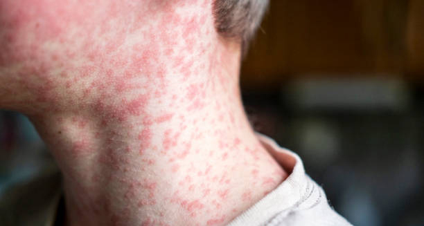
Red rash on the neck and jawline of a person with German Measles

German Measles

German Measles German measles is a contagious viral infection best known for its distinctive red-pink rash and mild flu-like symptoms. Also called rubella, German measles is caused by the rubella virus and typically results in a short, self-limiting illness in children and adults. However, when contracted during pregnancy—particularly in the first trimester—it can have devastating effects on the unborn baby, leading to congenital rubella syndrome (CRS), a serious condition with long-term consequences. Despite the name, German measles is not related to ordinary measles (rubeola), although the two diseases share similar symptoms. Rubella is usually milder and often goes unnoticed in healthy individuals. However, its ability to cause severe birth defects has made it a primary target for vaccination campaigns across the world. Thanks to widespread immunisation, the incidence of rubella has dropped dramatically in many countries, including South Africa. Nevertheless, outbreaks still occur, especially in communities with low vaccination rates or limited access to healthcare. This makes public awareness, early diagnosis, and continued vaccine coverage essential in preventing transmission and protecting vulnerable populations. What Is Rubella? Rubella is caused by the rubella virus, which belongs to the Togaviridae family. The virus spreads through respiratory droplets, meaning it can be passed on by coughing, sneezing, talking, or sharing drinks and utensils. Once inhaled, the virus multiplies in the nose and throat, then spreads through the bloodstream to other parts of the body. The incubation period is usually 14 to 21 days, during which time the person may not have symptoms but can still be contagious. Once symptoms appear, individuals are infectious from about 7 days before the rash appears to 7 days after. Who Is at Risk? Anyone who hasn’t been vaccinated or previously infected can contract rubella. Populations most at risk include: Unvaccinated children Adults who missed immunisation during childhood Pregnant women without immunity Healthcare workers in direct contact with patients Communities with vaccine hesitancy or poor access to healthcare Even in highly vaccinated countries, rubella remains a risk due to international travel and migration from regions where vaccination is less common. Signs and Symptoms Rubella often causes mild or no symptoms, especially in children. When symptoms do appear, they are usually more noticeable in adolescents and adults. Common signs include: Pink or red rash, starting on the face and spreading downward to the trunk and limbs Low-grade fever, typically under 38.5°C Swollen lymph nodes, especially behind the ears and at the back of the neck Mild joint pain, more common in adult women Headache Fatigue and malaise Mild conjunctivitis (red or irritated eyes) The rash usually lasts about three days, which is why rubella is sometimes referred to as “three-day measles.” The rash is not itchy in most cases and may fade without peeling or scabbing. In many cases, the illness is so mild that people do not realise they have it. However, the virus is still contagious during this time, making it important to stay cautious and informed, especially during outbreaks. Rubella in Pregnancy The most serious risk of German measles occurs when a pregnant woman contracts the virus, especially in the first 12 weeks of gestation. The rubella virus can cross the placenta and infect the developing foetus, leading to congenital rubella syndrome (CRS). CRS can cause a range of birth defects, including: Deafness Heart defects Cataracts and other eye abnormalities Intellectual disabilities Growth delays Liver and spleen damage Low birth weight or stillbirth The earlier in pregnancy the infection occurs, the greater the risk of severe outcomes. Because of this, rubella vaccination is strongly recommended for all individuals of childbearing age, and women are advised to wait at least one month after vaccination before conceiving. Global and Local Impact Worldwide, rubella has been largely eliminated in countries with robust immunisation programmes. In South Africa, rubella is not yet part of the routine Expanded Programme on Immunisation (EPI), although it may be introduced in future. Some private healthcare providers include the MMR vaccine (measles, mumps, and rubella) as part of childhood immunisation schedules. In regions where vaccination coverage is poor, rubella remains a leading cause of preventable congenital birth defects. Periodic outbreaks still occur, especially in communities with: Poor vaccination uptake Limited public health infrastructure High population movement Gaps in maternal screening Prevention The best protection against rubella is vaccination. The MMR vaccine is safe, effective, and provides long-lasting immunity. It is usually administered in two doses during childhood, but adults who are unsure of their immunisation status can receive a catch-up dose. Other prevention strategies include: Avoiding contact with people showing symptoms of rubella, especially if you are pregnant or immunocompromised Screening women of childbearing age for rubella immunity Staying home if you suspect you have rubella, to avoid spreading the virus Promoting vaccine education to reduce stigma and misinformation German Measles Rubella is a preventable disease. Ensuring high levels of immunity through public health education and access to vaccines is the key to eliminating outbreaks and protecting unborn babies. [Next: Causes of German Measles →]

German Measles Read More »

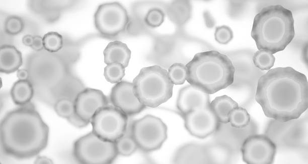
Microscopic view of the rubella virus causing German Measles

Causes of German Measles

Causes of German Measles The causes of German measles lie in the rubella virus — a contagious RNA virus that spreads through respiratory droplets and primarily affects the nose, throat, and lymph nodes. German measles, also known as rubella, is considered one of the most preventable childhood infections thanks to the availability of effective vaccines. However, it remains a risk in areas where immunisation coverage is low or inconsistent. Understanding how rubella spreads and what factors contribute to infection helps individuals and public health professionals take appropriate action to prevent outbreaks and reduce complications, especially for pregnant women and vulnerable populations. The Rubella Virus Rubella is caused by the rubella virus, a member of the Togaviridae family, under the genus Rubivirus. It is a single-stranded RNA virus that primarily targets epithelial cells in the upper respiratory tract. After initial replication, the virus enters the bloodstream and spreads throughout the body in a process called viraemia. This systemic spread allows the virus to reach: Lymphatic tissue Skin and mucous membranes Developing foetuses (via the placenta during pregnancy) The virus is highly contagious during the early stages of infection, even before symptoms become apparent. Transmission Pathways The rubella virus is spread from person to person through: Coughing or sneezing Close conversation or breathing shared air Touching contaminated surfaces, then touching the mouth or nose Vertical transmission from mother to baby during pregnancy In general, people with rubella are contagious from about 7 days before the rash appears until 7 days after. During this window, they can transmit the virus even if they don’t feel unwell or show visible symptoms. Incubation and Infectious Period After a person is exposed to the virus, there is typically an incubation period of 14 to 21 days. During this time, the virus replicates and spreads internally without any symptoms. A person becomes infectious about 7 days before symptoms emerge, particularly the rash, and may continue to shed the virus for a week afterward. Infants born with congenital rubella syndrome (CRS) can shed the virus in their urine or respiratory secretions for up to one year, making them a long-term source of infection if not carefully managed. Contributing Risk Factors While anyone who hasn’t been vaccinated or previously infected can contract rubella, some risk factors make transmission more likely or consequences more severe: 1. Lack of Immunisation Individuals who have not received the MMR vaccine or who missed childhood vaccinations are at greatest risk In many parts of Africa, including some regions of South Africa, rubella is not yet part of the public EPI, making vaccination status uneven 2. High Population Density Schools, universities, refugee camps, or overcrowded homes facilitate rapid spread Close contact in shared environments increases exposure to respiratory droplets 3. Travel and Migration Travellers from areas with low vaccination coverage may introduce rubella into communities with waning immunity Migrants or displaced populations may miss key childhood vaccinations 4. Pregnancy Without Immunity Women who are pregnant and not immune to rubella face the highest personal risk, as the infection can cause foetal malformations Prenatal screening is crucial to identify susceptibility before conception or early in pregnancy Congenital Rubella Transmission In pregnant individuals, the rubella virus can cross the placenta and infect the foetus. The earlier in pregnancy the infection occurs, the greater the chance of serious birth defects. Congenital rubella syndrome (CRS) is caused by: Direct viral damage to developing foetal organs Inflammation and reduced blood supply to foetal tissues Persistent infection that disrupts normal growth CRS remains one of the most serious consequences of rubella transmission and is entirely preventable through preconception screening and vaccination. Natural Immunity and Reinfection Once someone has had rubella, their body usually develops lifelong immunity. Reinfection is extremely rare, although mild reinfections have been documented, usually without significant symptoms. Immunity may come from: Natural infection MMR vaccination, which offers long-lasting protection Serology tests can check for rubella-specific IgG antibodies, which indicate immunity. This is especially useful for: Women planning pregnancy Healthcare workers Teachers and childcare providers Community and Global Spread Rubella tends to spread quietly because: Many cases are asymptomatic or very mild The contagious period begins before symptoms appear Individuals may not realise they are infected and continue their normal activities Global eradication efforts have made significant progress, but outbreaks still occur. The World Health Organization recommends maintaining vaccine coverage above 95% to prevent resurgence. In South Africa, there are ongoing discussions about incorporating rubella vaccination into the public programme to close existing gaps. Causes of German Measles Understanding the causes of German measles allows public health systems to build better vaccination strategies, inform at-risk populations, and prevent the devastating effects of congenital infection. [Next: Symptoms of German Measles→]

Causes of German Measles Read More »

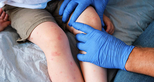
Child with rash on legs being examined by a doctor for symptoms of German measles

Symptoms of German Measles

Symptoms of German Measles The symptoms of German measles can vary significantly depending on age, immune status, and whether the person has previously been vaccinated. In many cases, especially in children, German measles—also known as rubella—presents with mild or even unnoticed symptoms. However, in adults, and particularly in pregnant women, symptoms can be more pronounced and carry serious consequences for the unborn child. Understanding the signs of rubella is critical for timely diagnosis, infection control, and—most importantly—prevention of transmission to those at risk. While often mistaken for other viral infections, rubella has distinct features that help differentiate it from illnesses such as measles, scarlet fever, or mild respiratory infections. Early Signs and General Onset The incubation period for rubella typically ranges from 14 to 21 days after exposure. During this time, the virus replicates in the respiratory tract and lymph nodes before entering the bloodstream. Individuals may begin to feel slightly unwell in the days leading up to visible symptoms. Common early symptoms include: Low-grade fever (usually under 38.5°C) Mild headache Sore throat Malaise or general fatigue Loss of appetite Red, irritated eyes (mild conjunctivitis) These early signs are non-specific and easily confused with other minor viral illnesses. However, they typically precede the hallmark rash by one or two days. The Rubella Rash One of the most identifiable symptoms of German measles is its characteristic rash, which usually appears first on the face, particularly around the hairline and behind the ears, before spreading downward to the trunk, arms, and legs. Key features of the rash include: Pink to red spots, often small and slightly raised Spreading in a downward pattern from the head Non-itchy or only mildly itchy in most cases Lasting approximately three days This is why rubella is sometimes referred to as “three-day measles.” The rash typically fades in the order it appeared and may disappear without any peeling or scabbing. Lymph Node Swelling A distinguishing symptom of German measles is lymphadenopathy, or swelling of the lymph nodes. This tends to affect: The back of the neck (posterior cervical nodes) Behind the ears (postauricular nodes) Under the jaw or in the armpits in some cases These swollen glands are often tender to the touch and may remain enlarged for several days after the rash resolves. Joint Pain and Arthritis In adolescents and adults, especially women, rubella may cause joint pain, swelling, and stiffness. This condition most often affects: Fingers and wrists Knees and ankles The joint symptoms can mimic mild arthritis and may last a week or more, even after the rash subsides. In some individuals, joint pain may be the only noticeable symptom of infection. Respiratory and Eye Involvement Other symptoms that may accompany rubella include: Mild cough Nasal congestion or runny nose Sore red eyes, which may be mistaken for conjunctivitis Photophobia, or light sensitivity, in more pronounced cases These upper respiratory symptoms are generally mild and self-limiting, resolving within a few days without treatment. Asymptomatic Infections It is estimated that up to 50% of people infected with the rubella virus may show no obvious symptoms at all, particularly in children. Despite the absence of symptoms, they can still spread the virus to others during the infectious period. This high rate of asymptomatic infection is one of the main reasons rubella can silently circulate within communities, making vaccination critical in controlling outbreaks. Duration and Recovery The typical rubella illness lasts around three to seven days, with the rash and fever usually resolving within that time frame. Fatigue or joint discomfort may linger slightly longer. Full recovery is expected in the vast majority of healthy individuals. There are rarely any lasting effects once the virus has run its course, provided the person is not pregnant or immunocompromised. Rubella in Adults Adults often experience a more pronounced illness than children. Symptoms such as: More noticeable joint involvement Greater fatigue Stronger immune responses, including swollen lymph nodes and fever Though still classified as a mild illness in most adults, the discomfort can be greater, and work or daily activities may be interrupted. Rubella in Pregnancy The most serious consequences of German measles occur when it infects a pregnant woman, particularly during the first trimester. Symptoms in the mother may be mild or even unnoticed, but the virus can cause: Miscarriage Stillbirth Congenital Rubella Syndrome (CRS) in the baby CRS can include a range of devastating outcomes, such as deafness, cataracts, heart defects, growth retardation, and intellectual disability. This is why rubella infection during pregnancy is considered a medical emergency, even if the symptoms seem trivial. Summary of Key Symptoms Symptom Frequency Facial/trunk rash Very common Swollen lymph nodes Common Low fever Common Sore throat Occasional Joint pain (adults) Fairly common Eye redness Mild and occasional Headache/fatigue Common Cough or runny nose Occasional     Symptoms of German Measles Recognising the symptoms of German measles allows for early intervention, helps reduce spread, and ensures that vulnerable individuals—particularly pregnant women—are protected. [Next: Diagnosis of German Measles →]

Symptoms of German Measles Read More »

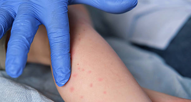
Close-up of a child's rash on the arm being examined for German measles diagnosis

Diagnosis of German Measles

Diagnosis of German Measles The diagnosis of German measles is based on a combination of clinical presentation, patient history, and—when necessary—laboratory testing. Since German measles (rubella) often presents with mild or non-specific symptoms, accurate diagnosis can be challenging, particularly in areas where the condition is rare due to widespread vaccination. However, in populations with lower vaccine coverage or among individuals with uncertain immunisation histories, healthcare providers must maintain a high index of suspicion—especially when exposed individuals include pregnant women. Clinical Diagnosis Rubella is most often diagnosed clinically, based on the typical progression of symptoms. Doctors will consider: Appearance and distribution of the rash Swollen lymph nodes, particularly behind the ears and in the neck Mild fever and joint pain, especially in adult women The timing of onset and how quickly symptoms spread Recent exposure to someone with a similar illness or history of travel to an outbreak area A thorough patient history is critical, particularly to determine: Vaccination status Possible contact with infected individuals Pregnancy status or plans to conceive The presence of postauricular and posterior cervical lymphadenopathy, combined with a facial rash that spreads to the body, is highly suggestive of rubella. Laboratory Confirmation Due to the overlap of rubella symptoms with other illnesses like measles, scarlet fever, roseola, and enteroviral rashes, laboratory testing is sometimes needed to confirm the diagnosis. This is especially true during outbreak investigations or in high-risk settings. 1. Rubella IgM Antibody Test Detects rubella-specific IgM antibodies in the blood IgM becomes detectable within 4–5 days after the rash appears A positive IgM result confirms recent infection 2. IgG Antibody Test Measures rubella-specific IgG antibodies Can distinguish between past infection or vaccination immunity Often used in prenatal screening or epidemiological studies A fourfold increase in IgG titres between paired samples collected two weeks apart can also indicate recent infection. 3. RT-PCR (Reverse Transcription Polymerase Chain Reaction) Detects rubella virus RNA in a throat swab, blood, or urine Used in settings where precise viral identification is required Most useful in early infection or in suspected congenital rubella cases RT-PCR is highly sensitive but may not be widely available outside specialised or reference laboratories. Differentiating Rubella from Other Conditions Many conditions present with similar symptoms to rubella, especially in children. A correct diagnosis must exclude: Measles – higher fever, more severe symptoms, and Koplik spots Scarlet fever – associated with streptococcal throat infection and “strawberry tongue” Roseola – fever precedes rash and typically affects infants Drug reactions – allergic rashes may resemble rubella but lack systemic signs In areas with widespread rubella vaccination, it is crucial to verify that a rash illness isn’t due to other more common viruses. Special Considerations for Pregnant Women If a pregnant woman presents with a rubella-like illness or is exposed to a confirmed case, she should undergo immediate testing to determine: Whether she is immune (IgG positive) Whether she has a current infection (IgM positive and/or viral RNA present) A confirmed rubella infection in pregnancy requires urgent obstetric and infectious disease consultation, as the risks to the developing foetus are significant. Even without symptoms, routine prenatal screening for rubella immunity is recommended to ensure protection before or during early pregnancy. Public Health Notification Rubella is a notifiable disease in many countries. Healthcare providers must report suspected or confirmed cases to public health authorities to: Prevent further transmission Track outbreaks Ensure protection of pregnant individuals who may have been exposed This surveillance allows for timely vaccination campaigns, educational efforts, and community containment measures. Screening and Immunity Testing Routine serological screening is recommended for: Women planning to conceive Healthcare workers and child-care staff People migrating from countries without universal vaccination programmes Seronegative individuals (those without protective antibodies) should be offered rubella vaccination unless contraindicated. Diagnosis of German Measles Knowing how to confirm the diagnosis of German measles helps clinicians act swiftly and protect high-risk populations from serious complications, particularly congenital infections. [Next: Treatment of German Measles→]

Diagnosis of German Measles Read More »

Young girl with cream on her face for German measles treatment

Treatment of German Measles

Treatment of German Measles The treatment of German measles is primarily supportive, as there is no specific antiviral medication that cures rubella. Since German measles (rubella) is usually a mild and self-limiting illness in children and adults, the focus of treatment is on alleviating symptoms, preventing complications, and reducing the spread of the virus — especially to pregnant individuals. Most people recover within a week with rest and basic care. However, special attention is required in certain cases, particularly among pregnant women, immunocompromised individuals, or those experiencing severe symptoms. General Treatment Approach Rubella is generally treated at home, and most individuals do not require hospitalisation unless complications arise. The goals of treatment are to: Reduce fever and discomfort Ease muscle aches or joint pain Encourage hydration and rest Prevent transmission to others Monitor for complications in high-risk groups Home Care and Symptom Relief The following strategies are commonly used to manage the symptoms of German measles: 1. Rest and Isolation Resting helps the body recover more efficiently Avoiding contact with others, especially pregnant women, during the infectious period is crucial Individuals should stay at home for at least 7 days after the rash appears to prevent further spread 2. Fever and Pain Management Paracetamol (acetaminophen) or ibuprofen can be used to relieve: Low-grade fever Headache Joint and muscle pain Aspirin should be avoided in children due to the risk of Reye’s syndrome 3. Fluids and Nutrition Encourage adequate fluid intake to prevent dehydration Light meals and comfort foods can help maintain energy In cases of sore throat or swollen glands, soft foods may be easier to consume 4. Eye Care If conjunctivitis or eye irritation is present: Use a clean, damp cloth to gently wipe discharge Avoid bright lights if photophobia is an issue Artificial tears may offer relief 5. Skin Comfort The rash typically causes minimal itching, but if discomfort occurs: A cool bath or non-perfumed moisturiser can soothe the skin Avoid scratching or using harsh soaps Treating Joint Pain Joint pain, particularly in adolescent girls and adult women, may persist for several days or even weeks. To manage this: Use non-steroidal anti-inflammatory drugs (NSAIDs) such as ibuprofen Apply warm compresses to affected joints Encourage gentle movement and stretching In rare cases, persistent arthritis-like symptoms may develop. Long-term joint problems are uncommon, but follow-up may be needed if pain continues beyond several weeks. When to Seek Medical Help While most rubella cases are mild, medical attention should be sought if: Fever exceeds 38.5°C and persists beyond a few days Severe headache, stiff neck, or vomiting occurs (signs of possible meningitis) Persistent joint swelling interferes with mobility Bleeding or bruising without injury occurs, which may indicate a rare complication such as thrombocytopaenia Immediate care is also essential if: A pregnant woman is exposed to or diagnosed with rubella An individual has a weakened immune system, such as those undergoing cancer treatment or living with advanced HIV Treatment During Pregnancy Rubella infection during pregnancy requires specialist management. Unfortunately, there is no treatment that can prevent the virus from affecting the foetus once maternal infection occurs. Steps include: Serological confirmation of infection Serial ultrasound scans to monitor foetal development In some cases, amniocentesis may be performed to detect the virus in the amniotic fluid A multidisciplinary team including obstetricians, neonatologists, and infectious disease specialists will guide ongoing care If the infection occurs in the first trimester, the risk of severe birth defects is highest. Counselling may be offered to discuss the prognosis and potential outcomes. Avoiding Unnecessary Medication Since rubella is caused by a virus: Antibiotics are not effective unless a secondary bacterial infection develops Unnecessary medications should be avoided, particularly during pregnancy Patients are advised to avoid self-medicating and to consult a healthcare provider for guidance. Preventing Spread During Treatment Even if symptoms are mild, individuals must take steps to prevent further transmission: Remain isolated for 7 days after the rash begins Practise good hand hygiene and cough etiquette Avoid sharing utensils, towels, or bedding Inform employers, schools, and close contacts as necessary Anyone caring for someone with rubella—particularly healthcare or childcare workers—should confirm their immunisation status and take appropriate precautions. Long-Term Management Most people recover from rubella without lasting effects. No specific long-term treatment is required unless complications occur. Individuals who have had rubella gain lifelong immunity, but documentation of the illness or vaccination may be required in certain professions or healthcare settings. Treatment of German Measles The best protection remains prevention through vaccination, which reduces the need for treatment and stops the virus from circulating in communities. The treatment of German measles focuses on comfort, containment, and monitoring for rare complications. With early detection and proper care, recovery is typically swift and complete. [Next: Complications of German Measles →]

Treatment of German Measles Read More »

Scroll to Top