Mental Matters

Author name: admin

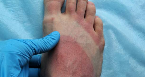
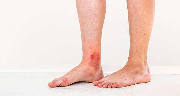
Medical professional examining red, inflamed foot consistent with erythromelalgia symptoms

Diagnosis of Erythromelalgia

Diagnosis of Erythromelalgia The diagnosis of erythromelalgia is based largely on symptoms and patient history. Since there are no specific blood tests or scans to confirm erythromelalgia. The condition is considered a clinical diagnosis—meaning doctors identify it based on what the patient describes and what is observed during a flare. Patient History Doctors will ask about: The nature and location of pain Colour and temperature changes in the skin How symptoms start, how long they last, and what makes them better or worse Whether symptoms are triggered by heat or relieved by cooling Any underlying conditions like autoimmune disease, diabetes, or blood disorders A history of repeated, painful redness in the hands or feet that worsens with heat and improves with cooling is highly suggestive of erythromelalgia. Physical Examination During an active flare, doctors may observe: Redness or a purplish rash Increased warmth of the skin Swelling Visible blood vessel changes When symptoms are not active, the skin may appear normal, making diagnosis more difficult. If needed, doctors may ask patients to keep a symptom diary or bring photographs of flare-ups. Blood and Laboratory Tests While there is no definitive blood test for erythromelalgia, several tests can help rule out underlying causes: Full blood count (FBC) – To check for polycythaemia vera or other myeloproliferative disorders Erythropoietin levels – To assess for secondary erythrocytosis Autoimmune markers – Including ANA and rheumatoid factor Blood glucose – To assess for diabetes-related neuropathy Thyroid function tests – To exclude thyroid dysfunction Nerve and Skin Testing If nerve involvement is suspected: Skin biopsy – May show small fibre neuropathy in some patients Quantitative sensory testing – Measures nerve response to stimuli Electromyography (EMG) and nerve conduction studies – Typically normal but may help exclude other conditions Differential Diagnosis Doctors must distinguish erythromelalgia from similar conditions: Raynaud’s phenomenon (which is cold-induced, not heat-triggered) Cellulitis or skin infection Complex regional pain syndrome (CRPS) Vasculitis or other vascular disorders Chilblains or pernio (cold-related vascular condition) Diagnosis of Erythromelalgia In South Africa In South Africa, diagnosis may be delayed due to low awareness and limited access to neurologists or dermatologists. Many patients are misdiagnosed with infections or allergic reactions. Telemedicine, symptom tracking apps, and patient support groups could improve recognition. The diagnosis of erythromelalgia requires a combination of careful history-taking, exclusion of other causes, and a high level of clinical awareness. Early recognition leads to better symptom control and prevents unnecessary testing or treatment. [Next: Treatment of Erythromelalgia →]
Diagnosis of Erythromelalgia Read More »

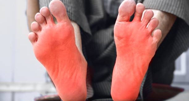
Person with visibly red, inflamed soles of feet raised for cooling, representing erythromelalgia treatment

Treatment of Erythromelalgia

Treatment of Erythromelalgia The treatment of erythromelalgia focuses on relieving symptoms, avoiding triggers, and addressing any underlying conditions. Because erythromelalgia is a chronic and often unpredictable condition, treatment must be personalised. No single therapy works for everyone, and a trial-and-error approach is often necessary. General Strategies Avoid heat exposure – Stay in cool environments, avoid hot showers, and reduce time in the sun Use cooling methods safely – Fans, cold water foot soaks (short periods only), cool packs, or air conditioning Wear loose, breathable clothing and open footwear Rest during flare-ups – Elevating the limbs can help improve symptoms Topical Treatments Some patients benefit from: Lidocaine creams or patches – Numbs the skin and may reduce burning Capsaicin cream – May desensitise overactive nerves (use cautiously, as it can cause irritation) Cooling gels or menthol-based creams – Provide short-term relief Oral Medications Treatment may include: Aspirin – Especially if symptoms are linked to high platelet counts or polycythaemia vera Anticonvulsants, such as gabapentin or pregabalin – For nerve-related pain Tricyclic antidepressants, like amitriptyline – Used for chronic neuropathic pain Selective serotonin-norepinephrine reuptake inhibitors (SNRIs) – May help in some patients Beta-blockers or calcium channel blockers – Occasionally used to regulate blood vessel dilation Medications must be started at low doses and increased slowly to monitor for side effects. Treatment of Underlying Conditions In secondary erythromelalgia, the best outcomes come from treating the root cause: Polycythaemia vera – Managed with phlebotomy or hydroxyurea Autoimmune diseases – May require steroids or immunosuppressants Neuropathy – Needs glucose control or treatment of the nerve condition Advanced Therapies For severe, treatment-resistant cases: Intravenous lidocaine infusions – Offered at specialist pain clinics Spinal cord stimulators – Rarely used, but may help in extreme cases Nerve blocks – Occasionally provide temporary relief Treatment of Erythromelalgia In South Africa Many patients in South Africa struggle to access pain specialists, especially within the public healthcare system. Increased training for primary care providers and access to neurologists can improve outcomes. The treatment of erythromelalgia requires a multidisciplinary, patient-focused approach. With the right combination of medication, cooling, and support, most patients can achieve meaningful symptom relief. [Next: Complications of Erythromelalgia →]

Treatment of Erythromelalgia Read More »

Lower leg with inflamed and broken skin showing complications of erythromelalgia

Complications of Erythromelalgia

Complications of Erythromelalgia The complications of erythromelalgia arise mainly from prolonged symptoms, poor symptom control, or unsafe attempts at self-treatment. Though erythromelalgia is not fatal, it can be physically and emotionally disabling without proper care. Skin Damage and Ulceration The urge to cool the skin during flare-ups can lead to: Cold burns, from ice packs applied directly to the skin Maceration, due to prolonged soaking in water Skin infections, if the skin barrier is broken Ulcers, particularly on the feet, from repeated trauma or overexposure to cold Educating patients about safe cooling methods is essential to avoid these problems. Reduced Mobility Frequent pain and flare-ups can result in: Avoidance of walking or standing, leading to deconditioning Dependence on mobility aids, especially in severe cases Increased fall risk, due to muscle weakness or sensory loss This may lead to isolation, weight gain, and worsening health over time. Sleep and Emotional Disturbances Many patients with erythromelalgia experience: Sleep disruption, especially from night-time flare-ups Chronic fatigue, due to unrelenting pain and poor rest Depression or anxiety, from coping with a rare and misunderstood illness Social withdrawal, due to embarrassment or mobility limitations Support groups, psychological counselling, and peer education can be invaluable. Work and Lifestyle Impact Absenteeism, due to pain and flare frequency Avoidance of social activities Strained relationships, especially if the condition is not well understood by family or friends Financial strain, from medication, travel to appointments, or loss of income Complications of Erythromelalgia In South Africa In South Africa, these complications are magnified by limited awareness and lack of specialist pain clinics in public health. Patients may be labelled as drug-seeking or misdiagnosed with psychiatric conditions. Community-level education and digital support platforms can help improve care and reduce stigma. The complications of erythromelalgia can be prevented or minimised with a proactive management plan. With consistent care, many people can live full lives despite the challenges posed by this complex condition. [Next: Back to Overview →]

Complications of Erythromelalgia Read More »

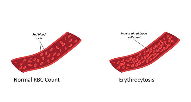
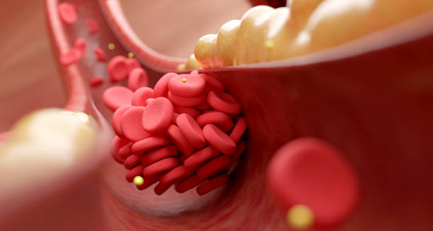
Comparison diagram showing normal red blood cell count versus erythrocytosis

Erythrocytosis

Erythrocytosis The condition, Erythrocytosis, is a condition in which the body produces too many red blood cells. Erythrocytosis causes the blood to become thicker than normal, which can increase the risk of blood clots, stroke, or heart attack. It is often discovered during routine blood tests showing elevated haemoglobin or haematocrit levels. Red blood cells are essential for carrying oxygen throughout the body. When there are too many of them, circulation may slow, and oxygen delivery can become less efficient. In some cases, erythrocytosis causes no symptoms and is picked up incidentally. In others, it may lead to headaches, dizziness, blurred vision, or high blood pressure. There are two main types: Primary erythrocytosis, often caused by bone marrow disorders like polycythaemia vera (a type of blood cancer) Secondary erythrocytosis, caused by external factors such as chronic lung disease, living at high altitude, or certain tumours Erythrocytosis In South Africa, erythrocytosis may be underdiagnosed due to limited routine screening in many rural areas. People living in mountainous regions or with undiagnosed respiratory illnesses may be at higher risk. Understanding erythrocytosis is essential for early intervention, especially in patients with cardiovascular risk factors or chronic health conditions. Recognising the Importance of Early Detection Erythrocytosis, though sometimes asymptomatic, can have serious consequences if left undetected or untreated. With increased blood viscosity comes a heightened risk of complications such as blood clots, strokes, and heart attacks. As such, recognising early warning signs and investigating elevated haemoglobin or haematocrit levels during routine medical checks can make a significant difference in outcomes. In regions like South Africa, where routine screenings may not be readily available in rural settings, awareness becomes an especially crucial tool in improving public health. Addressing Underlying Causes and Risk Factors Effective management of erythrocytosis hinges on correctly identifying whether it is primary or secondary in origin. While primary erythrocytosis often stems from bone marrow disorders like polycythaemia vera, secondary causes range from environmental factors such as high altitude to underlying diseases like chronic lung conditions. Understanding these distinctions allows for more tailored treatment strategies—whether that involves addressing a tumour, improving oxygenation, or managing chronic illnesses that contribute to the condition. Moving Forward with Awareness and Prevention Raising awareness of erythrocytosis and improving access to diagnostic resources are key steps toward reducing its impact. Especially in underserved communities. Healthcare providers should remain vigilant for signs of the condition, particularly in patients with risk factors such as smoking, cardiovascular issues, or high-altitude residence. With proactive monitoring and appropriate interventions. The complications associated with erythrocytosis can be mitigated, leading to better long-term health outcomes and enhanced quality of life for those affected. [Next: Causes of Erythrocytosis →]

Erythrocytosis Read More »

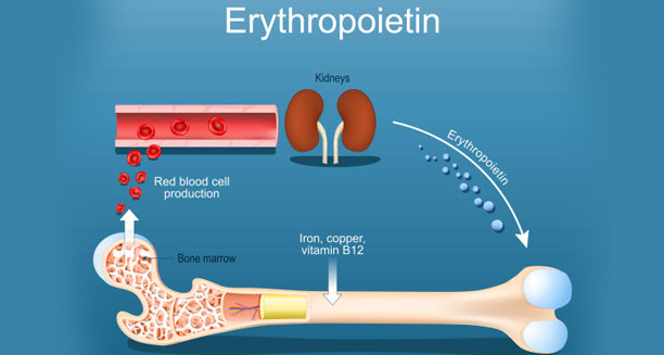
Diagram showing erythropoietin production by kidneys and its role in red blood cell formation

Causes of Erythrocytosis

Causes of Erythrocytosis The causes of erythrocytosis vary and are typically classified as either primary or secondary. Erythrocytosis can result from abnormal bone marrow function or be a natural response to conditions that limit oxygen delivery in the body. Primary Causes Primary erythrocytosis originates within the bone marrow itself. The most common cause is: Polycythaemia vera – A rare blood cancer in which the bone marrow produces too many red cells, often along with white cells and platelets. This is due to a mutation in the JAK2 gene. In this form, erythropoietin (EPO)—the hormone that stimulates red blood cell production—is usually low because the red cells are being overproduced without any external trigger. Secondary Causes These occur when another condition causes the body to produce extra erythropoietin in response to low oxygen levels or other stimuli: Chronic lung diseases, such as COPD or sleep apnoea Living at high altitudes, where oxygen levels are naturally lower Heart conditions, particularly congenital heart disease with low blood oxygen Kidney tumours, which may secrete excess EPO Liver disease, especially in those with cirrhosis or hepatocellular carcinoma Testosterone therapy, which can stimulate red blood cell production Anabolic steroid use, often seen in athletes or bodybuilders Apparent (Relative) Erythrocytosis In some cases, the red cell count appears high due to a reduction in plasma volume, not an actual increase in red cells. This is called relative erythrocytosis and may be caused by: Dehydration Severe burns Diuretic overuse Chronic stress or smoking Causes of Erythrocytosis In South Africa In South Africa, high-altitude erythrocytosis is more likely in populations living in the Drakensberg or Lesotho mountains. Smoking-related lung disease and chronic obstructive pulmonary conditions are also common contributors, particularly in older men. Kidney disease is a growing concern due to rising rates of hypertension and diabetes, and this may result in EPO overproduction in some individuals. Understanding the causes of erythrocytosis allows doctors to distinguish between dangerous forms that require treatment and benign types that may need only monitoring. [Next: Symptoms of Erythrocytosis →]

Causes of Erythrocytosis Read More »

Man holding his head and leaning on a pole, experiencing dizziness and headache—symptoms of erythrocytosis

Symptoms of Erythrocytosis

Symptoms of Erythrocytosis The symptoms of erythrocytosis depend on how elevated the red blood cell count becomes and whether complications arise. While erythrocytosis may be asymptomatic in its early stages, high red cell levels can cause blood to thicken, reducing oxygen flow and increasing the risk of clotting. Common Symptoms Headaches – Due to increased blood viscosity and reduced oxygen to the brain Dizziness or light-headedness, especially during exertion Blurred or double vision Flushed skin, particularly on the face Reddened eyes High blood pressure Fatigue, which may seem paradoxical despite the high oxygen-carrying capacity Shortness of breath, especially on exertion or when lying flat These symptoms may develop gradually or appear suddenly in cases of rapid red cell production. Serious Symptoms and Complications As blood thickens, the risk of clots increases. Patients may present with: Chest pain, which may signal a heart attack Leg swelling or pain, possibly due to deep vein thrombosis (DVT) Stroke-like symptoms, including slurred speech, weakness, or vision changes Bleeding or bruising, in conditions like polycythaemia vera, due to platelet abnormalities Clot-related complications are the most dangerous consequences of erythrocytosis and require immediate medical attention. Symptoms in Children Though rare, erythrocytosis in children may be linked to congenital heart disease or inherited mutations. Symptoms may include: Cyanosis (bluish skin) Poor growth Tiring easily Clubbing of fingers or toes Symptoms of Erythrocytosis In South Africa In South Africa, many patients with chronic lung conditions may develop secondary erythrocytosis without realising it. Symptoms such as breathlessness, fatigue, or dizziness may be wrongly attributed to ageing or poor fitness. Routine screening in high-risk groups could lead to earlier diagnosis. Public education on the signs of abnormal blood counts and routine health checks can play a vital role, particularly in regions where access to specialist care is limited. Recognising the symptoms of erythrocytosis early can prevent serious complications and guide timely intervention before a blood clot or stroke occurs. [Next: Diagnosis of Erythrocytosis→]

Symptoms of Erythrocytosis Read More »

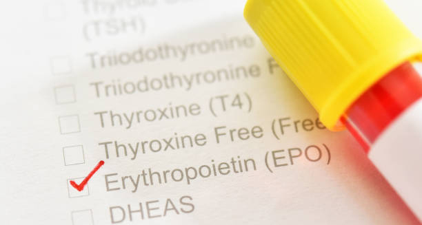
Erythropoietin blood test form with sample tube used to diagnose erythrocytosis

Diagnosis of Erythrocytosis

Diagnosis of Erythrocytosis The diagnosis of erythrocytosis begins with a full blood count (FBC). Doctors then perform tests to identify whether the condition is primary, secondary, or relative. Doctors use a step-by-step approach to determine whether to treat erythrocytosis or simply monitor it, since the condition has many causes. Step 1: Blood Tests The first clue comes from routine blood tests showing: Elevated red blood cell count (RBC) Increased haemoglobin (Hb) High haematocrit (Hct) In men, haematocrit levels above 52% and in women above 48% may prompt further investigation. Blood viscosity also increases at these levels, raising the risk of complications. Step 2: Rule Out Relative Erythrocytosis Doctors first determine whether the red cell count is truly elevated or whether reduced plasma volume is responsible. Furthermore, dehydration, diuretic use, or acute illness can cause relative erythrocytosis, which resolves with rehydration and correction of fluid balance. Step 3: Evaluate Oxygen Levels and EPO Pulse oximetry and arterial blood gases help assess oxygen levels in the blood Erythropoietin (EPO) levels are crucial to classify the erythrocytosis: Low EPO suggests a primary cause, like polycythaemia vera High EPO points to a secondary cause, such as lung disease or kidney tumours Step 4: Genetic and Bone Marrow Tests In suspected primary erythrocytosis: A JAK2 gene mutation test is performed. Thus, most cases of polycythaemia vera are JAK2-positive. Doctors may perform a bone marrow biopsy to assess cellular activity and rule out other haematological disorders. Imaging and Other Investigations Depending on suspected causes, doctors may request: Chest X-rays or CT scans – For lung disease or tumours Abdominal ultrasound or CT – To check the kidneys or liver Sleep studies, if sleep apnoea is suspected Diagnosis of Erythrocytosis In South Africa Some specialist centres in South Africa provide access to genetic testing. Additionally, bone marrow biopsies since availability can be limited in other areas. Primary care clinics often rely on basic blood tests and chest X-rays. Furthermore, healthcare providers maintain a high index of suspicion in areas with limited access to advanced diagnostics. The diagnosis of erythrocytosis should be thorough to prevent missed cases of blood cancer or secondary causes like TB-related lung damage. Finally, timely diagnosis allows patients to receive the correct treatment and avoid dangerous complications. [Next: Treatment of Erythrocytosis →]

Diagnosis of Erythrocytosis Read More »

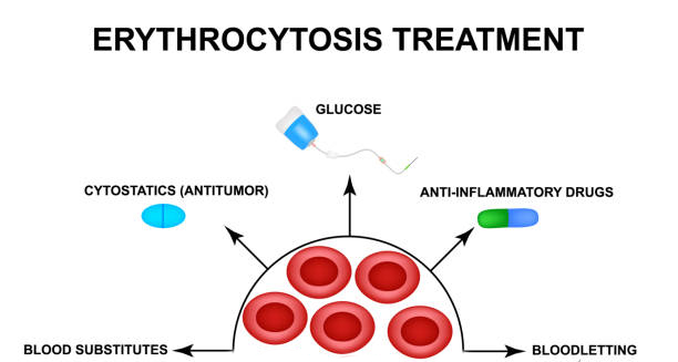
Diagram showing treatment options for erythrocytosis including bloodletting and medication

Treatment of Erythrocytosis

Treatment of Erythrocytosis The treatment of erythrocytosis depends on the cause. In most cases, the goal is to reduce the red cell mass, prevent blood clots, and manage underlying conditions. Erythrocytosis can often be controlled successfully with lifestyle changes, medication, or medical procedures. Treatment of Primary Erythrocytosis In polycythaemia vera (a form of primary erythrocytosis), treatment includes: 1. Phlebotomy Regular removal of blood (usually 500ml at a time) reduces red cell count and haematocrit Keeps blood viscosity within safe limits Can be done weekly at first, then less often 2. Low-dose Aspirin Helps prevent blood clots without increasing bleeding risk significantly Often prescribed long-term 3. Cytoreductive Therapy Hydroxyurea is the most commonly used medication to suppress bone marrow overproduction Interferon may be used in younger patients or those who cannot tolerate hydroxyurea Treatment of Secondary Erythrocytosis Treatment focuses on the root cause: Stop smoking, if tobacco use is the cause Oxygen therapy, for chronic lung disease or hypoxia Treat sleep apnoea, with CPAP machines or lifestyle changes Surgical removal of tumours, if a kidney or liver mass is producing excess EPO Control heart or kidney disease, using the appropriate medication Secondary cases may not require phlebotomy unless symptoms become severe or haematocrit is dangerously high. Lifestyle and Monitoring All patients are encouraged to: Stay well hydrated Avoid iron supplements unless specifically needed Monitor blood pressure and cardiovascular risk Reduce alcohol intake and avoid high altitudes if advised Treatment of Erythrocytosis In South Africa In South Africa, access to phlebotomy services may be limited in rural clinics. Some patients must travel to regional hospitals for procedures. Treatment of underlying causes like TB or COPD remains the priority in many secondary cases. The treatment of erythrocytosis is highly effective when personalised to the individual’s cause and risk profile. Early intervention prevents serious complications and improves quality of life. [Next: Complications of Erythrocytosis →]

Treatment of Erythrocytosis Read More »

Red blood cells blocking an artery due to erythrocytosis

Complications of Erythrocytosis

Complications of Erythrocytosis The complications of erythrocytosis mainly stem from the blood becoming too thick, which can impair circulation and increase the risk of dangerous clots. While erythrocytosis is manageable, untreated or poorly controlled cases can lead to severe, even fatal, outcomes. Blood Clots (Thrombosis) The most serious complication is the formation of blood clots, which can lead to: Stroke, if a clot blocks blood flow to the brain Heart attack, if a coronary artery becomes blocked Pulmonary embolism, a life-threatening clot in the lungs Deep vein thrombosis (DVT), often affecting the legs Thicker blood moves more slowly, allowing clots to form more easily, particularly in those who are inactive or have other cardiovascular risk factors. Bleeding Ironically, people with polycythaemia vera (a form of erythrocytosis) may also be at risk of bleeding, especially from: Nosebleeds Gum bleeding Gastrointestinal bleeding This is due to abnormalities in platelet function, even when platelet counts are high. High Blood Pressure Increased blood volume and thickness can raise blood pressure, straining the heart and blood vessels. This further raises the risk of stroke and heart disease. Gout and Kidney Stones High cell turnover can lead to increased uric acid levels, resulting in: Gout attacks, causing painful joint swelling Kidney stones, due to crystallised uric acid in the urine Progression to Myelofibrosis or Leukaemia In rare cases of polycythaemia vera, prolonged bone marrow stimulation can lead to: Myelofibrosis, where scar tissue replaces bone marrow Acute leukaemia, a serious blood cancer These complications are more likely in long-standing or poorly managed cases. Complications of Erythrocytosis In South Africa In South Africa, complications are more likely in undiagnosed or untreated patients. Those with limited healthcare access may present late with stroke or DVT. Public education and regular screening for high haematocrit levels can help reduce the burden of undiagnosed erythrocytosis. Patients with lung disease, especially in mining areas or high-altitude regions, should be monitored closely for signs of elevated red blood cell counts. The complications of erythrocytosis can be life-threatening, but most are preventable. With early diagnosis, proper monitoring, and appropriate treatment, patients can lead long, healthy lives. [Next: Back to Overview →]

Complications of Erythrocytosis Read More »

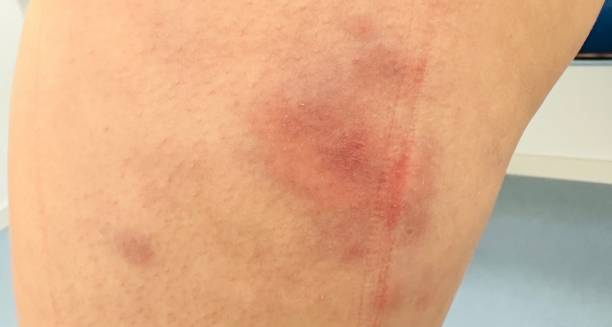
Red swollen patches on skin caused by erythema nodosum

Erythema Nodosum

Erythema Nodosum The condition, Erythema nodosum is an inflammatory condition that causes tender, red lumps to appear under the skin, most commonly on the shins. This condition affects the fatty layer of the skin (the subcutaneous fat) and is usually a reaction to an underlying condition, infection, or medication rather than a disease itself. The painful nodules typically start as firm, red bumps that slowly turn a bluish colour before fading away. Each bump may last up to two weeks, but the full episode can take four to six weeks to resolve. The condition affects more women than men and is most common between the ages of 20 and 40. It is considered a type of panniculitis—an inflammation of fat tissue beneath the skin. While often uncomfortable, erythema nodosum is not contagious and usually clears up on its own once the underlying cause is treated or resolves. Erythema Nodosum In South Africa, this condition is frequently seen in connection with infections like tuberculosis, streptococcal sore throat, and HIV-related conditions. Understanding the cause is key to proper management. Recognising this condition early helps reduce discomfort and ensures appropriate investigation into possible triggers. [Next: Causes of Erythema Nodosum →]

Erythema Nodosum Read More »

Scroll to Top