Mental Matters

Health Conditions

Find information on various health conditions affecting people of all ages. This category covers causes, symptoms, diagnosis, and management options for physical illnesses and chronic diseases. Get insights to help you understand different health challenges and make informed decisions about your wellbeing.

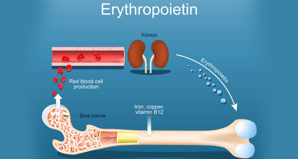
Diagram showing erythropoietin production by kidneys and its role in red blood cell formation

Causes of Erythrocytosis

Causes of Erythrocytosis The causes of erythrocytosis vary and are typically classified as either primary or secondary. Erythrocytosis can result from abnormal bone marrow function or be a natural response to conditions that limit oxygen delivery in the body. Primary Causes Primary erythrocytosis originates within the bone marrow itself. The most common cause is: Polycythaemia vera – A rare blood cancer in which the bone marrow produces too many red cells, often along with white cells and platelets. This is due to a mutation in the JAK2 gene. In this form, erythropoietin (EPO)—the hormone that stimulates red blood cell production—is usually low because the red cells are being overproduced without any external trigger. Secondary Causes These occur when another condition causes the body to produce extra erythropoietin in response to low oxygen levels or other stimuli: Chronic lung diseases, such as COPD or sleep apnoea Living at high altitudes, where oxygen levels are naturally lower Heart conditions, particularly congenital heart disease with low blood oxygen Kidney tumours, which may secrete excess EPO Liver disease, especially in those with cirrhosis or hepatocellular carcinoma Testosterone therapy, which can stimulate red blood cell production Anabolic steroid use, often seen in athletes or bodybuilders Apparent (Relative) Erythrocytosis In some cases, the red cell count appears high due to a reduction in plasma volume, not an actual increase in red cells. This is called relative erythrocytosis and may be caused by: Dehydration Severe burns Diuretic overuse Chronic stress or smoking Causes of Erythrocytosis In South Africa In South Africa, high-altitude erythrocytosis is more likely in populations living in the Drakensberg or Lesotho mountains. Smoking-related lung disease and chronic obstructive pulmonary conditions are also common contributors, particularly in older men. Kidney disease is a growing concern due to rising rates of hypertension and diabetes, and this may result in EPO overproduction in some individuals. Understanding the causes of erythrocytosis allows doctors to distinguish between dangerous forms that require treatment and benign types that may need only monitoring. [Next: Symptoms of Erythrocytosis →]

Causes of Erythrocytosis Read More »

Man holding his head and leaning on a pole, experiencing dizziness and headache—symptoms of erythrocytosis

Symptoms of Erythrocytosis

Symptoms of Erythrocytosis The symptoms of erythrocytosis depend on how elevated the red blood cell count becomes and whether complications arise. While erythrocytosis may be asymptomatic in its early stages, high red cell levels can cause blood to thicken, reducing oxygen flow and increasing the risk of clotting. Common Symptoms Headaches – Due to increased blood viscosity and reduced oxygen to the brain Dizziness or light-headedness, especially during exertion Blurred or double vision Flushed skin, particularly on the face Reddened eyes High blood pressure Fatigue, which may seem paradoxical despite the high oxygen-carrying capacity Shortness of breath, especially on exertion or when lying flat These symptoms may develop gradually or appear suddenly in cases of rapid red cell production. Serious Symptoms and Complications As blood thickens, the risk of clots increases. Patients may present with: Chest pain, which may signal a heart attack Leg swelling or pain, possibly due to deep vein thrombosis (DVT) Stroke-like symptoms, including slurred speech, weakness, or vision changes Bleeding or bruising, in conditions like polycythaemia vera, due to platelet abnormalities Clot-related complications are the most dangerous consequences of erythrocytosis and require immediate medical attention. Symptoms in Children Though rare, erythrocytosis in children may be linked to congenital heart disease or inherited mutations. Symptoms may include: Cyanosis (bluish skin) Poor growth Tiring easily Clubbing of fingers or toes Symptoms of Erythrocytosis In South Africa In South Africa, many patients with chronic lung conditions may develop secondary erythrocytosis without realising it. Symptoms such as breathlessness, fatigue, or dizziness may be wrongly attributed to ageing or poor fitness. Routine screening in high-risk groups could lead to earlier diagnosis. Public education on the signs of abnormal blood counts and routine health checks can play a vital role, particularly in regions where access to specialist care is limited. Recognising the symptoms of erythrocytosis early can prevent serious complications and guide timely intervention before a blood clot or stroke occurs. [Next: Diagnosis of Erythrocytosis→]

Symptoms of Erythrocytosis Read More »

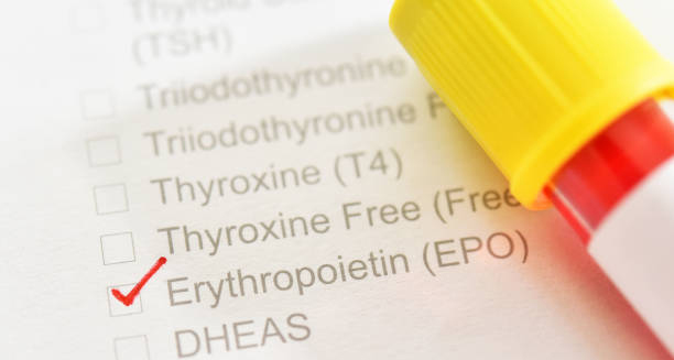
Erythropoietin blood test form with sample tube used to diagnose erythrocytosis

Diagnosis of Erythrocytosis

Diagnosis of Erythrocytosis The diagnosis of erythrocytosis begins with a full blood count (FBC). Doctors then perform tests to identify whether the condition is primary, secondary, or relative. Doctors use a step-by-step approach to determine whether to treat erythrocytosis or simply monitor it, since the condition has many causes. Step 1: Blood Tests The first clue comes from routine blood tests showing: Elevated red blood cell count (RBC) Increased haemoglobin (Hb) High haematocrit (Hct) In men, haematocrit levels above 52% and in women above 48% may prompt further investigation. Blood viscosity also increases at these levels, raising the risk of complications. Step 2: Rule Out Relative Erythrocytosis Doctors first determine whether the red cell count is truly elevated or whether reduced plasma volume is responsible. Furthermore, dehydration, diuretic use, or acute illness can cause relative erythrocytosis, which resolves with rehydration and correction of fluid balance. Step 3: Evaluate Oxygen Levels and EPO Pulse oximetry and arterial blood gases help assess oxygen levels in the blood Erythropoietin (EPO) levels are crucial to classify the erythrocytosis: Low EPO suggests a primary cause, like polycythaemia vera High EPO points to a secondary cause, such as lung disease or kidney tumours Step 4: Genetic and Bone Marrow Tests In suspected primary erythrocytosis: A JAK2 gene mutation test is performed. Thus, most cases of polycythaemia vera are JAK2-positive. Doctors may perform a bone marrow biopsy to assess cellular activity and rule out other haematological disorders. Imaging and Other Investigations Depending on suspected causes, doctors may request: Chest X-rays or CT scans – For lung disease or tumours Abdominal ultrasound or CT – To check the kidneys or liver Sleep studies, if sleep apnoea is suspected Diagnosis of Erythrocytosis In South Africa Some specialist centres in South Africa provide access to genetic testing. Additionally, bone marrow biopsies since availability can be limited in other areas. Primary care clinics often rely on basic blood tests and chest X-rays. Furthermore, healthcare providers maintain a high index of suspicion in areas with limited access to advanced diagnostics. The diagnosis of erythrocytosis should be thorough to prevent missed cases of blood cancer or secondary causes like TB-related lung damage. Finally, timely diagnosis allows patients to receive the correct treatment and avoid dangerous complications. [Next: Treatment of Erythrocytosis →]

Diagnosis of Erythrocytosis Read More »

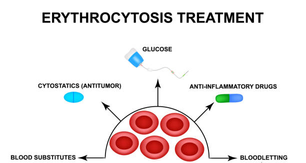
Diagram showing treatment options for erythrocytosis including bloodletting and medication

Treatment of Erythrocytosis

Treatment of Erythrocytosis The treatment of erythrocytosis depends on the cause. In most cases, the goal is to reduce the red cell mass, prevent blood clots, and manage underlying conditions. Erythrocytosis can often be controlled successfully with lifestyle changes, medication, or medical procedures. Treatment of Primary Erythrocytosis In polycythaemia vera (a form of primary erythrocytosis), treatment includes: 1. Phlebotomy Regular removal of blood (usually 500ml at a time) reduces red cell count and haematocrit Keeps blood viscosity within safe limits Can be done weekly at first, then less often 2. Low-dose Aspirin Helps prevent blood clots without increasing bleeding risk significantly Often prescribed long-term 3. Cytoreductive Therapy Hydroxyurea is the most commonly used medication to suppress bone marrow overproduction Interferon may be used in younger patients or those who cannot tolerate hydroxyurea Treatment of Secondary Erythrocytosis Treatment focuses on the root cause: Stop smoking, if tobacco use is the cause Oxygen therapy, for chronic lung disease or hypoxia Treat sleep apnoea, with CPAP machines or lifestyle changes Surgical removal of tumours, if a kidney or liver mass is producing excess EPO Control heart or kidney disease, using the appropriate medication Secondary cases may not require phlebotomy unless symptoms become severe or haematocrit is dangerously high. Lifestyle and Monitoring All patients are encouraged to: Stay well hydrated Avoid iron supplements unless specifically needed Monitor blood pressure and cardiovascular risk Reduce alcohol intake and avoid high altitudes if advised Treatment of Erythrocytosis In South Africa In South Africa, access to phlebotomy services may be limited in rural clinics. Some patients must travel to regional hospitals for procedures. Treatment of underlying causes like TB or COPD remains the priority in many secondary cases. The treatment of erythrocytosis is highly effective when personalised to the individual’s cause and risk profile. Early intervention prevents serious complications and improves quality of life. [Next: Complications of Erythrocytosis →]

Treatment of Erythrocytosis Read More »

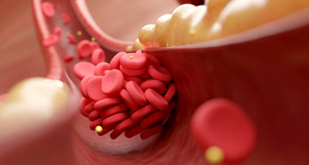
Red blood cells blocking an artery due to erythrocytosis

Complications of Erythrocytosis

Complications of Erythrocytosis The complications of erythrocytosis mainly stem from the blood becoming too thick, which can impair circulation and increase the risk of dangerous clots. While erythrocytosis is manageable, untreated or poorly controlled cases can lead to severe, even fatal, outcomes. Blood Clots (Thrombosis) The most serious complication is the formation of blood clots, which can lead to: Stroke, if a clot blocks blood flow to the brain Heart attack, if a coronary artery becomes blocked Pulmonary embolism, a life-threatening clot in the lungs Deep vein thrombosis (DVT), often affecting the legs Thicker blood moves more slowly, allowing clots to form more easily, particularly in those who are inactive or have other cardiovascular risk factors. Bleeding Ironically, people with polycythaemia vera (a form of erythrocytosis) may also be at risk of bleeding, especially from: Nosebleeds Gum bleeding Gastrointestinal bleeding This is due to abnormalities in platelet function, even when platelet counts are high. High Blood Pressure Increased blood volume and thickness can raise blood pressure, straining the heart and blood vessels. This further raises the risk of stroke and heart disease. Gout and Kidney Stones High cell turnover can lead to increased uric acid levels, resulting in: Gout attacks, causing painful joint swelling Kidney stones, due to crystallised uric acid in the urine Progression to Myelofibrosis or Leukaemia In rare cases of polycythaemia vera, prolonged bone marrow stimulation can lead to: Myelofibrosis, where scar tissue replaces bone marrow Acute leukaemia, a serious blood cancer These complications are more likely in long-standing or poorly managed cases. Complications of Erythrocytosis In South Africa In South Africa, complications are more likely in undiagnosed or untreated patients. Those with limited healthcare access may present late with stroke or DVT. Public education and regular screening for high haematocrit levels can help reduce the burden of undiagnosed erythrocytosis. Patients with lung disease, especially in mining areas or high-altitude regions, should be monitored closely for signs of elevated red blood cell counts. The complications of erythrocytosis can be life-threatening, but most are preventable. With early diagnosis, proper monitoring, and appropriate treatment, patients can lead long, healthy lives. [Next: Back to Overview →]

Complications of Erythrocytosis Read More »

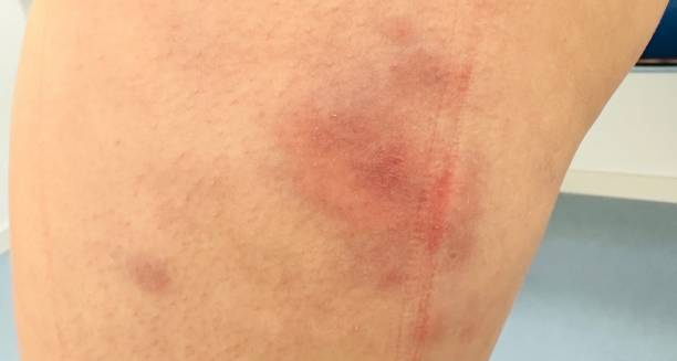
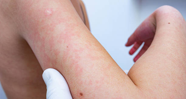
Red swollen patches on skin caused by erythema nodosum

Erythema Nodosum

Erythema Nodosum The condition, Erythema nodosum is an inflammatory condition that causes tender, red lumps to appear under the skin, most commonly on the shins. This condition affects the fatty layer of the skin (the subcutaneous fat) and is usually a reaction to an underlying condition, infection, or medication rather than a disease itself. The painful nodules typically start as firm, red bumps that slowly turn a bluish colour before fading away. Each bump may last up to two weeks, but the full episode can take four to six weeks to resolve. The condition affects more women than men and is most common between the ages of 20 and 40. It is considered a type of panniculitis—an inflammation of fat tissue beneath the skin. While often uncomfortable, erythema nodosum is not contagious and usually clears up on its own once the underlying cause is treated or resolves. Erythema Nodosum In South Africa, this condition is frequently seen in connection with infections like tuberculosis, streptococcal sore throat, and HIV-related conditions. Understanding the cause is key to proper management. Recognising this condition early helps reduce discomfort and ensures appropriate investigation into possible triggers. [Next: Causes of Erythema Nodosum →]

Erythema Nodosum Read More »

Woman coughing, possibly showing signs of infection linked to erythema nodosum

Causes of Erythema Nodosum

Causes of Erythema Nodosum The causes of erythema nodosum are varied, and in many cases, no single cause is ever found. However, erythema nodosum is often a reaction to infection, inflammation, or medication. Identifying the cause helps doctors decide whether further treatment or investigation is necessary. Infections Infectious causes are among the most common, particularly: Streptococcal throat infections – Often seen in children and young adults Tuberculosis (TB) – A major cause in regions like South Africa, especially in rural or high-prevalence areas HIV and associated infections – May trigger inflammatory reactions Fungal infections – Such as histoplasmosis or coccidioidomycosis in some regions Yersinia enterocolitica – A bacterial gut infection sometimes linked to erythema nodosum Inflammatory and Autoimmune Conditions Several chronic diseases can trigger the condition: Sarcoidosis – A common cause in adults, especially when combined with symptoms like cough and swollen lymph nodes Inflammatory bowel disease – Both Crohn’s disease and ulcerative colitis are linked Behçet’s disease – A rare condition that may present with ulcers, joint pain, and skin lesions Medications Some medications can provoke a hypersensitive reaction: Sulphonamides Oral contraceptives Penicillins Non-steroidal anti-inflammatory drugs (NSAIDs) Drug-related erythema nodosum often resolves once the medication is stopped. Pregnancy Hormonal changes during pregnancy may also trigger episodes of erythema nodosum, especially in the first trimester. Causes of Erythema Nodosum In South Africa In South Africa, TB is a leading cause. Erythema nodosum may be the first visible sign, especially in patients who have not yet shown respiratory symptoms. HIV-positive individuals are also more prone to skin-related inflammatory reactions. Doctors in South Africa should consider TB screening and HIV testing in patients presenting with unexplained erythema nodosum. Understanding the causes of erythema nodosum helps guide tests, treatment, and decisions about whether to investigate for underlying infections or autoimmune disorders. [Next: Symptoms of Erythema Nodosum →]

Causes of Erythema Nodosum Read More »

Illustration showing pain, redness, swelling, and numbness in hands

Symptoms of Erythema Nodosum

Symptoms of Erythema Nodosum The symptoms of erythema nodosum are mostly limited to the skin but may be accompanied by systemic signs depending on the underlying cause. Erythema nodosum typically begins with general flu-like symptoms, followed by the appearance of painful red lumps on the legs. Skin Symptoms Tender red nodules – Usually located on the front of the shins, but may also appear on the thighs, arms, or trunk Symmetrical distribution – Lesions typically appear on both legs Size – Nodules range from 1 to 5 cm in diameter Colour changes – Lesions start red, then become bluish or purple before fading to yellow or green like a bruise Pain or tenderness – Especially when touched or with pressure New nodules may appear for one to two weeks, and each lesion lasts about two weeks before resolving. General Symptoms Many people experience: Fever Fatigue Joint pain, especially in the knees or ankles Swollen ankles or legs Headache or malaise These systemic symptoms often occur before or alongside the skin eruption. They may be mistaken for a viral infection or flu in the early stages. Duration The full course of erythema nodosum usually lasts between three and six weeks. In some cases, the condition recurs, particularly if the trigger isn’t identified or resolved. In Children Children may have milder lesions, but they often present with fever, sore throat, and joint pain. A streptococcal infection is the most common cause in this age group. Symptoms of Erythema Nodosum In South Africa In South Africa, clinicians should be alert for TB in any patient presenting with erythema nodosum and constitutional symptoms like fever or weight loss. Patients with HIV may also show more severe symptoms or slower resolution. Prompt recognition of the symptoms of erythema nodosum allows doctors to manage discomfort while identifying any underlying medical cause. This ensures both symptom relief and appropriate long-term care. [Next: Diagnosis of Erythema Nodosum→]

Symptoms of Erythema Nodosum Read More »

Diagnosis of Erythema Nodosum

Diagnosis of Erythema Nodosum

Diagnosis of Erythema Nodosum The diagnosis of erythema nodosum is usually based on clinical examination and supported by investigations to identify the underlying cause. Because erythema nodosum is a skin reaction, not a disease on its own, doctors must determine what triggered it to offer appropriate treatment. Clinical Assessment Doctors begin with: A visual examination of the lesions Noting their size, colour, symmetry, and tenderness Asking about recent infections, medications, or travel Reviewing systemic symptoms such as fever, joint pain, sore throat, or weight loss The appearance of painful, red nodules on the shins in a symmetrical pattern is often enough to suspect erythema nodosum. However, identifying the cause is critical to prevent recurrence. Laboratory Investigations Several blood and diagnostic tests may be ordered to uncover a possible trigger: Full blood count (FBC) – Looks for elevated white cells or inflammation Erythrocyte sedimentation rate (ESR) / C-reactive protein (CRP) – Raised levels support the diagnosis of an inflammatory condition Anti-streptolysin O (ASO) titre – Detects a recent streptococcal infection HIV test – Especially important in South Africa due to the high prevalence Tuberculosis (TB) screening, including: Chest X-ray Sputum test or GeneXpert Tuberculin skin test (TST) or IGRA Additional Tests Depending on symptoms and suspected causes: Throat swab, if sore throat is present Stool cultures, if gastrointestinal symptoms are noted Chest CT scan, in cases where sarcoidosis is suspected Colonoscopy, if inflammatory bowel disease is suspected Pregnancy test, if relevant Skin Biopsy In uncertain or atypical cases, a skin biopsy may be performed. This can confirm panniculitis (inflammation of fat tissue) and help rule out other forms of skin disease such as vasculitis, cellulitis, or erythema induratum. Diagnosis of Erythema Nodosum In South Africa In South Africa, TB testing is often prioritised. Erythema nodosum may be the first symptom of a previously undiagnosed TB infection. In areas where access to biopsy or lab testing is limited, clinical diagnosis supported by chest X-ray and TB symptoms is commonly used. The diagnosis of erythema nodosum should be thorough, not just to confirm the condition but to uncover the underlying cause. This ensures faster recovery and reduces the risk of repeat episodes. [Next: Treatment of Erythema Nodosum →]

Diagnosis of Erythema Nodosum Read More »

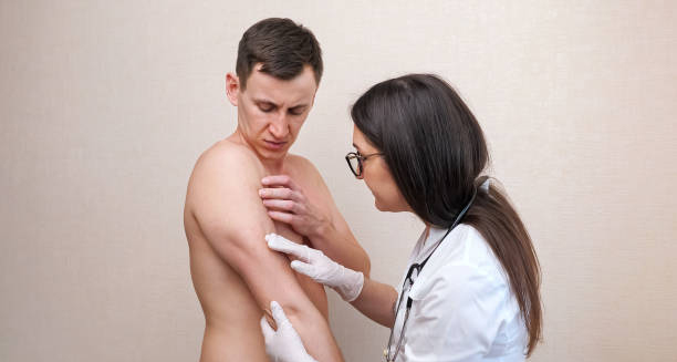
Medical professional examining erythema nodosum on a patient’s arm

Treatment of Erythema Nodosum

Treatment of Erythema Nodosum The treatment of erythema nodosum focuses on reducing inflammation, relieving pain, and addressing the underlying cause. Erythema nodosum usually resolves on its own within three to six weeks, but treatment helps manage symptoms and prevent recurrence. Rest and Supportive Care For most patients, the main treatment includes: Rest and leg elevation, especially during painful flare-ups Cool compresses, to reduce inflammation Compression stockings, to support circulation and minimise swelling Avoiding prolonged standing or walking, especially in the acute phase These basic steps can make a significant difference in comfort levels. Pain and Inflammation Relief To ease symptoms: Doctors commonly prescribe non-steroidal anti-inflammatory drugs (NSAIDs) such as ibuprofen or naproxen. Paracetamol can be used for fever or general discomfort In severe cases, doctors may prescribe oral corticosteroids like prednisone for short-term use—but only if there is no infection, such as tuberculosis Doctors should never start corticosteroids until they have ruled out infections like TB, as these medications can worsen the disease. Treating the Underlying Cause Identifying and addressing the trigger is crucial: Antibiotics, for streptococcal infections Anti-TB medication, if tuberculosis is confirmed Doctors stop the responsible medication if they suspect a drug-induced reaction Doctors prescribe immunosuppressants only when erythema nodosum is linked to autoimmune diseases like sarcoidosis or inflammatory bowel disease, and even then, only in rare cases. Recurrent Cases For patients with frequent episodes: Long-term NSAID therapy may be considered under supervision Colchicine or potassium iodide is occasionally used Antiviral or antimicrobial suppression may help if the cause is infectious Lifestyle adjustments, such as managing weight and avoiding known triggers, may also reduce recurrence. Treatment of Erythema Nodosum In South Africa In South Africa, TB-related erythema nodosum is common. Patients may start TB treatment based on chest X-ray and clinical suspicion. Public hospitals often provide NSAIDs and basic wound care supplies, while doctors reserve corticosteroids for non-infectious cases after thorough evaluation. Doctors often treat erythema nodosum straightforwardly, tailoring the treatment to each individual. Identifying and resolving the underlying cause leads to a faster recovery and prevents repeated flare-ups. [Next: Complications of Erythema Nodosum →]

Treatment of Erythema Nodosum Read More »

Scroll to Top