Mental Matters

Health Conditions

Find information on various health conditions affecting people of all ages. This category covers causes, symptoms, diagnosis, and management options for physical illnesses and chronic diseases. Get insights to help you understand different health challenges and make informed decisions about your wellbeing.

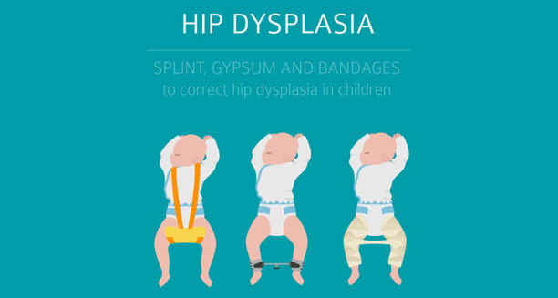
Illustration of infants with hip dysplasia braces

Symptoms of Developmental Dysplasia of the Hip

Symptoms of Developmental Dysplasia of the Hip Symptoms of developmental dysplasia of the hip can vary significantly depending on the age of the child and the severity of the condition. In many cases, particularly in newborns, developmental dysplasia of the hip does not cause any obvious signs and is only detected during routine screening. However, as a child grows, more noticeable symptoms may develop if the condition remains untreated. Symptoms in Newborns and Infants Hip instability: The hip may feel loose or dislocate with gentle movement during physical examination. Asymmetry in leg length: One leg may appear shorter than the other. Uneven skin folds: Extra or uneven folds may be noticeable on the thighs or buttocks. Limited range of motion: Difficulty in spreading the infant’s thighs apart during nappy changes. Audible ‘click’ or ‘clunk’: A sound or sensation during hip movement that may indicate dislocation. Routine screening by a healthcare provider includes the Ortolani and Barlow manoeuvres to detect any hip instability in the first few weeks of life. Symptoms in Older Infants and Toddlers Delayed walking: The child may begin walking later than expected. Limping or waddle: Walking may appear uneven or abnormal. Toe walking: The child may favour walking on their toes on the affected side. Exaggerated lumbar curve: An inward curve of the lower back (lordosis) may develop in bilateral DDH. In some cases, especially when the condition is mild, symptoms may be so subtle that they go unnoticed until a child begins walking or even later in childhood. Adolescent or Adult Symptoms | Symptoms of Developmental Dysplasia of the Hip If undiagnosed until adolescence or adulthood, individuals may experience: Hip or groin pain Stiffness and limited mobility Early onset osteoarthritis In conclusion, symptoms of developmental dysplasia of the hip are age-dependent and range from subtle instability in infants to noticeable gait problems in toddlers. Early screening and awareness of risk factors are essential for timely diagnosis and intervention. [Next: Diagnosis of Developmental Dysplasia of the Hip →]
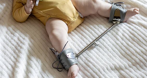

Symptoms of Developmental Dysplasia of the Hip Read More »

Infant wearing a Pavlik harness for hip dysplasia treatment

Diagnosis of Developmental Dysplasia of the Hip

Diagnosis of Developmental Dysplasia of the Hip Diagnosis of developmental dysplasia of the hip typically involves a combination of physical examination, imaging, and patient history. Since early detection dramatically improves outcomes, routine screening for developmental dysplasia of the hip is a standard part of newborn care in many countries. Clinical Examination In newborns and young infants, healthcare professionals perform specific manoeuvres during routine check-ups: Barlow test: The examiner attempts to gently dislocate the hip. Ortolani test: The examiner attempts to relocate a dislocated hip with a gentle ‘clunk.’ These tests are most effective within the first two to three months of life, as the infant’s muscles are still soft and flexible. As the child grows, the reliability of these tests decreases. Imaging Tests & Diagnosis of Developmental Dysplasia of the Hip When physical examination is inconclusive or the infant is at higher risk (e.g. breech birth, family history), imaging studies are used: Ultrasound: The preferred method for diagnosing DDH in infants under six months. It allows visualisation of the hip joint’s soft tissues and structure. X-ray: Used after six months of age, once the bones begin to ossify and become visible on radiographs. Screening Protocols Many countries follow national screening protocols, which may include: Newborn physical exams at birth and six-week check-ups Ultrasound screening for at-risk infants at 4–6 weeks Follow-up imaging at three to six months if abnormalities are detected Later Diagnosis In older children or adults presenting with gait issues or hip pain, diagnosis is based on clinical findings and X-rays. Delayed diagnosis often requires more invasive interventions. In summary, diagnosis of developmental dysplasia of the hip relies on early screening, clinical tests, and appropriate imaging. Timely diagnosis leads to more effective, less invasive treatment and significantly better long-term outcomes. [Next: Treatment of Developmental Dysplasia of the Hip →]

Diagnosis of Developmental Dysplasia of the Hip Read More »

Toddler receiving physical therapy in a standing support chair for hip dysplasia

Treatment of Developmental Dysplasia of the Hip

Treatment of Developmental Dysplasia of the Hip Treatment of developmental dysplasia of the hip depends on the child’s age at diagnosis and the severity of the condition. Early intervention is highly effective and often non-surgical. When developmental dysplasia of the hip is detected in newborns, conservative methods like bracing usually suffice. Delayed cases may require surgery. Newborns to 6 Months Pavlik harness: A soft brace that holds the hips in a flexed, abducted position. It allows the joint to develop correctly while still permitting some movement. Treatment duration usually lasts 6–12 weeks, with periodic monitoring using ultrasounds. Success rates are high if the condition is detected early. 6 Months to 2 Years If bracing is ineffective or diagnosis is delayed, a closed reduction under general anaesthesia may be necessary. The hip is gently manipulated into place and held in position with a hip spica cast. Additional imaging such as an arthrogram may be used to confirm hip positioning during the procedure. Over 2 Years Open reduction surgery may be required to realign the joint if non-surgical methods fail or if the hip has become rigid. This is often followed by casting and sometimes further surgical interventions to reshape the hip socket or femur. Rehabilitation and Monitoring Children require regular follow-ups with imaging to ensure proper hip development. Physiotherapy may be recommended to restore full mobility and strength, particularly after casting or surgery. Outcomes | Treatment of Developmental Dysplasia of the Hip Most children treated early develop normal, functional hips. Delay in diagnosis can lead to increased need for surgical intervention and long-term complications. In conclusion, the treatment of developmental dysplasia of the hip is most successful when started early. With the right care, many children go on to lead active, pain-free lives. [Next: Complications and Recovery from Developmental Dysplasia of the Hip →]

Treatment of Developmental Dysplasia of the Hip Read More »

Mother carrying child in hip-healthy baby carrier during recovery from hip dysplasia

Complications and Recovery from Developmental Dysplasia of the Hip

Complications and Recovery from Developmental Dysplasia of the Hip Complications and recovery from developmental dysplasia of the hip depend heavily on the timing of diagnosis and the type of treatment used. With early intervention, developmental dysplasia of the hip often resolves completely. However, delayed or inadequate treatment can lead to persistent issues and long-term joint damage. Potential Complications Residual dysplasia: Incomplete formation of the hip socket even after treatment, requiring further monitoring or surgery. Avascular necrosis: Damage to the blood supply of the femoral head, leading to bone death and joint collapse. Re-dislocation: The hip may become unstable again after bracing or surgery. Leg length discrepancy: One leg may grow shorter than the other due to changes in hip alignment. Osteoarthritis: Poorly aligned or damaged hips are at higher risk of developing arthritis in early adulthood. Post-Treatment Recovery | | Complications and Recovery from Developmental Dysplasia of the Hip Most children recover well with proper follow-up and physical therapy. Bracing generally allows full return to mobility within weeks after treatment ends. Surgery typically requires a longer recovery, including casting and gradual reintroduction to weight-bearing activities. Psychological and Developmental Support Long-term casting or hospitalisation may affect a child’s emotional well-being or developmental milestones. Parental support, early intervention services, and occupational therapy can help children stay on track. Adult Considerations Adults who were untreated or inadequately treated as children may require hip replacement surgery due to chronic pain or severe arthritis. Regular check-ups can detect early signs of degeneration. In summary, complications and recovery from developmental dysplasia of the hip are largely preventable with early diagnosis and proper management. Ongoing follow-up ensures that the joint continues to develop correctly and minimises long-term problems. [Next: Back to Overview →]

Complications and Recovery from Developmental Dysplasia of the Hip Read More »

Cartoon illustration showing symptoms and treatment of a detached retina

Detached Retina

Detached Retina – Overview A detached retina is a serious eye condition where the retina—the thin layer of tissue at the back of the eye—pulls away from its normal position. This separation cuts off the retina’s supply of oxygen and nutrients, which can result in permanent vision loss if not treated promptly. A detached retina is considered a medical emergency and requires immediate attention from an ophthalmologist. The retina plays a vital role in vision. It captures light and converts it into electrical signals sent to the brain via the optic nerve. When the retina detaches, this process is disrupted, causing blurred vision, flashes of light, floaters, or a shadow across the field of vision. If left untreated, the condition can lead to total blindness in the affected eye. Retinal detachment most commonly occurs due to ageing, injury, or underlying eye diseases. The condition affects about 1 in 10,000 people each year, often in those aged 50 and older. However, younger individuals with high myopia (short-sightedness), a family history of retinal problems, or those who have had previous eye surgery are also at increased risk. Detached Retina There are three main types of retinal detachment: rhegmatogenous (caused by a tear or hole in the retina), tractional (where scar tissue pulls the retina away), and exudative (caused by fluid build-up beneath the retina without a tear). Each type requires different treatment approaches. In summary, a detached retina is a potentially sight-threatening condition that demands swift diagnosis and surgical intervention. Early recognition of symptoms and access to professional care significantly improve the chances of preserving vision. [Next: Causes of Detached Retina →]

Detached Retina Read More »

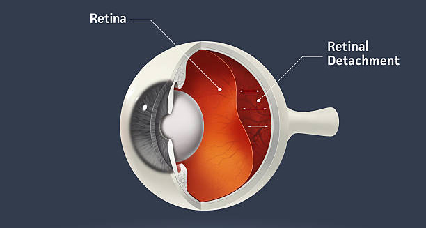
Cross-section diagram showing retinal detachment inside the eye

Causes of Detached Retina

Causes of Detached Retina Causes of detached retina are varied, but they all lead to the separation of the retina from the underlying layers that supply it with oxygen and nutrients. A detached retina can occur suddenly or gradually, and understanding the underlying causes can aid in prevention and early treatment. Rhegmatogenous Retinal Detachment This is the most common type and is caused by a tear or hole in the retina: Ageing: As people age, the vitreous gel inside the eye shrinks and may pull away from the retina, causing a tear Myopia (short-sightedness): People with severe myopia have longer eyeballs, which stretches the retina and makes it thinner and more prone to tearing Eye injury or trauma: Blunt or penetrating injuries can cause tears or breaks in the retina Previous eye surgery: Procedures like cataract removal increase the risk of retinal detachment, especially in the first year after surgery Once a tear occurs, fluid from the vitreous gel can seep through the opening and collect underneath the retina, causing it to lift off. Tractional Retinal Detachment | Causes of Detached Retina This form is often seen in people with: Diabetic retinopathy: Scar tissue from abnormal blood vessels pulls the retina away from its base Retinal vein occlusion: Blocked blood vessels may lead to fibrosis and traction on the retina Inflammatory conditions: Diseases like uveitis can result in scarring and traction This type develops slowly and is typically painless. Exudative Retinal Detachment This type occurs when fluid builds up beneath the retina without any tears or breaks: Tumours: Ocular melanoma or metastatic cancers can leak fluid under the retina Inflammation: Conditions like scleritis or Coats’ disease can cause abnormal fluid accumulation Systemic diseases: High blood pressure or kidney disorders may contribute to fluid leakage in the eye Other Risk Factors Family history of retinal detachment Lattice degeneration: Thinning of the peripheral retina, increasing vulnerability Extreme sports or sudden head movements: Though rare, can increase risk In conclusion, causes of detached retina span age-related degeneration, trauma, disease, and anatomical predisposition. Knowing the risk factors allows at-risk individuals to monitor their eye health and seek urgent care when symptoms arise. [Next: Symptoms of Detached Retina →]

Causes of Detached Retina Read More »

Symptoms of Detached Retina

Symptoms of Detached Retina

Symptoms of Detached Retina Symptoms of detached retina often appear suddenly and can range from subtle visual changes to dramatic vision loss. A detached retina itself is painless, but the signs are typically noticeable and should never be ignored. Timely recognition is critical, as delays in treatment may result in permanent vision impairment. Flashes of Light (Photopsia) Often one of the first symptoms Appears as brief, flickering lights or lightning streaks Caused by the vitreous gel pulling on the retina Flashes may be more noticeable in dim light or when moving the eyes quickly. Floaters Small dark shapes or specks drifting across the vision May look like threads, cobwebs, or blobs A sudden increase in floaters may signal a retinal tear or bleeding Floaters are common with ageing but become concerning when they appear suddenly and in large numbers. Shadow or Curtain Over Vision A dark shadow or curtain may spread from the side, top, or bottom of the visual field Indicates the retina is detaching further, affecting peripheral vision If untreated, the shadow can expand and result in complete central vision loss Blurred or Distorted Vision Straight lines may appear wavy Objects may appear smaller or distorted in shape Reading or recognising faces becomes difficult Loss of Vision | Symptoms of Detached Retina Central or peripheral vision loss in the affected eye Can progress rapidly within hours or days Vision loss is typically irreversible without surgery Symptoms by Type of Retinal Detachment Rhegmatogenous: Sudden and dramatic onset Tractional: Gradual visual decline without acute symptoms Exudative: Often starts with blurring and progresses to distortion When to Seek Emergency Care Any new flashes, floaters, or vision changes should prompt immediate eye examination Even if symptoms disappear, the risk of retinal detachment remains In summary, symptoms of detached retina are visual warning signs that require urgent attention. Early intervention offers the best chance of restoring or preserving sight. [Next: Diagnosis of Detached Retina →]

Symptoms of Detached Retina Read More »

Eye exam for retinal issues

Diagnosis of Detached Retina

Diagnosis of Detached Retina Diagnosis of detached retina relies on a detailed eye examination by an optometrist or ophthalmologist. When someone presents with symptoms of a detached retina, swift and accurate diagnosis is critical to prevent irreversible vision loss. The evaluation includes a combination of clinical observation, imaging tests, and assessment of visual function. Initial Clinical Evaluation The doctor will ask about the onset and nature of symptoms, such as floaters, flashes, or shadowed vision The eye care specialist takes a thorough medical history, including any previous eye surgeries, trauma, or conditions like diabetes or high myopia Visual Acuity Testing Measures how clearly a person can see at various distances The specialist checks whether central or peripheral vision is affected Establishes a baseline for monitoring changes before and after treatment Dilated Fundus Examination Special eye drops dilate the pupils to allow a clear view of the retina Using a slit-lamp microscope with a bright light and lens, the doctor can examine the entire retina Signs of retinal tears, detachment, or bleeding are carefully assessed Indirect Ophthalmoscopy A head-mounted light and lens are used to view the peripheral retina in detail This technique provides a wide field of vision to detect small or hidden retinal breaks Imaging Tests Optical Coherence Tomography (OCT): Produces cross-sectional images of the retina Identifies fluid accumulation, retinal layers, and subtle detachment Ultrasound (B-scan): Used when the view of the retina is obscured by bleeding (vitreous haemorrhage) Detects full or partial detachment and measures the extent of separation Fundus Photography: Captures detailed images for documentation and comparison over time Differential Diagnosis The following conditions may mimic or accompany retinal detachment: Posterior vitreous detachment (PVD) Retinal vein occlusion Macular degeneration Ocular tumours Urgency of Diagnosis of Detached Retina A suspected detached retina is a medical emergency The faster the diagnosis is made, the better the prognosis for preserving vision In conclusion, diagnosis of detached retina depends on clinical skill and advanced imaging tools. Prompt detection allows for timely surgical intervention and maximises the chances of vision recovery. [Next: Treatment of Detached Retina →]

Diagnosis of Detached Retina Read More »

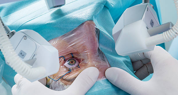
Retinal detachment eye surgery

Treatment of Detached Retina

Treatment of Detached Retina Treatment of detached retina aims to reattach the retina to its normal position and preserve or restore vision. Because a detached retina will not heal on its own, surgical intervention is almost always necessary. The specific type of surgery depends on the cause, extent, and location of the detachment. Laser Photocoagulation Used for small retinal tears or holes before detachment occurs A laser creates burns around the tear to form scar tissue The scar seals the retina to the underlying tissue, preventing fluid from entering This treatment is usually quick, performed in a clinic, and requires no hospital stay. Cryopexy (Freezing Therapy) Involves freezing the area around the retinal tear Causes inflammation and scarring to secure the retina Often combined with other surgical techniques Vitrectomy Treatment of Detached Retina A common procedure for larger or more complex detachments Removes the vitreous gel pulling on the retina The surgeon then fills the eye with a gas bubble or silicone oil to hold the retina in place Performed under local or general anaesthetic, with a recovery period of several weeks The body slowly reabsorbs gas bubbles, while the surgeon may need to remove silicone oil in a second surgery. Scleral Buckle The surgeon places a silicone band around the white part of the eye (sclera) This indents the wall of the eye, relieving traction on the retina Often used in rhegmatogenous detachment, especially in younger patients Pneumatic Retinopexy A gas bubble is injected into the vitreous cavity The patient must maintain a specific head position to keep the bubble over the retinal tear The bubble pushes the retina back into place, followed by laser or cryopexy Recovery and Follow-Up Most procedures are successful after one operation Vision may take weeks or months to improve Follow-up visits are essential to monitor healing and manage complications In summary, treatment of detached retina involves specialised surgery tailored to the type and severity of detachment. Early intervention dramatically increases the chance of visual recovery. [Next: Complications and Recovery from Detached Retina →]

Treatment of Detached Retina Read More »

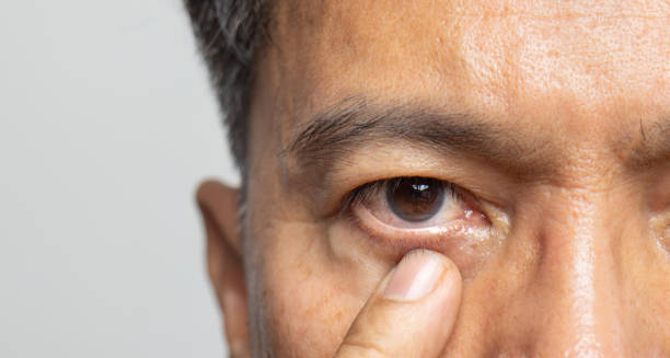
Close-up of eye after retinal surgery

Complications and Recovery from Detached Retina

Complications and Recovery from Detached Retina Complications and recovery from detached retina vary depending on the extent of the detachment, the timing of treatment, and the surgical method used. While most people regain useful vision, a detached retina can lead to long-term issues, especially if treatment is delayed. Understanding the possible outcomes helps manage expectations and supports better aftercare. Common Post-Surgical Complications Re-detachment: The retina may detach again, especially if new tears form Additional surgery may be needed to secure the retina Cataracts: Especially common after vitrectomy May develop within months of surgery and require cataract removal Increased Eye Pressure (Glaucoma): Gas bubbles or silicone oil may raise intraocular pressure Requires medication or surgical management Macular Pucker or Epiretinal Membrane: Scar tissue can form on the retina’s surface, causing distortion and blurred vision Infection (Endophthalmitis): Rare but serious complication Presents with pain, redness, and worsening vision Visual Recovery Timeline Visual improvement typically begins a few weeks post-surgery Full recovery may take up to six months Some vision loss may be permanent, especially if the macula was involved Long-Term Outlook on Complications and Recovery from Detached Retina Many patients regain functional vision, though some may experience: Slight distortion Reduced colour sensitivity Night vision difficulties Post-Surgical Care Eye drops are prescribed to reduce inflammation and prevent infection Patients may be instructed to avoid heavy lifting or strenuous activity Specific head positioning is crucial after gas bubble procedures Preventing Recurrence Regular eye exams, especially for high-risk individuals Prompt attention to new symptoms such as flashes or floaters Wearing protective eyewear during sports or hazardous activities Emotional and Lifestyle Impact Sudden vision loss can affect independence and mental wellbeing Support from low-vision specialists, counsellors, or peer groups can be beneficial In conclusion, complications and recovery from detached retina underscore the importance of early detection and careful follow-up. With expert care, most patients achieve stable vision and can return to normal daily activities. [Next: Back to Overview →]

Complications and Recovery from Detached Retina Read More »

Scroll to Top