Mental Matters

Health Conditions

Find information on various health conditions affecting people of all ages. This category covers causes, symptoms, diagnosis, and management options for physical illnesses and chronic diseases. Get insights to help you understand different health challenges and make informed decisions about your wellbeing.

Cancer patient undergoing chemotherapy for chronic lymphocytic leukaemia

Complications and Recovery from Chronic Lymphocytic Leukaemia

Complications and Recovery from Chronic Lymphocytic Leukaemia Complications and recovery from chronic lymphocytic leukaemia vary based on disease stage, treatment response, and overall patient health. While chronic lymphocytic leukaemia can remain stable for years, it may eventually cause systemic complications that require close management. 1. Infections Because CLL weakens the immune system, infections are a leading cause of complications. Patients may experience frequent respiratory, urinary tract, or skin infections. Even minor infections can become severe, particularly after chemotherapy or monoclonal antibody therapy. Preventive antibiotics, vaccinations (excluding live vaccines), and immunoglobulin replacement therapy may be used to reduce risk. 2. Anaemia and Thrombocytopenia As cancerous lymphocytes crowd out normal bone marrow function, patients may develop low red blood cell counts (anaemia) and low platelet counts (thrombocytopenia). This leads to fatigue, weakness, easy bruising, and prolonged bleeding. These conditions may be managed with medication, transfusions, or changes in treatment. 3. Autoimmune Complications CLL can trigger autoimmune disorders where the immune system mistakenly attacks red blood cells or platelets. This leads to autoimmune haemolytic anaemia (AIHA) or immune thrombocytopenia (ITP). These complications may require steroids, rituximab, or immunosuppressants. 4. Richter’s Transformation In 2–10% of cases, CLL can transform into an aggressive form of lymphoma called Richter’s syndrome. This complication is associated with sudden worsening of symptoms, including rapid lymph node enlargement, high fevers, and weight loss. Prognosis is poor, and treatment typically involves aggressive chemotherapy or clinical trial options. 5. Second Cancers | Complications and Recovery from Chronic Lymphocytic Leukaemia Patients with CLL are at increased risk of developing other cancers, including skin cancer, lung cancer, and gastrointestinal malignancies. Regular screenings and sun protection are important preventative measures. 6. Treatment Side Effects While targeted therapies have fewer side effects than traditional chemotherapy, they can still cause issues like atrial fibrillation (with BTK inhibitors), low blood counts, diarrhoea, or fatigue. Monitoring and adjusting treatment is vital to maintain quality of life. Recovery and Monitoring | Complications and Recovery from Chronic Lymphocytic Leukaemia Recovery from CLL is a relative term. Many patients achieve long-term remission, especially with modern therapies. Recovery focuses on maintaining quality of life, preventing complications, and monitoring for relapse. Regular follow-up appointments involve: Emotional support and mental health care are also essential, as living with a chronic cancer diagnosis can be psychologically taxing. Patient support groups and counselling can greatly aid in coping with uncertainty. In summary, complications and recovery from chronic lymphocytic leukaemia require a multidisciplinary approach. With vigilant monitoring, tailored treatment, and supportive care, many people can manage their condition successfully and maintain a good quality of life for years. [Next: Back to Overview →]

Complications and Recovery from Chronic Lymphocytic Leukaemia Read More »
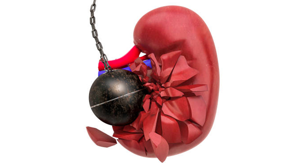
Kidney being struck by a wrecking ball symbolising chronic kidney disease damage

Chronic Kidney Disease

Chronic Kidney Disease – Overview Chronic kidney disease is a long-term condition in which the kidneys lose function progressively over time. Although the early stages often present no symptoms. This kidney disease can eventually lead to kidney failure, necessitating dialysis or transplantation. This condition affects approximately 10% of the global population, with numbers rising due to increasing rates of diabetes and hypertension. The kidneys are essential organs that filter waste and excess fluid from the blood. Maintain the balance of salts and minerals, regulate blood pressure, and produce hormones that influence red blood cell production and bone health. When kidney function deteriorates, these vital processes are compromised, resulting in a wide range of health issues. CKD is classified into five stages, based on the glomerular filtration rate (GFR). Which estimates how efficiently the kidneys filter blood. Stage 1 signifies mild kidney damage with normal GFR. While stage 5 represents end-stage renal disease (ESRD), where kidney function drops below 15%, and renal replacement therapy is typically required. Chronic Kidney Disease – Overview Diabetes and high blood pressure are the most common causes of CKD. Other risk factors include glomerulonephritis, polycystic kidney disease, chronic urinary tract obstruction, and prolonged use of nephrotoxic medications. CKD can remain undetected until late stages due to its insidious onset. Which is why routine screening is critical in at-risk individuals. Managing CKD involves controlling the underlying causes, such as regulating blood sugar and blood pressure. Furthermore, adjusting diet and avoiding medications that may further impair renal function. Early detection and treatment can significantly slow the progression of the disease. Additionally, reduce the likelihood of complications like cardiovascular disease, anaemia, and bone disorders. In summary, this condition is a progressive yet manageable condition. With regular monitoring, timely medical intervention, and lifestyle adjustments, many individuals can lead full, productive lives while maintaining kidney function. [Next: Causes of Chronic Kidney Disease →]

Chronic Kidney Disease Read More »

3D illustration of kidney gradually disintegrating, symbolising chronic kidney disease causes

Causes of Chronic Kidney Disease

Causes of Chronic Kidney Disease Causes of chronic kidney disease are primarily linked to conditions that damage the kidneys’ filtering units over time. The most prevalent contributors to chronic kidney disease are diabetes and hypertension, but several other diseases and lifestyle factors also play a significant role. 1. Diabetes Mellitus Diabetic nephropathy is the leading cause of CKD worldwide. Prolonged high blood sugar damages the delicate filtration system in the kidneys, allowing protein to leak into the urine and eventually impairing kidney function. Strict glycaemic control is essential to prevent or delay kidney damage in diabetics. 2. Hypertension | Causes of Chronic Kidney Disease High blood pressure can damage the tiny blood vessels in the kidneys, making them less efficient. Over time, untreated or poorly controlled hypertension accelerates kidney decline and increases the risk of heart disease, another major CKD complication. 3. Glomerulonephritis This condition involves inflammation of the glomeruli — the small filters in the kidneys. It can be acute or chronic and may result from infections, autoimmune diseases, or unknown causes. Chronic glomerulonephritis is a significant contributor to ESRD in younger individuals. 4. Polycystic Kidney Disease An inherited condition, PKD causes numerous fluid-filled cysts to develop in the kidneys, enlarging them and damaging surrounding tissue. Although the disease often remains asymptomatic for years, it typically progresses to CKD and, in many cases, ESRD. 5. Urinary Tract Obstruction Obstruction of urine flow — due to kidney stones, tumours, an enlarged prostate, or congenital defects — increases pressure within the kidney and leads to scarring. Chronic obstruction without proper treatment often results in permanent damage. 6. Recurrent Infections | Causes of Chronic Kidney Disease Frequent kidney infections, or pyelonephritis, can cause long-term inflammation and scarring. In children, recurrent UTIs due to vesicoureteral reflux may also result in CKD if left untreated. 7. Autoimmune Disorders Conditions such as lupus and vasculitis can directly target kidney tissue, leading to inflammation, fibrosis, and eventually kidney failure if not aggressively treated. 8. Prolonged Medication Use Overuse of NSAIDs (like ibuprofen), certain antibiotics, and some chemotherapy agents are known to be nephrotoxic. Regular monitoring of renal function is advised in patients requiring long-term use of such medications. 9. Lifestyle and Environmental Factors | Causes of Chronic Kidney Disease Obesity, smoking, poor diet, and exposure to environmental toxins (e.g., lead, mercury) can accelerate kidney damage and worsen the effects of other risk factors. By identifying the causes of chronic kidney disease, healthcare providers can develop targeted prevention and management plans. Public health initiatives aimed at controlling diabetes and hypertension are especially critical for reducing the global burden of CKD. [Next: Symptoms of Chronic Kidney Disease →]

Causes of Chronic Kidney Disease Read More »

Elderly woman holding lower back in pain, showing symptoms of chronic kidney disease

Symptoms of Chronic Kidney Disease

Symptoms of Chronic Kidney Disease Symptoms of chronic kidney disease often do not appear until the disease is advanced. For this reason, chronic kidney disease is sometimes referred to as a “silent killer.” By the time symptoms emerge, significant damage may already have occurred. Knowing the symptoms enables early detection, which is key to slowing disease progression. 1. Fatigue and Tiredness As kidney function declines, waste builds up in the blood, making people feel weak and tired. Anaemia caused by decreased erythropoietin production further exacerbates fatigue, making even routine activities challenging. 2. Changes in Urination | Symptoms of Chronic Kidney Disease Changes may include increased urination at night (nocturia), foamy or bubbly urine (indicating protein), reduced urine output, or visible blood. These symptoms reflect the kidneys’ impaired filtering capacity and may be among the earliest signs noticed. 3. Oedema (Swelling) Sodium and fluid retention cause swelling in the legs, ankles, hands, and face. This is due to the kidneys’ reduced ability to remove excess salt and water. In severe cases, fluid may accumulate in the lungs, causing breathlessness. 4. High Blood Pressure As kidneys regulate blood pressure through salt and fluid balance and hormone release, dysfunction often results in hypertension. This becomes both a symptom and a contributor to further kidney damage. 5. Skin Issues Dry, itchy skin may occur due to mineral imbalances or toxin accumulation. In advanced CKD, calcium and phosphate dysregulation can lead to severe itching and skin discolouration. 6. Nausea and Appetite Loss The build-up of urea and other toxins leads to gastrointestinal symptoms such as nausea, vomiting, and a metallic taste in the mouth. These contribute to weight loss and malnutrition. 7. Cognitive Changes | Symptoms of Chronic Kidney Disease Toxin build-up can affect brain function, causing poor concentration, memory lapses, and sleep disturbances. These are common but often overlooked symptoms. 8. Muscle Cramps Electrolyte imbalances, especially low calcium and high phosphorus, lead to cramps, particularly at night. These are not only painful but also indicate worsening kidney function. Because these symptoms can resemble other health issues, CKD is frequently underdiagnosed. Routine screening, especially in people with diabetes or hypertension, is essential. Recognising the symptoms of chronic kidney disease early improves treatment outcomes and can delay the need for dialysis or transplantation. [Next: Diagnosis of Chronic Kidney Disease →]

Symptoms of Chronic Kidney Disease Read More »

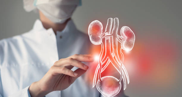
Doctor examining a digital diagram of kidneys to diagnose chronic kidney disease

Diagnosis of Chronic Kidney Disease

Diagnosis of Chronic Kidney Disease Diagnosis of chronic kidney disease involves a combination of blood tests, urine analysis, and imaging studies. Since chronic kidney disease can be silent for years, proactive screening — especially in at-risk populations — is essential for early detection and management. 1. Glomerular Filtration Rate (GFR) The GFR is the most reliable indicator of kidney function. It is calculated using blood creatinine levels, age, sex, and body size. A GFR below 60 for more than three months confirms a diagnosis of CKD. GFR also helps stage the disease from Stage 1 (normal) to Stage 5 (end-stage). 2. Serum Creatinine and Urea Elevated creatinine and urea levels indicate the kidneys are not efficiently clearing waste. These are among the earliest biochemical markers of declining renal function. 3. Urine Albumin-to-Creatinine Ratio (ACR) This test detects protein (mainly albumin) in the urine, a key sign of kidney damage. Persistent proteinuria is a strong predictor of disease progression and cardiovascular risk. 4. Urinalysis | Diagnosis of Chronic Kidney Disease This general test screens for red blood cells, white blood cells, protein, glucose, and cellular casts. It can identify infections, glomerulonephritis, or tubular dysfunction. 5. Blood Pressure Monitoring Hypertension is both a cause and effect of CKD. Ongoing blood pressure measurement helps in diagnosis, staging, and treatment monitoring. 6. Kidney Ultrasound An ultrasound provides images of the kidneys, detecting abnormalities such as cysts, small or shrunken kidneys, tumours, or obstructions. 7. Electrolyte Panel | Diagnosis of Chronic Kidney Disease Abnormal levels of potassium, calcium, phosphorus, and sodium reflect impaired renal function and guide dietary and medical interventions. 8. Kidney Biopsy In certain cases, especially where autoimmune disease or glomerulonephritis is suspected, a biopsy may be performed. This involves removing a small sample of kidney tissue for microscopic examination. By combining laboratory, imaging, and clinical data, a thorough diagnosis can be established, allowing for a comprehensive and individualised treatment approach. [Next: Treatment and Recovery from Chronic Kidney Disease →]

Diagnosis of Chronic Kidney Disease Read More »

Patient receiving dialysis treatment for chronic kidney disease at home

Treatment and Recovery from Chronic Kidney Disease

Treatment and Recovery from Chronic Kidney Disease Treatment and recovery from chronic kidney disease depend on the stage and underlying cause of the condition. Most people cannot reverse chronic kidney disease onceMost people cannot reverse chronic kidney disease once it develops. However, timely intervention and lifestyle changes can slow or even halt its progression in some cases. The goal is to preserve existing kidney function, manage symptoms, and prevent complications. 1. Managing Underlying Conditions Controlling diabetes and hypertension is critical. This may involve medications like ACE inhibitors or angiotensin receptor blockers (ARBs), which help lower blood pressure and protect the kidneys. You must keep your blood sugar within target ranges by following a healthy diet, exercising regularly, and using medications or insulin as needed. 2. Dietary Modifications Diet plays a key role in slowing CKD progression. Doctors usually advise people to: 3. Medication Management Nephrotoxic drugs (like NSAIDs) should be avoided. Doctors may prescribe medications to control cholesterol, treat anaemia, strengthen bones, and manage acidosis. In advanced CKD, phosphate binders, erythropoietin injections, and vitamin D analogues are commonly used. 4. Lifestyle Changes Patients should stop smoking, reduce alcohol, maintain a healthy weight, and exercise regularly. These changes improve overall cardiovascular health, which is closely tied to CKD outcomes. 5. Monitoring and Follow-Up Regular check-ups include GFR tests, urine protein checks, blood pressure monitoring, and medication reviews. Early intervention based on these results allows timely adjustments to treatment plans. 6. Preparing for Renal Replacement | Treatment and Recovery from Chronic Kidney Disease In Stage 4 or 5, planning for dialysis or transplantation becomes necessary. Education about options such as haemodialysis, peritoneal dialysis, and donor kidney transplant is important to make informed decisions. 7. Emotional and Psychological Support Chronic illness impacts mental health. Patients may experience anxiety, depression, or stress related to lifestyle changes or the prospect of dialysis. Counselling and support groups offer valuable help. In conclusion, treatment and recovery from chronic kidney disease require a multidisciplinary approach. With proactive care and support, individuals with CKD can enjoy improved quality of life and significantly delay the onset of kidney failure. [Next: Back to Overview →]

Treatment and Recovery from Chronic Kidney Disease Read More »

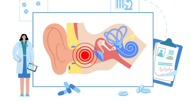
Diagram of the human ear showing cholesteatoma inside the middle ear

Cholesteatoma

Cholesteatoma – Overview Cholesteatoma is a destructive yet non-cancerous growth of skin cells that occurs in the middle ear, behind the eardrum. Although it is not a tumour in the traditional sense, a cholesteatoma can cause serious complications if left untreated. It expands slowly but progressively, damaging the delicate bones of hearing, and in severe cases, spreading into the skull base or brain. Cholesteatomas form when the skin lining of the outer ear canal invades the middle ear space. This often happens due to repeated infections or Eustachian tube dysfunction. When the Eustachian tube (which connects the middle ear to the back of the throat) does not function properly, negative pressure can develop in the middle ear. This pulls the eardrum inward, forming a pocket that traps dead skin and debris. Over time, this pocket expands into a cholesteatoma. There are two main types: congenital cholesteatoma, which is present at birth due to developmental abnormalities, and acquired cholesteatoma, which occurs later in life and is much more common. The acquired type typically affects people with a history of chronic middle ear infections, grommet insertion, or perforated eardrums. Despite being benign, the growth can erode the ossicles (the tiny bones that transmit sound), the mastoid bone, and even reach the inner ear or brain in rare cases. It can cause permanent hearing loss, facial nerve paralysis, dizziness, and life-threatening infections if not treated. For this reason, early diagnosis and surgical removal are critical. Cholesteatoma Cholesteatomas usually affect only one ear, although in rare instances, both ears may be involved. They can affect both children and adults, with slightly higher prevalence in individuals who have poor access to medical care or are immunocompromised. The condition is often identified due to persistent ear discharge, a sense of ear fullness, and progressive hearing loss. Sometimes, more severe symptoms like facial muscle weakness or vertigo may be the first clues to deeper complications. Although cholesteatoma is rare, it is considered serious due to its aggressive nature. Surgical removal is the only effective treatment, and ongoing monitoring is needed to prevent recurrence. Recurrence is possible even after successful surgery, which makes long-term follow-up essential. In summary, cholesteatoma is a dangerous but manageable ear condition that requires surgical treatment. It is not cancerous, but it behaves in a locally invasive way and must be dealt with promptly to avoid long-term damage. [Next: Causes of Cholesteatoma →]

Cholesteatoma Read More »

Woman using cotton swab in ear with discomfort, indicating poor ear hygiene

Causes of Cholesteatoma

Causes of Cholesteatoma Causes of cholesteatoma fall into two main categories: congenital and acquired. In both cases, the condition results from the build-up of squamous epithelium (skin cells) in the middle ear, which is a space normally lined with respiratory epithelium. Over time, this abnormal skin collection enlarges, becomes infected, and begins to erode surrounding structures. While congenital cases are rare, the majority of cholesteatoma diagnoses are acquired. The most common mechanism involves Eustachian tube dysfunction. When the Eustachian tube fails to equalise air pressure in the middle ear, negative pressure builds up. This can pull part of the eardrum inward, forming a retraction pocket. These pockets collect dead skin and keratin, which are normally shed and expelled in a healthy ear. In a blocked or retracted pocket, they become trapped and begin to accumulate. Chronic otitis media, or long-term middle ear infection, is another leading cause. Repeated infections weaken the structure of the eardrum and create an environment in which skin migration into the middle ear becomes more likely. This allows a cholesteatoma to form gradually over time, usually without pain in the early stages. Causes of Cholesteatoma Perforated eardrums, whether caused by trauma, infection, or surgical intervention (such as grommet insertion), may also play a role. Through these perforations, skin cells can invade the middle ear and begin forming a cholesteatoma sac. In rare cases, cholesteatoma is congenital, meaning a small nest of skin cells becomes trapped in the middle ear during foetal development. These cases are usually diagnosed in children, often when they present with hearing loss despite no history of infections or trauma. Other contributing factors include cleft palate, craniofacial abnormalities, and immunosuppression, all of which are linked to higher risks of Eustachian tube malfunction and chronic infections. Even severe allergies and sinus problems may indirectly contribute by creating nasal congestion and affecting tube drainage. Causes of Cholesteatoma Some researchers also suggest that a family history of chronic ear disease may slightly increase susceptibility, though cholesteatoma is not considered a genetic condition. Understanding the causes of cholesteatoma is important because early intervention in chronic ear problems can prevent it from forming. Treating underlying infections promptly, monitoring children with ear abnormalities, and educating high-risk groups about symptoms can reduce incidence and recurrence. [Next: Symptoms of Cholesteatoma →]

Causes of Cholesteatoma Read More »

Elderly man holding his ear in pain, indicating symptoms of cholesteatoma

Symptoms of Cholesteatoma

Symptoms of Cholesteatoma Symptoms of cholesteatoma may be subtle at first but worsen as the growth expands. Since cholesteatoma can erode bone and surrounding structures, its symptoms range from simple hearing issues to more serious neurological signs. Recognising these early signs allows for quicker diagnosis and reduces the risk of permanent complications. The most common symptom is chronic ear discharge, often foul-smelling and continuous. Unlike occasional fluid drainage from an ear infection, cholesteatoma discharge tends to persist for weeks or months and doesn’t respond to ordinary ear drops. This is due to the trapped, infected keratin debris constantly draining through a hole or retraction in the eardrum. Another hallmark symptom is hearing loss in the affected ear. Initially, this hearing loss is conductive in nature, caused by the blockage of sound transmission through the ossicles. As the cholesteatoma erodes these small bones, the hearing loss becomes more severe and may also involve sensorineural components if the inner ear is affected. Symptoms of Cholesteatoma Many patients report a feeling of fullness or pressure in the ear. This may be accompanied by mild discomfort or the sense that something is stuck. While pain is not a typical early feature, some individuals do report dull aching or sharp episodes, especially if infection is present. Tinnitus, or ringing in the ear, is another frequent complaint. This may present as a high-pitched whistle or low-frequency humming and can be constant or intermittent. The presence of tinnitus often indicates irritation of the auditory nerve or inner ear. In advanced cases, the cholesteatoma may extend into the balance organs, leading to dizziness or vertigo. This is particularly concerning as it implies erosion beyond the middle ear into the labyrinth, which may affect spatial awareness and movement coordination. More severe symptoms include facial muscle weakness or twitching. The facial nerve runs through the middle ear, and cholesteatomas that invade this area may compress or damage it, resulting in drooping or paralysis of facial muscles. Symptoms of Cholesteatoma Other signs can include recurrent ear infections, frequent need for antibiotics, and a visible white mass behind the eardrum during an ear exam. In children, delayed speech or poor academic performance may be linked to undiagnosed hearing loss from a growing cholesteatoma. Not all symptoms occur at once, and some patients experience only one or two signs. Regardless, persistent discharge or progressive hearing changes should always trigger evaluation by an ENT specialist. [Next: Diagnosis of Cholesteatoma →]

Symptoms of Cholesteatoma Read More »

ENT specialist examining a diagram of the inner and middle ear for cholesteatoma diagnosis

Diagnosis of Cholesteatoma

Diagnosis of Cholesteatoma Diagnosis of cholesteatoma relies on careful clinical evaluation, supported by hearing tests and imaging. Because a cholesteatoma can silently erode bone and spread into surrounding structures, early detection is crucial to avoid permanent damage. Many patients present with long-standing symptoms like ear discharge or hearing loss, which can be overlooked or misdiagnosed without proper assessment. The process begins with a full medical history. The ENT specialist will ask about chronic ear infections, surgeries like grommet insertion, childhood ear disease, or symptoms like ear discharge, hearing changes, dizziness, or facial weakness. The history helps differentiate cholesteatoma from common middle ear infections or conditions like otitis media with effusion. The next step is a detailed otoscopic examination. Using a handheld otoscope or, preferably, a microscope or endoscope, the clinician inspects the ear canal and eardrum. Signs suggestive of cholesteatoma include: Diagnosis of Cholesteatoma Advanced examination with otoendoscopy provides a clearer and more magnified view of hidden recesses, making it easier to detect small or early cholesteatomas. An audiogram or hearing test is essential to assess the extent of hearing loss. Most patients show conductive hearing loss, where sound cannot pass effectively through the middle ear. In some cases, particularly if the inner ear is involved, there may also be sensorineural hearing loss. Speech discrimination scores may also be reduced. Tympanometry is a valuable test that measures eardrum compliance and middle ear pressure. It can confirm eustachian tube dysfunction, fluid build-up, or abnormal movement of the tympanic membrane. High-resolution CT scanning of the temporal bones is the gold standard for evaluating the extent of the disease. It provides detailed images of: In specific cases, an MRI may be requested to identify soft tissue extension or complications like brain abscess, meningitis, or sigmoid sinus thrombosis. Post-operatively, MRI with diffusion-weighted imaging is also used to monitor recurrence. Diagnosis of Cholesteatoma In infected cases, a microbial culture may be taken to guide antibiotic therapy. However, bacterial identification does not confirm or exclude the presence of a cholesteatoma. A confirmed diagnosis of cholesteatoma warrants surgical referral. Early identification allows for conservative and less extensive surgery, while delayed diagnosis may result in more complex and high-risk procedures. [Next: Treatment of Cholesteatoma →]

Diagnosis of Cholesteatoma Read More »

Scroll to Top