Mental Matters

Author name: admin

Man holding lower back in pain, indicating possible urinary tract or reproductive infection

Causes of Epididymitis

Causes of Epididymitis The causes of epididymitis vary depending on a person’s age, sexual activity, and overall health. Epididymitis most commonly results from infections—either sexually transmitted or related to the urinary tract—but it can also develop without infection in some cases. Sexually Transmitted Infections (STIs) In men under 35, epididymitis is often caused by sexually transmitted bacteria, including: Chlamydia trachomatis Neisseria gonorrhoeae These infections can travel through the urethra and into the reproductive tract, inflaming the epididymis. STIs are a leading cause among sexually active young men, particularly in areas with limited access to regular screening and sexual health education. Urinary Tract Infections (UTIs) In older men, epididymitis is commonly associated with: Escherichia coli (E. coli) and other common UTI-causing bacteria Enlarged prostate (BPH), which can lead to urine retention and increased risk of infection Catheter use or recent urological procedures Infections from the bladder or prostate may spread backward to the epididymis, causing inflammation and pain. Non-Infectious causes of Epididymitis Less common causes include: Trauma or injury to the groin Heavy lifting or straining, which may cause urine to flow backward (chemical epididymitis) Certain medications, such as amiodarone, a heart drug known to irritate the epididymis in high doses Tuberculosis (TB), which can cause chronic epididymitis in rare cases—more commonly seen in South Africa due to the high TB burden Risk Factors Several factors increase the risk of developing epididymitis: Having multiple sexual partners or unprotected sex History of urinary tract infections Recent prostate or bladder surgery Use of urinary catheters Prolonged sitting (such as long road trips or desk jobs) In South Africa, men in rural communities may delay seeking treatment due to stigma or poor access to healthcare, which increases the risk of complications and further spread of infection. Understanding the causes of epididymitis helps doctors identify the most effective treatment and helps individuals take steps to prevent future episodes. [Next: Symptoms of Epididymitis →]
Causes of Epididymitis Read More »

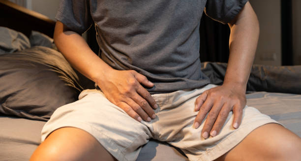
Man sitting on bed holding his groin in pain, indicating symptoms of epididymitis

Symptoms of Epididymitis

Symptoms of Epididymitis The symptoms of epididymitis typically develop quickly and can cause significant discomfort. Since epididymitis affects the testicles and surrounding structures, it often leads to both local and general symptoms that should never be ignored. Early treatment can prevent severe complications, including infertility or long-term pain. Common Symptoms Scrotal pain – Usually starts on one side and may spread to the groin or lower abdomen. Swelling and redness – The affected side of the scrotum may become visibly larger, red, and warm to the touch. Tenderness – Even gentle touch or movement may cause significant discomfort. Painful urination – A burning sensation or discomfort when passing urine. Increased urinary frequency or urgency – The urge to urinate more often, especially with accompanying discomfort. Discharge from the penis – May be present if the cause is a sexually transmitted infection. Blood in semen – Though uncommon, some men notice pink or red streaks in ejaculate. Fever and chills – Indicate that the infection is more widespread or severe. In Chronic Cases Some men may develop chronic epididymitis, lasting more than six weeks. Symptoms include: Persistent dull pain or heaviness in the scrotum Occasional swelling Discomfort during or after ejaculation Anxiety or distress about fertility or sexual performance Chronic cases may occur after incomplete treatment of an initial infection or due to non-infectious inflammation. Symptoms of Epididymitis in Children In prepubescent boys, epididymitis is rare but can happen due to trauma, urinary reflux, or structural abnormalities. Symptoms may be mistaken for testicular torsion, which is a medical emergency, so urgent evaluation is essential. In South Africa Delayed treatment is a concern in many rural areas. Men may be reluctant to speak up about testicular symptoms due to cultural stigma or fear of being judged. As a result, many present late with advanced infection or complications. Public education and sexual health awareness campaigns can help reduce this stigma and encourage early diagnosis and treatment. Recognising the symptoms of epididymitis and seeking medical attention promptly can prevent further discomfort and long-term damage. [Next: Diagnosis of Epididymitis→]

Symptoms of Epididymitis Read More »

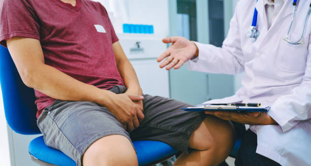
Man consulting with urologist about groin pain, indicating possible epididymitis

Diagnosis of Epididymitis

Diagnosis of Epididymitis The diagnosis of epididymitis is usually based on a physical examination, patient history, and laboratory tests. Because epididymitis can mimic other serious conditions—such as testicular torsion—accurate and timely diagnosis is crucial, especially in younger patients. Medical History and Symptom Review Doctors begin by asking about: The onset and duration of pain Urinary symptoms, such as burning or frequency Any discharge from the penis Recent sexual activity or known STI exposure History of urinary tract infections, catheter use, or trauma This information helps narrow down the likely cause—whether it’s an STI, UTI, or a non-infectious trigger. Physical Examination During the exam, the doctor will: Check for swelling, tenderness, and warmth in the scrotum Examine both testicles and the epididymis Assess for signs of inflammation or discharge Rule out testicular torsion, a condition where the spermatic cord twists and cuts off blood supply—a surgical emergency that requires immediate attention The Prehn’s sign may be used: lifting the scrotum slightly relieves pain in epididymitis but not in testicular torsion. However, this test alone is not always reliable. Urine and Swab Tests Urinalysis – Detects white blood cells, bacteria, or blood in the urine Urine culture – Identifies the bacteria causing infection NAAT (nucleic acid amplification tests) – Detect chlamydia and gonorrhoea Urethral swab – In cases of discharge, swabs help identify the STI organism These tests help guide antibiotic selection and confirm whether the infection is sexually transmitted or related to a urinary cause. Blood Tests | Diagnosis of Epididymitis A full blood count (FBC) may reveal elevated white cells, indicating infection. CRP or ESR levels can also confirm inflammation. Scrotal Ultrasound Ultrasound with Doppler imaging is the most reliable test to confirm epididymitis and rule out torsion. It shows: Blood flow to the testicles Swelling of the epididymis Fluid build-up around the testicles (hydrocele) In South Africa, access to ultrasound may be limited in rural clinics, causing delays in diagnosis. In such cases, clinical assessment becomes even more important. The diagnosis of epididymitis should be made quickly and confidently. Early treatment prevents complications and offers rapid symptom relief. [Next: Treatment of Epididymitis →]

Diagnosis of Epididymitis Read More »

Close-up of antibiotics and pills used in the treatment of epididymitis

Treatment of Epididymitis

Treatment of Epididymitis The treatment of epididymitis depends on the underlying cause—whether it’s bacterial, sexually transmitted, or non-infectious. Epididymitis is highly treatable, especially when caught early. The main goals are to eliminate infection, relieve pain, and prevent complications. Antibiotics Doctors should begin antibiotic treatment as soon as they suspect a bacterial infection. The choice of antibiotic depends on the patient’s age and likely cause: For sexually active men under 35: Ceftriaxone (single injection) for gonorrhoea Plus doxycycline (100 mg twice daily for 7–14 days) for chlamydia For older men or men with UTIs: Ciprofloxacin or levofloxacin (oral fluoroquinolones) Treatment usually lasts 10–14 days Patients should complete the full course of antibiotics even if symptoms improve early. Supportive Care Pain relief – Paracetamol or ibuprofen helps reduce pain and inflammation Scrotal support – Wearing snug underwear or using a scrotal support sling can relieve pressure Rest – Bed rest for the first few days can speed up healing Cold packs – Reduce swelling and provide pain relief Doctors advise patients to avoid sexual activity until they complete treatment and all symptoms have fully resolved. Treating Sexual Partners If a sexually transmitted infection is the cause, doctors must test and treat the patient’s sexual partners as well. Re-infection is common if partners are untreated. In South Africa, public clinics offer free STI treatment and counselling services. Follow-Up | Treatment of Epididymitis Most patients improve within 48–72 hours of starting antibiotics. If symptoms persist or worsen, additional testing may be needed to rule out: Abscess formation Testicular involvement (orchitis) Tuberculosis-related epididymitis In chronic or recurring cases, referral to a urologist is recommended. The treatment of epididymitis is usually successful with antibiotics and self-care. Early intervention helps avoid long-term issues like infertility or chronic pain. [Next: Complications of Epididymitis →]

Treatment of Epididymitis Read More »

Man sitting in emotional distress with partner comforting him, representing complications of epididymitis

Complications of Epididymitis

Complications of Epididymitis The complications of epididymitis can arise when the condition is left untreated, treated too late, or does not respond to medication. Although epididymitis is usually curable, ignoring symptoms can lead to long-term pain and even reproductive health issues. Chronic Epididymitis In some cases, inflammation persists after the infection has cleared. This is called chronic epididymitis and may involve: Ongoing testicular pain or discomfort Occasional swelling Pain during ejaculation or prolonged sitting Emotional distress due to chronic symptoms Chronic forms are harder to treat and may need long-term anti-inflammatories, nerve pain medication, or physical therapy. Abscess Formation Severe bacterial infections can lead to an abscess—a pocket of pus within the epididymis or testicle. Symptoms include: Severe, localised pain High fever Visible swelling that doesn’t improve with antibiotics Surgical drainage may be required if the abscess does not respond to medication. Infertility Long-standing or severe epididymitis can cause scarring in the epididymal ducts, blocking sperm flow. This may result in: Reduced sperm count Poor sperm motility Male factor infertility Bilateral epididymitis (affecting both testicles) increases the risk. Prompt treatment reduces the chances of fertility problems later. Testicular Involvement (Epididymo-orchitis) If the infection spreads to the testicle, the condition becomes epididymo-orchitis. This is more serious and can cause: Intense pain Swelling of the entire testicle Possible damage to testicular tissue In some cases, this can lead to testicular atrophy (shrinkage) or the need for surgical removal. Recurrent Infections Men who do not complete their antibiotic course or who are re-exposed to STIs are at risk of recurring episodes. This can cause cumulative damage and increase the risk of chronic symptoms. Complications of Epididymitis In South Africa In South Africa, complications often arise due to delayed presentation. Many men avoid seeking help for genital symptoms due to embarrassment or lack of access to affordable care. Education campaigns and improved access to male health services are essential to reduce the burden of this condition. Proper hygiene, STI prevention, and early treatment can stop most complications before they begin. Understanding the complications of epididymitis empowers men to act early, complete treatment, and protect their reproductive health and overall wellbeing. [Next: Back to Overview →]

Complications of Epididymitis Read More »

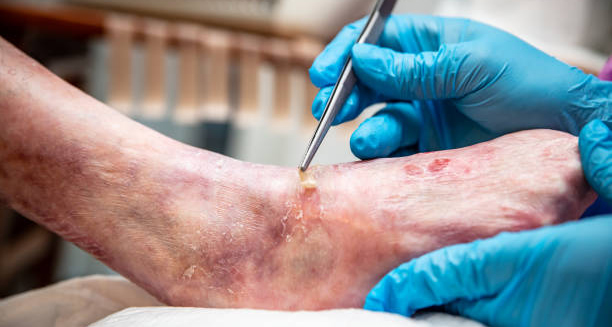
Close-up of wound care on a leg with Epidermolysis Bullosa.

Epidermolysis Bullosa

Epidermolysis Bullosa Epidermolysis bullosa is a group of rare, inherited skin disorders that cause the skin to become extremely fragile. Even the slightest friction or trauma can lead to painful blisters, open wounds, and skin infections. Epidermolysis bullosa often begins at birth or in early infancy and can range from mild to life-threatening. The condition occurs because the skin layers do not bind together properly. In healthy skin, the outer layer (epidermis) is firmly attached to the layer beneath (dermis). In people with this condition, the anchoring proteins or collagen fibres that hold these layers together are missing or faulty. This results in skin that tears or blisters with minimal pressure or rubbing. There are several types of epidermolysis bullosa (EB), with varying severity: EB simplex – the most common and usually mild, affecting mainly the hands and feet Junctional EB – more severe, often beginning at birth, and may involve internal organs Dystrophic EB – causes scarring and can lead to long-term disability Kindler syndrome – a rare form with features of all other types Epidermolysis Bullosa In South Africa, access to diagnosis and specialist care for EB can be limited, especially in rural areas. As a result, many families struggle with wound management, nutrition, and mobility challenges without adequate medical guidance. Though EB is rare, the impact on quality of life is immense. Children often live with daily pain, limited mobility, and a high risk of infections. With early diagnosis, specialised wound care, and supportive therapies, many individuals can live longer, more comfortable lives. Understanding epidermolysis bullosa is the first step toward offering proper care and reducing unnecessary suffering. [Next: Causes of Epidermolysis Bullosa →] Causes and Types of Blisters

Epidermolysis Bullosa Read More »

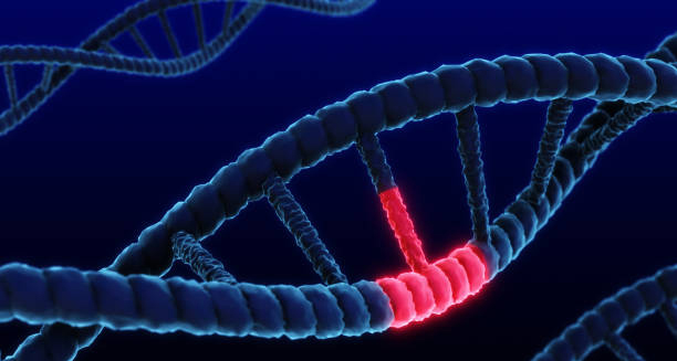
DNA strand showing genetic mutation, representing the causes of Epidermolysis Bullosa

Causes of Epidermolysis Bullosa

Causes of Epidermolysis Bullosa The causes of epidermolysis bullosa are genetic mutations that affect the proteins responsible for holding skin layers together. Epidermolysis bullosa does not result from infection, allergy, or poor hygiene. Parents pass it down through families, either from one or both, depending on the type. Genetic Mutations Mutations in genes that provide instructions for making the proteins that bind the layers of skin cause EB. The most commonly affected genes include: KRT5 and KRT14 in EB simplex COL17A1 and LAMA3, LAMB3, LAMC2 in junctional EB COL7A1 in dystrophic EB These mutations interfere with the skin’s ability to maintain integrity when exposed to stress or friction. Without strong connections between layers, even minor trauma can cause blistering and peeling. Inheritance Patterns | Causes of Epidermolysis Bullosa The condition is usually inherited in one of two ways: Autosomal dominant – one copy of the mutated gene (from one parent) is enough to cause the condition Autosomal recessive – both parents must pass on a faulty gene; each parent is usually a carrier without symptoms Dystrophic and junctional types are often inherited recessively and tend to be more severe. EB simplex is typically inherited in a dominant pattern and may cause milder symptoms. Spontaneous mutations can also occur, meaning a child may be affected even with no family history. Furthermore, these cases still result in lifelong EB, though the risk of passing it on to future children may vary. Risk Factors There are no environmental or lifestyle risk factors for EB. However, consanguinity (marriage between close relatives) increases the risk of passing on recessive conditions like EB, especially in rural or traditional communities. In South Africa, genetic testing is not widely available in the public health system, making diagnosis more difficult. As a result, doctors often rely on clinical signs and skin biopsy to identify the type of EB. Understanding the causes of epidermolysis bullosa helps families make informed decisions, consider genetic counselling, and plan for long-term care. Accurate diagnosis also helps direct treatment and support. [Next: Symptoms of Epidermolysis Bullosa →]

Causes of Epidermolysis Bullosa Read More »

Symptoms of Epidermolysis Bullosa

Symptoms of Epidermolysis Bullosa

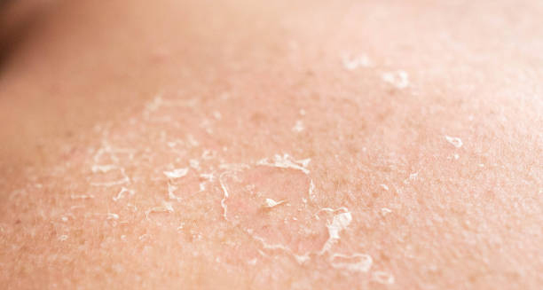
Symptoms of Epidermolysis Bullosa The symptoms of epidermolysis bullosa vary depending on the type and severity of the condition. However, all forms share one core characteristic: fragile skin that blisters and tears with little or no friction. Epidermolysis bullosa can affect not just the outer skin but also internal linings like the mouth, oesophagus, and urinary tract. Common Skin Symptoms Blistering from minor friction, such as rubbing clothing or gentle pressure Skin peeling or open sores after blister rupture Slow-healing wounds that may become infected Scarring, especially in dystrophic EB Thickened skin on the palms and soles Loss of fingernails and toenails Blisters may appear anywhere on the body but are especially common on the hands, feet, knees, elbows, and areas under pressure from medical devices or footwear. Internal Symptoms In more severe forms, EB affects the inner body: Mouth and throat blisters that make eating painful Difficulty swallowing, due to scarring in the oesophagus Voice changes, if blisters form on the vocal cords Urinary issues, from damage to the lining of the bladder or urethra Dental problems, such as tooth decay and fragile enamel Long-Term Effects Anaemia, from chronic blood loss and poor nutrition Malnutrition, due to pain while eating or limited food choices Joint contractures, where scarring pulls joints into fixed positions Reduced mobility, from pain and damage to feet or legs Increased risk of skin cancer, especially in older children and adults with severe EB Symptoms of Epidermolysis Bullosa in South Africa In South Africa, children with EB may face additional challenges due to poor access to specialised wound care, nutritional support, and physiotherapy. School attendance can be limited, and families may struggle to afford dressing supplies or pain medication. Despite these challenges, many families find ways to adapt through strong support networks, education, and community health initiatives. NGOs and advocacy groups are increasingly active in providing resources and raising awareness. Recognising the symptoms of epidermolysis bullosa early can lead to better pain management, fewer infections, and more effective long-term care. [Next: Diagnosis of Epidermolysis Bullosa→]

Symptoms of Epidermolysis Bullosa Read More »

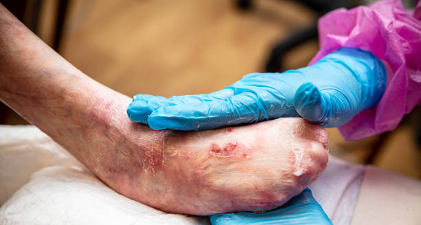
Medical professional examining blistered foot for Epidermolysis Bullosa diagnosis

Diagnosis of Epidermolysis Bullosa

Diagnosis of Epidermolysis Bullosa The diagnosis of epidermolysis bullosa is based on clinical observation, family history, and specialist testing. Because epidermolysis bullosa is rare and mimics other skin conditions in early stages, diagnosis often requires input from dermatologists and genetic specialists. Clinical Assessment Doctors first examine: The location, shape, and severity of blisters How easily the skin peels or tears Healing patterns (with or without scarring) Nail, hair, and tooth abnormalities History of blisters at birth or in infancy A full family medical history is vital. If close relatives have EB or unexplained skin fragility, this raises suspicion of a genetic condition. Skin Biopsy and Immunofluorescence Mapping To confirm EB, doctors perform a skin biopsy. A small piece of affected skin is removed and tested in a laboratory to determine: The layer of skin where blistering occurs Which skin proteins are missing or abnormal Immunofluorescence mapping (IFM) uses special dyes to highlight defective proteins under a microscope. This test helps classify EB into one of its main types: simplex, junctional, dystrophic, or Kindler syndrome. Genetic Testing | Diagnosis of Epidermolysis Bullosa Genetic testing identifies the exact mutation causing the condition. This confirms the diagnosis, predicts the severity, and helps with family planning. In developed countries, genetic testing is routine for EB patients and can even be done before birth in high-risk pregnancies. However, in South Africa, public health facilities rarely offer genetic testing due to cost and limited access to genetic laboratories. Diagnosis in most cases relies on clinical examination and biopsy. Prenatal and Carrier Testing For families with a known history of EB, prenatal testing can detect the condition during pregnancy using procedures like chorionic villus sampling (CVS) or amniocentesis. Carrier screening helps parents understand their risk before conceiving. Counselling is recommended for affected families to help them understand the diagnosis, inheritance pattern, and implications for future children. Accurate diagnosis of epidermolysis bullosa ensures early intervention, correct classification, and better long-term management. It also allows families to access specialised resources and emotional support. [Next: Treatment of Epidermolysis Bullosa →]

Diagnosis of Epidermolysis Bullosa Read More »

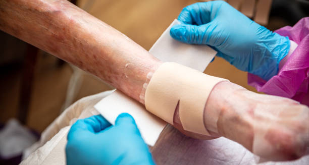
Medical professional applying bandages to fragile skin affected by Epidermolysis Bullosa

Treatment of Epidermolysis Bullosa

Treatment of Epidermolysis Bullosa The treatment of epidermolysis bullosa focuses on wound care, pain relief, infection prevention, and support for mobility and nutrition. While epidermolysis bullosa has no cure, early and consistent treatment can reduce complications and improve quality of life. Wound Care Daily skin care is essential. Steps include: Sterile dressings over open wounds to prevent infection Non-stick bandages to avoid damaging fragile skin Antiseptic washes to cleanse blisters and reduce bacteria Moisturisers and barrier creams to soothe dry, irritated skin Parents and caregivers must be trained to manage wounds at home. Supplies such as silicone dressings, foam padding, and adhesive-free tapes are preferred, though they are often expensive or hard to source in South Africa. Pain Management Children and adults with EB often live in constant discomfort. Pain relief may include: Paracetamol or ibuprofen Oral opioids for severe pain Topical anaesthetics during dressing changes Anti-itch medications like antihistamines Psychological support and relaxation techniques are also important for coping with daily pain. Nutritional Support Feeding difficulties due to mouth or oesophageal blisters can lead to poor weight gain or anaemia. Dietitians may recommend: High-calorie, soft foods Vitamin and iron supplements Feeding tubes in severe cases to support growth Infection Control Wounds can easily become infected. Caregivers should watch for: Increased redness, swelling, or discharge Fever or signs of sepsis Strong odour from wounds Prompt treatment with antibiotics is crucial to prevent complications. Physiotherapy and Occupational Therapy To maintain mobility and prevent contractures, children with EB often need: Gentle stretching and movement exercises Custom shoes or splints Mobility aids like wheelchairs or walkers as needed Emotional and Family Support | Treatment of Epidermolysis Bullosa Living with EB can be emotionally draining. Counselling, support groups, and community outreach help reduce isolation. Parents may need support to manage burnout, grief, or anxiety. In South Africa, some NGOs and charities assist with wound care supplies and education. However, much of the burden still falls on families, especially in rural or low-income settings. The treatment of epidermolysis bullosa is lifelong. Multidisciplinary care from doctors, nurses, nutritionists, and therapists provides the best outcomes. [Next: Complications of Epidermolysis Bullosa →]

Treatment of Epidermolysis Bullosa Read More »

Scroll to Top