Mental Matters

Author name: admin

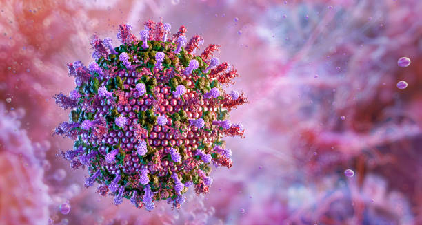
3D illustration of virus particles that cause encephalitis.

Causes of Encephalitis

Causes of Encephalitis The causes of encephalitis are most often viral, but bacterial, fungal, autoimmune, or parasitic infections can also be responsible. In all cases, encephalitis results from inflammation of the brain, which disrupts normal nerve function and leads to potentially life-threatening symptoms. Viral Causes Viruses are the leading cause. Common culprits include: Herpes simplex virus (HSV-1) – one of the most dangerous and common causes of severe encephalitis in adults Enteroviruses, including coxsackievirus and echovirus Arboviruses, such as West Nile virus, Japanese encephalitis virus, and tick-borne encephalitis virus Measles, mumps, and rubella – particularly in unvaccinated individuals Rabies virus, although rare, is almost always fatal if not treated before symptoms appear HIV-related encephalitis, due to weakened immunity or co-infections In South Africa, mosquito-borne viruses, HIV complications, and opportunistic infections like cytomegalovirus (CMV) in people with AIDS can also trigger encephalitis. Bacterial and Other Infections Bacteria like Mycoplasma pneumoniae or Borrelia burgdorferi (Lyme disease) may rarely cause encephalitis. In infants, group B streptococcus and Listeria infections can be responsible. Tuberculosis is another known cause in South Africa, especially in rural or high-burden areas. Fungal encephalitis is uncommon but can occur in people with compromised immunity. Fungi such as Cryptococcus or Aspergillus may spread to the brain in such cases. Autoimmune Encephalitis | Causes of Encephalitis In autoimmune encephalitis, the immune system mistakenly attacks the brain. This type may be triggered by cancer, infections, or may occur without a clear cause. Anti-NMDA receptor encephalitis is one recognised form, especially in young women. Post-Infectious or Parainfectious Encephalitis This occurs when the body’s immune response to an infection—such as measles or influenza—causes inflammation in the brain after the initial infection has cleared. It is more common in children and is sometimes referred to as acute disseminated encephalomyelitis (ADEM). Understanding the causes of encephalitis helps doctors choose the right tests and treatments quickly. Early identification is vital, especially in regions like South Africa where TB and HIV may complicate the clinical picture. [Next: Symptoms of Encephalitis →]

Causes of Encephalitis Read More »

Man holding his head in pain with red highlight indicating headache from encephalitis.

Symptoms of Encephalitis

Symptoms of Encephalitis The symptoms of encephalitis vary widely depending on the cause, severity, and area of the brain affected. In mild cases, encephalitis may resemble a cold or flu, while in more severe cases, symptoms can progress quickly and become life-threatening. Recognising these warning signs early is essential for seeking urgent medical care. Early Symptoms Initial signs often resemble a viral illness and may include: Fever Headache Fatigue or weakness Nausea and vomiting Stiff neck Sensitivity to light (photophobia) These may go unnoticed or be mistaken for a common infection. However, symptoms often progress over hours or days. Neurological Symptoms As the infection worsens, brain function becomes affected, leading to: Confusion or disorientation Difficulty speaking or understanding speech Behavioural changes or irritability Memory loss or inability to concentrate Seizures Hallucinations Drowsiness or loss of consciousness Children may show symptoms differently. They may become extremely irritable, cry persistently, refuse food or milk, or have seizures. In infants, a bulging fontanelle (soft spot) may signal increased pressure in the brain. Severe and Emergency Symptoms Difficulty waking Coma Sudden paralysis or weakness on one side of the body Severe seizures that don’t stop Breathing difficulty | Symptoms of Encephalitis In South Africa, where HIV and TB are prevalent, these symptoms may overlap with other infections, making diagnosis harder. However, the rapid onset of altered mental state or seizures should always prompt emergency medical care. Symptoms of encephalitis are serious and may progress quickly. Early hospital admission, brain imaging, and lab testing are key to identifying the cause and starting treatment. [Next: Diagnosis of Encephalitis→]

Symptoms of Encephalitis Read More »

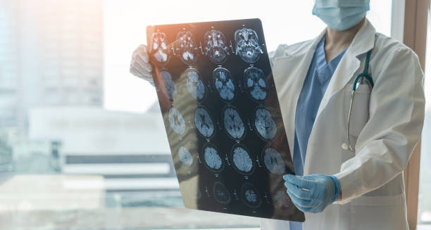
Doctor examining brain scans for signs of encephalitis.

Diagnosis of Encephalitis

Diagnosis of Encephalitis The diagnosis of encephalitis can be challenging because the symptoms often resemble other conditions such as meningitis, stroke, or severe flu. However, accurate and early diagnosis is critical, as encephalitis can progress quickly and cause permanent brain damage or death if not treated promptly. Doctors begin by reviewing the patient’s symptoms, medical history, and possible exposures. They will ask about recent infections, vaccination status, travel history, insect bites, and whether the person has a weakened immune system. In South Africa, TB, HIV, and vector-borne illnesses are also considered when diagnosing encephalitis. Physical and Neurological Examination A detailed examination checks for signs of brain inflammation. This may include: Level of consciousness Reflexes and coordination Muscle strength and tone Eye movements and speech Any change in mental state, seizures, or confusion suggests possible brain involvement. Tests to Confirm Diagnosis Lumbar puncture (spinal tap) – This is one of the most important tests. A small sample of cerebrospinal fluid (CSF) is taken from the spine and analysed for: White blood cell count (indicating infection or inflammation) Glucose and protein levels Presence of viruses or bacteria using PCR or cultures Tuberculosis testing in TB-prone regions Blood tests – Used to detect infections, autoimmune markers, or inflammation. In HIV-positive individuals, opportunistic infections like CMV or toxoplasmosis may be checked. Brain imaging (CT or MRI) – These scans help identify inflammation, swelling, bleeding, or masses in the brain. MRI is especially helpful in viral encephalitis cases. Electroencephalogram (EEG) – Records electrical activity in the brain. It may show patterns suggestive of encephalitis, particularly in those with seizures or altered consciousness. Viral and bacterial PCR testing – Detects genetic material of pathogens such as herpes simplex virus (HSV), enteroviruses, or TB. This allows for targeted treatment. HIV testing – In South Africa, many patients with encephalitis also undergo routine HIV screening to identify immunosuppression and guide care. Diagnosis of Encephalitis Diagnosis can take time, particularly when the cause is unknown or the patient is severely ill. However, treatment is often started before test results return if clinical suspicion is high. The diagnosis of encephalitis relies on a combination of tests, imaging, and clinical judgement. Early recognition and hospital-based investigation are essential for protecting brain function and improving outcomes. [Next: Treatment of Encephalitis →]

Diagnosis of Encephalitis Read More »

Woman holding head in distress due to encephalitis complications.

Complications of Encephalitis

Complications of Encephalitis The complications of encephalitis can be serious and long-lasting, particularly if treatment is delayed or if the infection is severe. Since encephalitis causes inflammation in the brain, complications often affect memory, movement, speech, and personality. Some people recover fully, but others may experience lifelong challenges. Neurological Complications Seizures – Brain inflammation can trigger seizures during the illness and increase the risk of epilepsy later. Cognitive problems – Memory loss, confusion, attention deficits, and poor problem-solving skills may remain after recovery. Speech and language difficulties – Some people struggle to speak, understand, or write clearly. Movement disorders – Tremors, weakness, or difficulty with balance and coordination can persist. Vision or hearing loss – Inflammation near sensory nerves may lead to partial or complete loss of sight or hearing. Behavioural and Emotional Changes Brain inflammation may lead to: Depression Anxiety or irritability Changes in personality Difficulty with social interaction These symptoms can affect relationships, employment, and independence. Physical Disabilities In severe cases, encephalitis may result in: Paralysis of one side of the body (hemiparesis) Chronic fatigue Dependence on carers for daily tasks Rehabilitation can help manage these symptoms, but some individuals may need long-term care. Death Though not common in mild cases, encephalitis can be fatal—especially in very young children, elderly people, or those with weak immune systems. Mortality rates are higher in untreated herpes simplex encephalitis, TB-related encephalitis, and cases involving rabies. In South Africa Complications of Encephalitis In South Africa, late diagnosis and limited access to advanced imaging or ICU support can increase the risk of complications. Infections such as HIV, TB, and malaria also contribute to more complex cases of encephalitis. Awareness among community health workers and faster referral systems can reduce the burden of complications. Access to rehabilitation and counselling services must also be improved—especially for those recovering in rural areas. Understanding the complications of encephalitis encourages early action and highlights the importance of continued care beyond hospital discharge. With support and therapy, many people regain independence and live fulfilling lives, even after severe illness. [Next: Back to Overview →]

Complications of Encephalitis Read More »

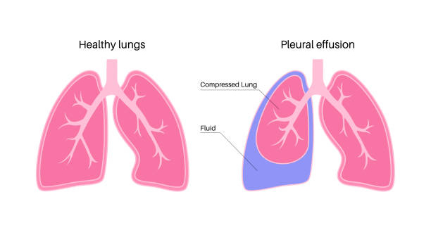
Diagram comparing healthy lungs and pleural effusion in empyema

Empyema

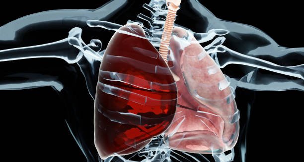
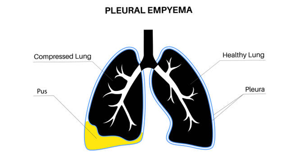
Empyema Empyema is a serious medical condition in which pus collects in the space between the lungs and the chest wall, known as the pleural cavity. This often happens as a complication of pneumonia when infection spreads from the lungs to the pleural space. This can make breathing difficult and may cause permanent lung damage if not treated promptly. The condition begins with fluid build-up in the pleural space—known as a pleural effusion. If bacteria infect that fluid, it becomes thick with pus and white blood cells, turning into empyema. This infected fluid restricts lung movement and causes inflammation, leading to chest pain, fever, and breathing difficulties. Empyema can occur at any age but is more common in: Children with underdeveloped immune systems Older adults People with weakened immunity due to HIV, diabetes, or cancer Patients recovering from chest trauma or surgery Empyema In South Africa, empyema remains a public health concern, particularly in communities with limited access to early treatment for respiratory infections. Tuberculosis (TB) is also a common cause of this condition in regions where TB rates are high. Early treatment is essential. Without it, the condition can progress, leading to lung scarring, trapped lungs, or widespread infection. Treatment often includes antibiotics and drainage of the infected fluid, and in some cases, surgery. Understanding empyema is crucial for recognising complications of chest infections and ensuring early medical care—especially in areas with high TB and pneumonia rates. [Next: Causes of Empyema →]

Empyema Read More »

Causes of Empyema

Causes of Empyema

Causes of Empyema The causes of empyema usually involve an infection that spreads into the pleural space, the thin gap between the lungs and the chest wall. Empyema develops when pus-forming bacteria invade this space, creating a thick, infected fluid that prevents the lungs from expanding properly. Common Causes of Empyema Pneumonia – The most frequent cause. Bacteria like Streptococcus pneumoniae, Staphylococcus aureus, and Klebsiella pneumoniae are often involved. Tuberculosis (TB) – In countries like South Africa, TB is a leading cause of empyema, particularly in patients with HIV or poor access to healthcare. Lung abscess – A pocket of pus in the lung may burst and leak infection into the pleural cavity. Chest trauma – Penetrating chest injuries or broken ribs can introduce bacteria into the pleural space. Surgery complications – Operations involving the lungs or heart may allow bacteria to enter and cause infection. Oesophageal rupture – Rare, but serious; it allows food and bacteria to leak into the chest cavity, causing empyema. Risk Factors Some people are more likely to develop empyema. Risk factors include: A weakened immune system (due to HIV, cancer, or long-term steroid use) Diabetes Poor nutrition Alcohol misuse Smoking or chronic lung disease (like COPD) Delayed or incomplete treatment of pneumonia Causes of Empyema In children, empyema often follows a bacterial chest infection. In South Africa, overcrowding, poor nutrition, and limited access to early antibiotics contribute to higher rates in young patients. Empyema usually develops in stages: Exudative stage – Clear fluid builds up in the pleural space. Fibrinopurulent stage – The fluid becomes cloudy and infected, forming pus. Organising stage – Scar tissue forms, trapping the lung and making breathing harder. Understanding the causes of empyema allows for early recognition and better prevention of serious complications. Treating chest infections promptly and monitoring high-risk patients are key strategies in avoiding this condition. [Next: Symptoms of Empyema →]

Causes of Empyema Read More »

Symptoms of Empyema

Symptoms of Empyema

Symptoms of Empyema The symptoms of empyema often develop after a respiratory infection like pneumonia. As the infection spreads into the pleural cavity, the build-up of pus creates pressure on the lungs, causing pain and breathing difficulty. Recognising the symptoms of empyema early can prevent serious complications and long-term lung damage. Common Symptoms Chest pain – Sharp pain that worsens with deep breathing, coughing, or movement. Often felt on one side of the chest. Shortness of breath – Caused by reduced lung expansion due to the infected fluid pressing against the lungs. Fever and chills – Indicate the body is fighting an infection. Persistent cough – May be dry or produce phlegm. Fatigue and weakness – Ongoing infection can drain energy and make daily activities difficult. Loss of appetite and weight loss – Common in longer-lasting or severe infections. Children may present differently. They may become irritable, breathe rapidly, refuse to eat, or show signs of chest retraction—where the chest appears to suck inwards with each breath. Advanced Symptoms As empyema progresses, additional signs may appear: Night sweats Thick or foul-smelling sputum Swelling or tenderness over the affected side of the chest Bluish skin (cyanosis) in severe cases where oxygen levels are low In South Africa, where TB is common, empyema may develop more slowly, with low-grade fever and gradual weight loss. This “cold empyema” is harder to detect without imaging like chest X-rays or ultrasound. Symptoms of Empyema Some patients also show signs of sepsis—a widespread body infection—which includes very high fever, confusion, rapid heartbeat, and dangerously low blood pressure. This is a medical emergency. Symptoms can vary depending on the stage of empyema and the patient’s immune status. People with chronic illness or HIV may have fewer noticeable symptoms but still suffer from significant infection. Recognising the symptoms of empyema—especially after pneumonia or TB—is critical. Prompt medical evaluation can lead to early drainage, effective antibiotic treatment, and full recovery before permanent damage occurs.[Next: Diagnosis of Empyema→]

Symptoms of Empyema Read More »

Diagnosis of Empyema

Diagnosis of Empyema

Diagnosis of Empyema The diagnosis of empyema starts with a clinical evaluation, especially in patients who have recently had pneumonia or other lung infections. Since empyema involves pus collecting in the pleural space, doctors must confirm the presence of fluid and determine whether it’s infected. Early and accurate diagnosis is key to preventing serious complications. The doctor begins by asking about symptoms such as chest pain, shortness of breath, cough, and fever. A physical examination may reveal reduced breath sounds, dullness to percussion, or decreased chest expansion on one side—all signs of fluid build-up. Imaging Tests | Diagnosis of Empyema Chest X-ray – May show pleural effusion (fluid around the lung), but not whether it’s infected. Ultrasound – Useful for detecting fluid and guiding drainage. It can differentiate between clear fluid and thick pus. CT scan – Provides a detailed view of the chest, helping identify pockets of pus, loculated fluid, or lung collapse. This is especially helpful when empyema is advanced or complex. Diagnostic Thoracentesis This is a key step. The doctor uses a needle to remove a sample of pleural fluid from the chest. The fluid is then analysed in the lab for: Appearance – cloudy or foul-smelling fluid suggests empyema Cell count – high white blood cells indicate infection Protein and glucose levels – help distinguish between types of effusions Culture – to identify the bacteria responsible, allowing targeted antibiotic treatment In TB-endemic areas like South Africa, fluid may also be tested for Mycobacterium tuberculosis using specialised tests like GeneXpert or TB culture. Diagnosis of Empyema If empyema is suspected in a child, imaging and thoracentesis are often done at the same time, under sedation or anaesthesia. In South Africa, delays in diagnosis are often due to limited access to imaging or specialist care in rural settings. Mobile X-ray units and ultrasound machines in clinics are helping to bridge this gap. The diagnosis of empyema involves combining clinical signs, imaging, and fluid analysis. Once confirmed, treatment can begin immediately—reducing the risk of long-term lung damage and life-threatening complications. [Next: Treatment of Empyema →]

Diagnosis of Empyema Read More »

Treatment of Empyema

Treatment of Empyema

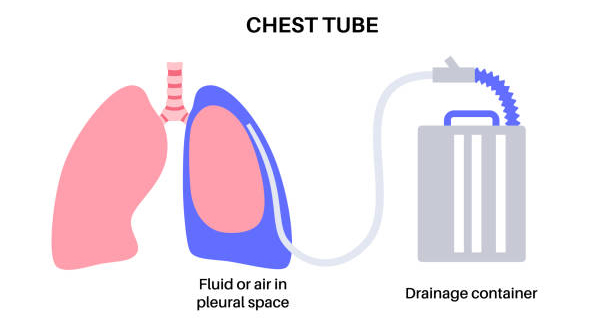
Treatment of Empyema The treatment of empyema focuses on removing the infected fluid, eliminating the infection, and restoring lung function. Since empyema can become life-threatening if untreated, early intervention is essential. Treatment varies depending on the stage and severity of the condition. Antibiotics Doctors immediately start broad-spectrum intravenous antibiotics to fight the infection. Once lab results identify the specific bacteria, doctors adjust the antibiotics accordingly. In TB-related empyema, doctors prescribe a full course of anti-tuberculosis medication, typically lasting six to nine months. Common antibiotics used for bacterial empyema include: Ceftriaxone or piperacillin-tazobactam Clindamycin or metronidazole (for anaerobic bacteria) Vancomycin (if MRSA is suspected) Patients often remain on IV antibiotics for several days, followed by oral antibiotics for 2–4 weeks. Drainage Doctors must remove the infected fluid to allow the lung to re-expand. This is done using: Chest tube insertion (intercostal drain) – Doctors insert a flexible tube into the chest to drain pus. Doctors often use ultrasound to guide the procedure. Image-guided drainage – In some cases, CT or ultrasound helps place the drain in the best location. Intrapleural fibrinolytics – Doctors may use medications like alteplase and DNase through the drain to break up thick pus and improve drainage. Surgery | Treatment of Empyema If drainage and antibiotics do not improve empyema, doctors may need to perform surgery: Video-assisted thoracoscopic surgery (VATS) – Minimally invasive; used to break up loculated pus and remove infected tissue Open thoracotomy – In severe cases, especially with organised empyema and lung entrapment, surgeons perform full chest surgery Children usually respond well to antibiotics and chest tube drainage. Surgery is less often required in paediatric cases unless there is extensive loculation or no improvement. In South Africa, treatment access may vary. Urban hospitals typically offer surgical options and intensive care, while rural clinics may rely on antibiotics and basic drainage. Outreach programmes and hospital referrals help ensure continuity of care. The treatment of empyema requires a team effort—combining infection control, fluid removal, and lung rehabilitation. When addressed early, most patients recover well without long-term issues. [Next: Complications of Empyema →]

Treatment of Empyema Read More »

Complications of Empyema

Complications of Empyema

Complications of Empyema The complications of empyema can be severe if the condition is not recognised and treated early. Because empyema causes infected pus to build up in the chest cavity, it can lead to long-term lung damage, infection spread, and even death in serious cases. Lung-Related Complications Trapped lung – Pus and inflammation can scar the pleura (lung lining), causing the lung to stick and collapse. The lung cannot re-expand fully, resulting in long-term shortness of breath. Fibrothorax – A thick fibrous layer forms around the lung, limiting its movement permanently. Bronchopleural fistula – An abnormal connection between the lung and pleural space can form, leading to continuous air leakage and prolonged recovery. Chronic empyema – If not fully drained, the infection may become long-standing and more difficult to treat. Infection Spread Empyema can lead to systemic infection (sepsis), a life-threatening condition in which bacteria spread through the bloodstream. Signs include high fever, rapid heart rate, low blood pressure, confusion, and organ failure. This is a medical emergency requiring intensive care. Respiratory Failure Severe cases can lead to reduced oxygen levels and the need for mechanical ventilation. This is especially dangerous in the elderly or people with pre-existing lung disease. Recurrent Infections Patients who recover from empyema may still be at risk for recurrent pneumonia or pleural effusions. Preventative care, such as vaccines for pneumococcus and influenza, is highly recommended after recovery. In Children In children, long-term complications are rare if empyema is treated promptly. However, delayed treatment may lead to growth delays, reduced lung function, or chronic respiratory problems. Complications of Empyema In South Africa In TB-endemic areas, empyema may occur alongside drug-resistant tuberculosis, complicating treatment and increasing the risk of chronic illness. Delays in accessing surgical care in rural provinces can worsen outcomes. Strengthening referral pathways and early chest X-rays are key in avoiding advanced complications. With the right care, many complications are preventable. Chest physiotherapy, good nutrition, and follow-up imaging help ensure the lung heals fully. Education on early symptoms of pneumonia or TB also plays a role in preventing empyema in the first place. Understanding the complications of empyema highlights the importance of early diagnosis, complete treatment, and post-recovery care. With timely intervention, most patients can make a full and lasting recovery. [Next: Back to Overview →]

Complications of Empyema Read More »

Scroll to Top