Mental Matters

Author name: admin

Physiotherapist assisting patient with joint support during treatment for Ehlers-Danlos Syndromes.

Treatment of Ehlers-Danlos Syndromes

Treatment of Ehlers-Danlos Syndromes The treatment of Ehlers-Danlos syndromes focuses on managing symptoms, preventing injuries, and supporting long-term quality of life. As Ehlers-Danlos syndromes are genetic, there is currently no cure—but with early intervention and the right support, many people live active, fulfilling lives. Doctors tailor treatment plans to each individual’s specific needs. They depend on the type of EDS and the severity of symptoms. Most patients benefit from a multi-disciplinary approach, involving physiotherapists, pain specialists, occupational therapists, and psychologists. Physiotherapy plays a central role. A structured exercise programme helps stabilise joints, strengthen muscles, and reduce the risk of dislocations. Doctors avoid stretching because excessive flexibility can worsen joint instability. Doctors often recommend core strengthening, low-impact activities, and proprioception exercises to improve balance and coordination. Pain management is crucial. Many people with EDS experience chronic pain due to joint instability, muscle fatigue, or previous injuries. Treatment may include: Paracetamol and NSAIDs Low-dose antidepressants or anti-epileptic medications for nerve pain Physical therapy and massage Cognitive behavioural therapy (CBT) to help manage the emotional impact of long-term pain Braces or supports may be used to stabilise vulnerable joints, especially during flare-ups. Custom orthotics or mobility aids may be helpful in cases with severe joint laxity or fatigue. Occupational therapy can assist with adapting daily tasks and preventing joint strain. Techniques include joint protection strategies, activity pacing, and ergonomic support for work or study. Mental health support is vital. Living with EDS can lead to anxiety, depression, or frustration—especially when diagnosis is delayed or misunderstood. Support groups, therapy, and community awareness programmes can make a big difference. Treatment of Ehlers-Danlos Syndromes For rarer types like vascular EDS, more urgent medical oversight is needed. Patients must avoid high-risk activities and may require routine imaging to monitor blood vessels. Any sudden chest, abdominal, or limb pain must be treated as a medical emergency due to the risk of arterial rupture. In South Africa, access to comprehensive treatment depends on location and medical coverage. While larger urban hospitals may offer rheumatology and physiotherapy services, rural patients often face long travel distances and limited care options. Non-profits and advocacy groups are working to increase awareness and support for those living with EDS across all provinces. The treatment of Ehlers-Danlos syndromes requires long-term planning and ongoing support. Although it cannot reverse the condition, it empowers individuals to manage symptoms, protect their bodies, and improve daily functioning. [Next: Complications of Ehlers-Danlos Syndromes →]

Treatment of Ehlers-Danlos Syndromes Read More »

Man sitting on bed with hand on face, experiencing pain from Ehlers-Danlos Syndromes complications.

Complications of Ehlers-Danlos Syndromes

Complications of Ehlers-Danlos Syndromes The complications of Ehlers-Danlos syndromes can be wide-ranging and vary depending on the type and severity of the condition. Because Ehlers-Danlos syndromes affect connective tissue throughout the body, complications may arise in the joints, skin, internal organs, and vascular system. Early diagnosis and proper management reduce the risk of serious long-term damage. The most common complications include: Joint dislocations and subluxations, particularly in hypermobile EDS. These can occur during daily activities and cause pain, instability, and soft tissue injury. Chronic pain, which often worsens over time due to muscle fatigue, nerve strain, and joint instability. Delayed healing, thin scarring, and skin that bruises easily or splits with minor trauma—especially in classical EDS. Fatigue, which may stem from poor sleep, deconditioning, and constant pain. In vascular EDS, complications are far more serious. The most dangerous risks include: Arterial rupture, often affecting the aorta or medium-sized arteries Organ rupture, especially in the intestines or uterus (during childbirth) Internal bleeding, which may be life-threatening and require emergency care Pregnancy in women with vascular EDS requires high-risk monitoring due to the risk of uterine rupture or vascular events. Women with other EDS types may also face challenges such as joint pain, pelvic instability, and premature delivery. Digestive complications can also occur. These include irritable bowel syndrome, gastroparesis, and acid reflux, which are common in hypermobile EDS. Constipation and bladder dysfunction may result from weak pelvic support structures. Other complications include: Dental issues, like fragile teeth or gum disease Frequent injuries, such as sprains, strains, and tendon ruptures Depression and anxiety, often due to long-term pain, disability, or isolation Adverse reactions to surgery, including poor healing or excessive bleeding Complications of Ehlers-Danlos Syndromes In South Africa, the complications of EDS are often compounded by delayed diagnosis, limited access to physical therapy, and a shortage of specialists. As a result, many people live with untreated symptoms or worsening disability. Education for healthcare workers and community awareness can help close the gap in care. While the complications of EDS can be overwhelming, they are not inevitable. With proper care, most people can reduce injuries, manage their pain, and maintain independence. Support from family, health professionals, and the broader community plays a key role in this journey. Understanding the complications of Ehlers-Danlos syndromes helps healthcare teams and families create realistic care plans and empower those affected to live more comfortably and safely. [Next: Back to Overview →]

Complications of Ehlers-Danlos Syndromes Read More »

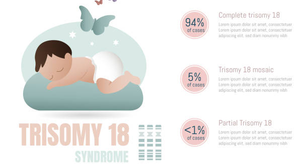
Illustration explaining Edwards’ Syndrome with baby and trisomy 18 chromosome breakdown.

Edwards’ Syndrome

Edwards’ Syndrome Edwards’ syndrome, also known as trisomy 18, is a serious genetic condition caused by the presence of an extra copy of chromosome 18 in a baby’s cells. This additional genetic material disrupts normal development, leading to severe physical and intellectual disabilities. This syndrome affects around 1 in 5,000 live births, but many pregnancies with this condition end in miscarriage or stillbirth. The condition was first described by Dr. John H. Edwards in 1960. It occurs randomly during cell division in the early stages of embryo development. Most cases are not inherited and are instead linked to errors during the formation of egg or sperm cells. The risk of having a baby with Edwards’ syndrome increases with maternal age, particularly in women over 35. Babies born with Edwards’ syndrome often have a low birth weight and a range of physical abnormalities. These may include a small head, clenched fists with overlapping fingers, heart defects, and severe developmental delays. Many also experience breathing difficulties and feeding problems shortly after birth. Sadly, the condition is associated with a high mortality rate. Most babies with this syndrome die before or shortly after birth. Of those who are born alive, only a small percentage survive past their first year, and those who do face complex medical challenges. Edwards’ Syndrome In South Africa, the availability of prenatal screening and diagnostic services varies. In urban areas, pregnant women may be offered blood tests and ultrasound scans that can detect markers for Edwards’ syndrome. However, in rural or under-resourced regions, limited access to these services may delay or prevent early diagnosis. Despite the severe nature of the condition, some families choose to continue with the pregnancy and provide palliative care after birth. Others may opt for medical intervention where appropriate, depending on the child’s condition and prognosis. Understanding Edwards’ syndrome helps expectant parents make informed decisions and prepares families and healthcare providers to offer the best support possible—both emotionally and medically. [Next: Causes of Edwards’ Syndrome →]

Edwards’ Syndrome Read More »

Chromosomal diagram showing Trisomy 18 as the cause of Edwards’ Syndrome.

Causes of Edwards’ Syndrome

Causes of Edwards’ Syndrome The causes of Edwards’ syndrome lie in a genetic abnormality known as trisomy 18. In this condition, a baby has three copies of chromosome 18 instead of the usual two. This extra chromosome affects nearly every organ and system in the developing body, leading to a wide range of health problems. Edwards’ syndrome is not usually inherited—it occurs as a random event during the formation of reproductive cells. During fertilisation, an egg from the mother and a sperm from the father combine to form a new life with 46 chromosomes—23 from each parent. In trisomy 18, one of the parents’ cells contributes an extra chromosome 18. This typically happens due to a process called nondisjunction, where chromosomes fail to separate properly during cell division. There are three types of Edwards’ syndrome: Full trisomy 18 – where every cell in the body contains an extra chromosome 18. This is the most common and most severe form. Mosaic trisomy 18 – where only some cells have the extra chromosome. The symptoms may be milder depending on the number and type of affected cells. Partial trisomy 18 – where only a part of chromosome 18 is present in triplicate. This is rare and often caused by a piece of the chromosome attaching to another during cell formation. The exact reason for these chromosomal errors is not well understood, but they are more likely to happen as a woman gets older. The risk increases significantly for mothers aged 35 and above. Causes of Edwards’ Syndrome In South Africa, many women may not be aware of these risks, especially in areas with limited access to antenatal education. Routine prenatal screenings may not always be offered, particularly in underfunded clinics. Increasing awareness of maternal age-related risks and improving access to prenatal diagnostics could help identify Edwards’ syndrome earlier. Although the condition usually occurs by chance, parents who have had one child with Edwards’ syndrome may be offered genetic counselling. This can help them understand their risk in future pregnancies and explore available options. Understanding the causes of Edwards’ syndrome allows for better screening and emotional preparation. It also supports more informed conversations between parents and healthcare providers when faced with difficult decisions. [Next: Symptoms of Edwards’ Syndrome →]

Causes of Edwards’ Syndrome Read More »

Symptoms of Edwards’ Syndrome

Symptoms of Edwards’ Syndrome

Symptoms of Edwards’ Syndrome The symptoms of Edwards’ syndrome are wide-ranging and often visible before or shortly after birth. The condition affects growth, development, and organ function. Babies with Edwards’ syndrome usually show multiple physical abnormalities, as well as severe intellectual and developmental delays. These signs help doctors suspect and confirm the diagnosis early. During pregnancy, ultrasound scans may show: Low birth weight Excess amniotic fluid (polyhydramnios) A small placenta Reduced foetal movement Congenital heart defects Once the baby is born, common physical features include: A small, misshapen head (microcephaly) A prominent back part of the head (occiput) Low-set ears A small jaw and mouth Clenched fists with overlapping fingers Rocker-bottom feet Short breastbone Narrow eyelid openings Many babies also have structural problems in the heart, kidneys, lungs, and digestive system. Heart defects are particularly common and include holes between heart chambers (ventricular or atrial septal defects) or valve abnormalities. These can lead to severe breathing and circulation problems. Feeding difficulties are also a major symptom. Many babies struggle to suck or swallow effectively, leading to poor weight gain and increased risk of aspiration or infections. Breathing may be irregular due to underdeveloped lungs or nerve dysfunction. Developmentally, most children with Edwards’ syndrome do not reach typical milestones. They may not sit, crawl, or speak. Some may respond to sound or touch, but progress is extremely limited. Seizures and neurological impairments are also common. Symptoms of Edwards’ Syndrome In South Africa, detection of symptoms may be delayed if the baby is born in a setting without trained staff or access to specialised care. Community healthcare workers play an essential role in identifying signs early and referring families for support. While the symptoms of Edwards’ syndrome are often severe, the degree can vary—especially in mosaic or partial trisomy 18. Some children may live longer than expected and show small signs of progress with the right medical and emotional care. Recognising the symptoms of Edwards’ syndrome allows for early intervention, accurate diagnosis, and compassionate care. Families facing this diagnosis deserve access to honest, supportive information that helps them make the best decisions for their unique circumstances. [Next: Diagnosis of Edwards’ Syndrome→]

Symptoms of Edwards’ Syndrome Read More »

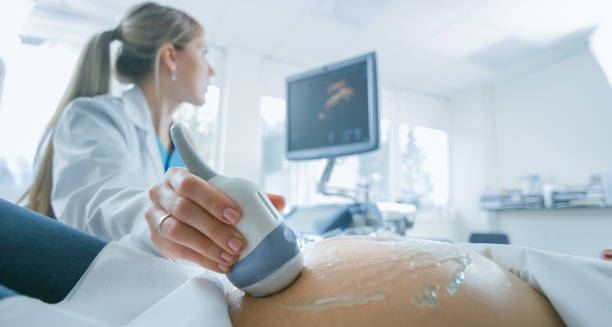
Pregnant woman undergoing ultrasound screening to detect chromosomal abnormalities like Edwards’ Syndrome.

Diagnosis of Edwards’ Syndrome

Diagnosis of Edwards’ Syndrome The diagnosis of Edwards’ syndrome can happen during pregnancy or shortly after birth. Since Edwards’ syndrome leads to a wide range of physical abnormalities, doctors may suspect the condition early, especially when routine pregnancy scans show signs of abnormal growth or development. Early diagnosis gives families more time to understand the condition and prepare for possible outcomes. In pregnancy, screening tests are often the first step. These include: Ultrasound scans, which may detect physical features such as low foetal weight, heart defects, or clenched hands Maternal blood tests, which check for abnormal levels of proteins and hormones that may signal a chromosomal condition Non-invasive prenatal testing (NIPT), which analyses foetal DNA in the mother’s blood to detect extra chromosomes These screenings cannot confirm the condition but can indicate a higher risk. If results show a high chance of Edwards’ syndrome, doctors may recommend diagnostic tests such as: Chorionic villus sampling (CVS) – done around 10–13 weeks, where a small piece of placental tissue is tested Amniocentesis – done around 15–20 weeks, where a sample of amniotic fluid is tested for chromosomal abnormalities Both tests carry a small risk of miscarriage but provide a definitive diagnosis by checking the baby’s chromosomes directly. After birth, a baby showing typical signs—such as clenched fists, small jaw, or heart problems—may be tested using a karyotype analysis, which examines the number and structure of chromosomes. This test confirms whether full, mosaic, or partial trisomy 18 is present. Diagnosis of Edwards’ Syndrome In South Africa, access to these prenatal tests may vary widely depending on whether a woman uses public or private healthcare. While private clinics in urban areas often offer NIPT and early ultrasounds, many rural facilities may only detect the condition after birth. This highlights the importance of strengthening prenatal services across the country. Once a diagnosis is confirmed, genetic counselling is usually offered to help parents understand the condition, possible outcomes, and future reproductive choices. Counsellors provide information in an empathetic way, helping families process complex emotions during a difficult time. The diagnosis of Edwards’ syndrome gives families a clearer understanding of their child’s health and life expectancy. It also allows healthcare professionals to plan the most appropriate care and support. [Next: Treatment of Edwards’ Syndrome →]

Diagnosis of Edwards’ Syndrome Read More »

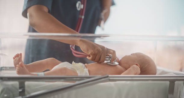
Newborn with Edwards’ Syndrome receiving medical care in a neonatal unit.

Treatment of Edwards’ Syndrome

Treatment of Edwards’ Syndrome The treatment of Edwards’ syndrome focuses on easing symptoms and improving the quality of life. Since Edwards’ syndrome has no cure, treatment is based on the baby’s specific needs, the severity of the condition, and the family’s preferences. Medical teams aim to provide either supportive or palliative care, depending on the baby’s condition. At birth, babies with Edwards’ syndrome often have serious health challenges. Treatment in the neonatal intensive care unit (NICU) may be required for: Breathing difficulties, supported by oxygen or mechanical ventilation Feeding problems, managed with feeding tubes or special bottles Heart defects, which may require medication or surgery in rare cases Infections, treated with antibiotics Some babies may not need all these interventions, especially in cases of mosaic or partial trisomy 18, where symptoms are milder. In these situations, treatment plans may involve developmental support, physical therapy, and regular medical monitoring. In many cases, families and doctors focus on palliative care. This approach prioritises comfort, dignity, and love—supporting the baby with gentle care while avoiding aggressive medical treatments that may cause distress or have limited benefit. Palliative care includes pain management, warm clothing, skin-to-skin contact, and emotional support for the family. Medical teams work closely with parents to create a care plan that respects their wishes and beliefs. In South Africa, palliative care services are growing, though access remains uneven. Some families in public hospitals may not receive the same level of specialised care as those in private facilities. Advocacy is helping improve access to compassionate, community-based support in more regions. For babies who survive beyond infancy, long-term treatment may involve: Ongoing heart monitoring Nutritional support Management of seizures or reflux Speech and physiotherapy Assistive devices for movement and communication Treatment of Edwards’ Syndrome Support for the family is vital. Parents may need counselling, respite care, or financial assistance. In South Africa, several non-profit organisations help families living with trisomy conditions, offering guidance, equipment, and emotional support. The treatment of Edwards’ syndrome is highly individualised. Some babies may need intensive support early on, while others may surprise doctors with longer-than-expected survival. Every child is different, and every family deserves support in making informed, compassionate decisions. [Next: Complications of Edwards’ Syndrome →]

Treatment of Edwards’ Syndrome Read More »

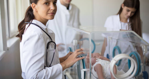
Neonatal doctors attending to a newborn in intensive care for complications of Edwards’ Syndrome.

Complications of Edwards’ Syndrome

Complications of Edwards’ Syndrome The complications of Edwards’ syndrome are often severe and affect multiple organs and systems. Because Edwards’ syndrome involves an extra chromosome in every cell (in full trisomy 18), it leads to widespread developmental challenges that begin before birth and continue throughout life. The most serious complications include: Congenital heart defects – present in over 90% of affected babies, these include holes in the heart or valve abnormalities. These can cause breathlessness, poor circulation, and failure to thrive. Breathing difficulties – due to weak muscles, irregular brain signals, or structural lung abnormalities. Some babies require constant oxygen or assisted breathing. Feeding problems – poor sucking reflexes, low energy, and underdeveloped digestive systems make feeding difficult. Malnutrition and dehydration are common. Neurological issues – including seizures, intellectual disability, and abnormal brain structure. Kidney and urinary problems, which can affect body chemistry and growth. Care From Birth Many babies are born very small and have weak immune systems, making them vulnerable to infections such as pneumonia and urinary tract infections. Repeated hospital stays are common, and some babies may need long-term medical care from birth. In those who survive past the newborn stage, delayed development is expected. Children may not walk, talk, or feed independently. Many cannot sit up or hold their head without assistance. Some can recognise familiar voices or respond to music or touch, but overall development remains severely limited. Emotional complications also affect families. Parents face uncertainty, difficult decisions, and grief—whether after a miscarriage, stillbirth, or the early death of a newborn. Mental health support for parents, siblings, and extended families is essential for healing and adjustment. In South Africa, complications are often made worse by limited access to paediatric specialists, ventilators, or early intervention services. Rural hospitals may not have NICU facilities, leaving families with fewer treatment options. Public health education and improved transport for critical cases can help reduce preventable losses. Complications of Edwards’ Syndrome Healthcare professionals must also be trained to recognise signs of Edwards’ syndrome and offer appropriate care options that respect cultural values and family preferences. Although the condition is life-limiting, not all children are the same. A small number of children with mosaic or partial trisomy 18 survive into their teens. Their lives may be complex, but they bring meaning and joy to their families. These rare cases remind us of the importance of individualised care and ongoing research. Understanding the complications of Edwards’ syndrome helps parents, doctors, and communities provide informed, respectful, and supportive care—ensuring every child, no matter their condition, is treated with dignity. [Next: Back to Overview →]

Complications of Edwards’ Syndrome Read More »

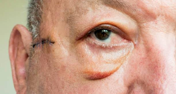
Close-up of an elderly man's eye showing signs of ectropion with visible eyelid turning outward.

Ectropion

Ectropion Ectropion is a condition in which the lower eyelid turns outward, away from the eye. This outward turning exposes the inner surface of the eyelid, which normally rests against the eyeball. Ectropion can cause discomfort, dryness, and irritation because the exposed surface is not properly protected or lubricated by tears. This condition most often affects older adults, especially those over 60, as the muscles and tissues supporting the eyelid naturally weaken with age. However, ectropion can also result from facial nerve damage, trauma, scarring, or certain skin conditions. It may affect one or both eyes, depending on the underlying cause. When the eyelid is not in contact with the eye, it disrupts the normal tear drainage process. Tears may not spread evenly, leading to dryness. Additionally, tears may overflow continuously down the cheek, a condition known as epiphora. This can cause social discomfort and increase the risk of eye infections or damage to the cornea. Ectropion In South Africa and other parts of the world, access to eye care varies. Older adults in rural or under-resourced communities may not receive early diagnosis or treatment, which can lead to worsening symptoms and complications. Public awareness campaigns around age-related eye conditions like ectropion can help improve outcomes. Fortunately, ectropion is treatable. In mild cases, artificial tears and ointments may provide relief. For more severe cases or those involving scarring or nerve damage, surgical correction is usually recommended to restore the eyelid to its normal position. Understanding ectropion and its symptoms can lead to earlier diagnosis, better eye health, and improved quality of life—especially for ageing populations. [Next: Causes of Ectropion →]

Ectropion Read More »

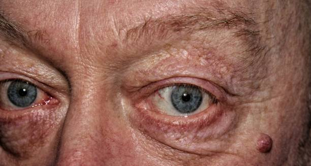
Close-up of a man's eyes showing sagging lower eyelids, a common sign of ectropion causes.

Causes of Ectropion

Causes of Ectropion The causes of ectropion are varied, but the condition most commonly results from age-related changes in the tissues and muscles around the eye. As we age, the connective tissues supporting the eyelids lose elasticity and tone, causing the lower eyelid to sag and turn outward. This form is known as involutional ectropion and is the most frequent type seen in clinical practice. Another significant cause is facial nerve paralysis, particularly of the seventh cranial nerve, also known as the facial nerve. When this nerve is damaged—due to Bell’s palsy, stroke, or head trauma—it affects the muscles that hold the eyelid in place. As a result, the eyelid can droop outward, leading to paralytic ectropion. Scarring from previous surgeries, burns, trauma, or chronic eye infections can pull the eyelid outward. This is known as cicatricial ectropion. Inflammation or damage to the skin and tissues around the eye alters the normal structure, preventing the eyelid from sitting properly against the eye. Chronic allergic reactions or infections can also cause the eyelid to swell and lose its normal positioning over time. In rare cases, tumours near the eye can push the eyelid outwards, creating mechanical ectropion. Certain congenital conditions—present from birth—may also lead to ectropion. Although rare, babies born with eyelid deformities may experience symptoms early in life, requiring specialist care and sometimes surgery. Causes of Ectropion In South Africa, trauma and burns are significant contributors to ectropion, particularly in rural or low-income areas where open cooking fires and untreated skin infections are more common. Lack of access to prompt treatment can result in scar formation, increasing the risk of cicatricial ectropion. Long-term use of certain eye medications can also contribute to tissue changes around the eyelid. Prolonged irritation may weaken the eyelid’s structure and lead to eventual outward turning. Regardless of the cause, early recognition and treatment are key. Left untreated, ectropion can lead to chronic discomfort, tearing, and even permanent damage to the eye surface. Understanding the causes of ectropion allows for prevention in some cases and supports faster intervention in others. [Next: Symptoms of Ectropion →]

Causes of Ectropion Read More »

Scroll to Top