Mental Matters

Author name: admin

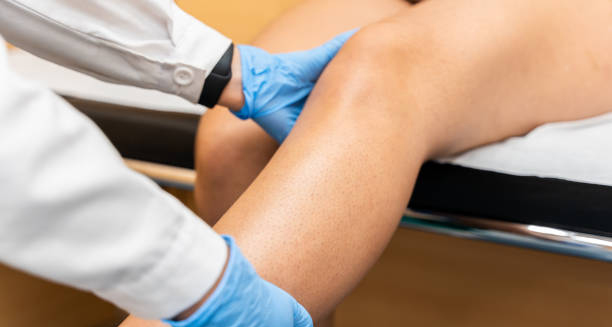
Doctor examining patient’s knee for dislocation treatment on clinic bed

Treatment of Dislocated Kneecap

Treatment of Dislocated Kneecap The treatment of dislocated kneecap starts with reducing the dislocation—repositioning the kneecap back into its proper place. A trained medical professional typically performs this, often in an emergency or orthopaedic setting. Once the kneecap is back in place, the focus shifts to managing pain, reducing swelling, and preventing future dislocations. Initially, the RICE protocol—Rest, Ice, Compression, and Elevation—is recommended. Applying ice to the knee for 15–20 minutes every few hours helps reduce swelling. A medical professional usually immobilises the knee with a brace or splint for a short period to allow the soft tissue around the joint to begin healing. Crutches may be provided to keep weight off the injured leg. Pain relief is an important part of the treatment of dislocated kneecap. Non-steroidal anti-inflammatory drugs (NSAIDs) such as ibuprofen help manage both pain and inflammation. Once swelling subsides, physiotherapy becomes the cornerstone of recovery. Rehabilitation exercises focus on strengthening the quadriceps, especially the vastus medialis oblique (VMO) muscle, which plays a key role in stabilising the patella. Physical therapy also includes range-of-motion exercises, balance training, and gradual return-to-sport drills. Strengthening the muscles around the hip and thigh is just as important as focusing on the knee. These efforts help prevent future dislocations by improving joint stability. In some cases, especially with recurrent dislocations or structural abnormalities, a surgeon may recommend surgery. Surgical options include tightening or realigning the tissues around the kneecap, reconstructing torn ligaments, or deepening the groove in which the kneecap sits (trochleoplasty). Arthroscopy may also be used to remove loose fragments or repair cartilage. Treatment of Dislocated Kneecap In South Africa, surgical treatment of dislocated kneecap is available through both public and private healthcare systems, though waiting times and access to post-operative rehabilitation may differ significantly between the two sectors. Urban hospitals often have quicker access to orthopaedic care, while rural areas may face delays in specialist treatment. Education is a key part of recovery. Patients are advised on proper movement patterns, footwear, and activity modification. Athletes may need to modify training routines temporarily and use supportive braces during high-risk movements. With the right treatment of dislocated kneecap, most people regain full function and return to their normal activities. However, ignoring rehabilitation or returning to sport too early increases the risk of repeat injuries and long-term knee instability. [Next: Complications of Dislocated Kneecap →]
Treatment of Dislocated Kneecap Read More »

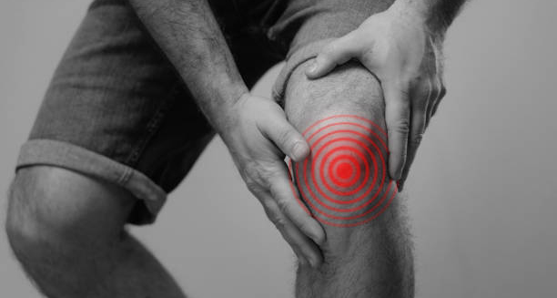
Man holding knee in pain with red target zone indicating dislocation complication

Complications of Dislocated Kneecap

Complications of Dislocated Kneecap The complications of dislocated kneecap can range from mild, short-term setbacks to serious, long-lasting damage if the injury is not properly managed. While most people recover with conservative treatment, some experience repeated dislocations, chronic pain, or reduced knee function over time. One of the most common complications of dislocated kneecap is patellar instability. After an initial dislocation, the ligaments and tissues that hold the kneecap in place may remain stretched or weakened. This increases the likelihood of future dislocations, even during everyday activities like walking or climbing stairs. Each subsequent dislocation causes more wear and tear on the joint and makes recovery harder. Cartilage damage is another major concern. When the kneecap slides out of place, it can scrape against the femur, damaging the cartilage that cushions the joint. This can lead to osteochondral injuries—where both bone and cartilage are damaged. In the long term, this wear can contribute to patellofemoral arthritis, causing chronic pain and stiffness. Another possible outcome is a fracture. Sometimes, the force of the dislocation causes small pieces of bone to break off, especially around the kneecap or femoral groove. These fragments may require surgical removal or fixation. If missed, they can float in the joint, causing catching or locking sensations, and further damage the cartilage. Recovering People recovering from a dislocated kneecap may also experience ongoing muscle weakness or atrophy, particularly in the quadriceps. If rehabilitation is delayed or incomplete, it can affect gait, balance, and the ability to return to sport. Scar tissue from previous injuries or surgeries can also restrict movement. Many people often overlook the psychological effects. After a traumatic injury like a dislocated kneecap, individuals—especially athletes—may develop fear of re-injury. This hesitation can interfere with recovery and reduce performance, particularly if the person avoids full engagement in rehab or physical activity. Complications of Dislocated Kneecap In South Africa, disparities in access to specialist care and physiotherapy can increase the risk of complications, particularly in public hospitals serving rural communities. Without consistent follow-up, patients may not complete rehabilitation, making recurrence and chronic knee instability more likely. Surgery can lead to complications such as infection, stiffness, or incorrect alignment if the surgeon does not properly stabilize the kneecap. That’s why patients should always follow surgical interventions with supervised rehabilitation and regular monitoring. Avoiding the complications of dislocated kneecap depends on early diagnosis, proper reduction, comprehensive physiotherapy, and ongoing patient education. With careful management, most individuals recover well and maintain strong, stable knees for the long term. [Next: Back to Overview →]

Complications of Dislocated Kneecap Read More »

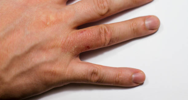
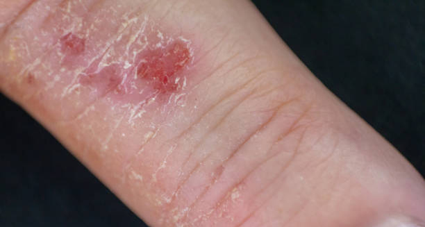
Close-up of red inflamed discoid eczema patch on the wrist

Discoid Eczema

Discoid Eczema Discoid eczema is a chronic skin condition characterised by coin-shaped patches of inflamed, itchy, and often scaly skin. These circular lesions usually appear on the arms, legs, or torso and may become infected if scratched excessively. While this eczema can affect people of any age. It is more commonly seen in adults and tends to be more severe during colder months or in dry climates. Unlike other forms of eczema that can appear more widespread, discoid eczema typically presents in well-defined patches. The condition is not contagious. However, its appearance can be distressing and cause discomfort. Especially when the lesions ooze, crust over, or leave behind dark or light pigmentation after healing. Although the exact cause of discoid eczema remains unknown. It is believed to result from a combination of genetic, environmental, and lifestyle factors. Common triggers include dry skin, stress, changes in weather, and the use of harsh soaps or skin irritants. People with a history of atopic eczema, asthma, or allergies may be more susceptible. In South Africa, the drier regions such as the Karoo or parts of the Free State can make managing this eczema more challenging due to the lack of humidity. Similarly, limited access to dermatological care in rural clinics can delay diagnosis and treatment, which in turn increases the risk of infection and scarring. Discoid Eczema Internationally, discoid eczema affects a significant number of individuals, particularly during autumn and winter when central heating and cold air can strip the skin of its natural oils. The condition is known by various names, including nummular eczema and nummular dermatitis, but all refer to the same circular pattern of inflamed skin. With early diagnosis and consistent treatment, most people can manage discoid eczema effectively. Moisturising regularly, avoiding known irritants, and using prescribed anti-inflammatory creams can reduce flare-ups and improve skin appearance. Though the condition may recur, long-term management strategies can make a significant difference in quality of life. [Next: Causes of Discoid Eczema →]

Discoid Eczema Read More »

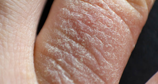
Dry, cracked skin on the hand indicating possible discoid eczema flare-up

Causes of Discoid Eczema

Causes of Discoid Eczema The causes of discoid eczema are not fully understood, but several risk factors and triggers are known to contribute to its development. The condition appears to result from an interaction between genetic predisposition, environmental exposure, and skin barrier dysfunction. When the skin becomes dry or damaged, it is more vulnerable to irritation and inflammation, leading to the characteristic round patches of discoid eczema. One of the primary causes of discoid eczema is excessively dry skin. When skin loses moisture, small cracks can form, allowing irritants or allergens to penetrate the outer layer. These substances can trigger an immune response, leading to inflammation, redness, and itching. Harsh soaps, alcohol-based sanitizers, and frequent washing with hot water can exacerbate this process. Skin trauma also plays a role. Even minor injuries like insect bites, scratches, or burns can spark a flare-up in people predisposed to discoid eczema. This response is called the Koebner phenomenon, where trauma leads to lesions forming at the site of injury. Other contributing causes of discoid eczema include emotional stress, hormonal changes, and climate. Stress is a well-known factor in many inflammatory conditions, and discoid eczema is no exception. Individuals under pressure—whether emotional or physical—often experience more frequent and intense flare-ups. In South Africa, extreme weather changes between hot dry seasons and damp winters can make managing skin hydration more difficult, especially in rural or informal communities with limited access to skincare products. Allergic contact dermatitis is another trigger. Exposure to allergens like nickel, fragrances, preservatives, or certain topical antibiotics can lead to or worsen discoid eczema. Identifying and avoiding these allergens is a key part of long-term management. Causes of Discoid Eczema Genetics also play a part. People with a family history of eczema, asthma, or hay fever are more likely to develop discoid eczema. This suggests a link between allergic tendencies and skin barrier weakness. Finally, infection can turn mild eczema into a more serious case. Scratching the itchy patches can break the skin, creating an entry point for bacteria like Staphylococcus aureus. Once infection sets in, symptoms become more severe and may include weeping sores, yellow crusts, and increased pain. Understanding the causes of discoid eczema can empower individuals to take proactive steps in prevention. By keeping skin moisturised, reducing exposure to irritants, and managing stress, many flare-ups can be avoided or significantly reduced. [Next: Symptoms of Discoid Eczema →]

Causes of Discoid Eczema Read More »

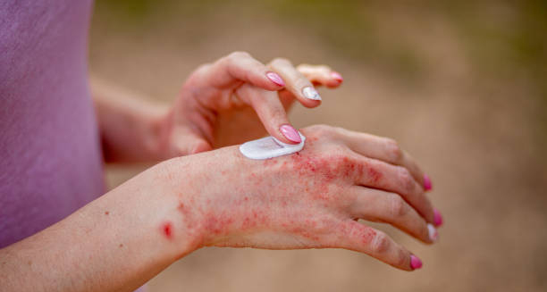
Close-up of red, inflamed patches on a hand showing symptoms of discoid eczema

Symptoms of Discoid Eczema

Symptoms of Discoid Eczema The symptoms of discoid eczema are usually easy to identify due to the distinctive round or oval-shaped patches that form on the skin. These lesions often start as small red spots or bumps that gradually enlarge and merge into larger discs. The affected areas become inflamed, scaly, itchy, and sometimes blistered or cracked. In some cases, the skin may ooze fluid, crust over, and later become thickened or discoloured. Early symptoms of discoid eczema include intense itchiness and irritation, which can quickly lead to scratching and skin damage. The round patches often have a well-defined border and may appear pink, red, or brown, depending on the individual’s skin tone. In lighter skin, the redness is more pronounced, while in darker skin, patches may look grey, dark brown, or purplish. Lesions can appear anywhere on the body, but they are most commonly found on the arms, legs, hands, and torso. The face and scalp are rarely affected. The symptoms of discoid eczema tend to appear symmetrically, often affecting both arms or both legs at the same time. Without treatment, the patches may last for weeks or even months and often recur in the same spots. Infected eczema is a serious concern. When bacteria enter the broken skin caused by scratching, additional symptoms develop. These include yellow crusting, swelling, warmth, and increased pain. Fever or feeling generally unwell may follow, indicating a systemic infection. In such cases, prompt medical attention is necessary, and antibiotics may be required. The symptoms of discoid eczema can significantly impact quality of life. Constant itchiness disrupts sleep, while visible patches may lead to embarrassment or social anxiety. This emotional toll can further worsen the condition, creating a cycle of stress and flare-ups. Symptoms of Discoid Eczema In South Africa, hot and dry weather, particularly in the inland provinces, can aggravate symptoms due to rapid skin dehydration. In contrast, humid climates may cause excessive sweating, which can irritate the skin and worsen the itch. Identifying symptoms of discoid eczema early is crucial. When treatment begins promptly, the risk of infection and long-term scarring is reduced. With the right skincare routine and medical guidance, many individuals manage the condition well and experience long periods without symptoms. [Next: Diagnosis of Discoid Eczema →]

Symptoms of Discoid Eczema Read More »

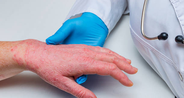
Doctor examining a patient’s inflamed hand for diagnosis of discoid eczema

Diagnosis of Discoid Eczema

Diagnosis of Discoid Eczema The diagnosis of discoid eczema is based largely on the appearance of the skin and a detailed medical history. Most healthcare providers can identify the condition by examining the coin-shaped patches, especially when they appear on the arms, legs, or trunk. However, because the lesions resemble other skin conditions, additional steps may be necessary to rule out similar diseases. During the initial consultation, doctors usually ask about the duration of symptoms, any known triggers, past episodes of eczema or allergies, and use of skincare products or medications. The patient’s personal and family history of atopic conditions—such as asthma or hay fever—can provide important clues. In many cases, the diagnosis of discoid eczema is clinical, meaning it is made without laboratory tests. Nevertheless, in uncertain cases, a skin biopsy may be done. This involves removing a small piece of affected skin under local anaesthetic and examining it under a microscope. A biopsy helps distinguish discoid eczema from other conditions like fungal infections, psoriasis, or skin cancers, which may present similarly. Doctors might also perform a swab test to check for bacterial infection, especially if the patches are weeping, crusted, or unusually painful. If infection is confirmed, antibiotics are often added to the treatment plan. In South Africa, limited access to dermatologists in public clinics may delay diagnosis, particularly in rural areas. As a result, general practitioners and nurses are trained to recognise common signs and begin early treatment. Another useful tool in the diagnosis of discoid eczema is patch testing. This method checks for allergic reactions to various substances that may be causing or aggravating the condition. It’s especially useful if contact dermatitis is suspected, and the patient reports exposure to nickel, fragrances, or other common allergens. Diagnosis of Discoid Eczema Photographs may also be taken to monitor progress over time. This is particularly helpful in chronic cases where flare-ups come and go. Regular check-ins with a healthcare provider ensure that treatment remains appropriate and effective. An accurate diagnosis of discoid eczema is essential to prevent unnecessary treatments, avoid worsening the condition, and offer proper guidance. Misdiagnosis can lead to delays in healing, especially if steroid creams are avoided or misused based on mistaken assumptions. Once diagnosed, a comprehensive plan is developed to manage symptoms, reduce triggers, and prevent future outbreaks. Patients are encouraged to keep a symptom diary, maintain a regular skincare routine, and follow up if symptoms persist or worsen. Early and accurate diagnosis can make a significant difference in controlling discoid eczema and improving overall skin health. [Next: Treatment of Discoid Eczema →]

Diagnosis of Discoid Eczema Read More »

Treatment of Discoid Eczema

Treatment of Discoid Eczema

Treatment of Discoid Eczema The treatment of discoid eczema involves a combination of lifestyle changes, topical medications, and in some cases, oral therapies. Because the condition is chronic and tends to recur, ongoing management is often necessary. The goal is to relieve symptoms, reduce inflammation, heal the skin, and prevent flare-ups. The first and most important step in the treatment of discoid eczema is restoring and maintaining the skin barrier. This means keeping the skin well-hydrated using emollients or moisturisers multiple times a day. Fragrance-free, thick creams or ointments are best, as they provide a strong protective layer. In South Africa, emollients are available over the counter, but affordability can be a barrier in underprivileged communities. Some clinics offer subsidised skincare products through public health programmes. Topical corticosteroids are commonly prescribed to reduce inflammation and itching. These are applied directly to the affected patches and can range in strength depending on severity. Mild eczema may respond to low-potency steroids, while more stubborn patches may need medium or high-potency creams. It is important to use these under medical guidance, especially on delicate areas or for prolonged periods. Seconday Infections If secondary infection is suspected, topical or oral antibiotics may be added. Crusting, weeping, or a yellowish appearance of lesions often indicates bacterial involvement, which doctors must address to allow healing. In some cases, doctors prescribe oral antihistamines to help reduce itching and improve sleep, especially for patients who scratch during the night. For more persistent or widespread cases, other options may be considered. These include topical calcineurin inhibitors, such as tacrolimus or pimecrolimus, which reduce inflammation without the side effects of steroids. In severe cases, dermatologists may use phototherapy or systemic immunosuppressants under close supervision. Lifestyle adjustments are also essential in the treatment of discoid eczema. Avoiding hot baths, using gentle cleansers, and wearing soft, breathable clothing all help reduce skin irritation. Identifying and managing triggers—such as allergens, stress, or certain fabrics—can greatly reduce the frequency and severity of flare-ups. Treatment of Discoid Eczema Education plays a key role in successful management. Patients must understand how and when to use medications, the importance of daily moisturising, and how to monitor for signs of infection. In South Africa, community health workers are often the first line of support in helping patients adhere to their skincare plans, especially in areas with limited access to dermatologists. With a tailored treatment plan, many individuals see significant improvement. Although the condition can be long-lasting, effective treatment of discoid eczema can reduce discomfort, improve appearance, and restore confidence. [Next: Complications of Discoid Eczema →]

Treatment of Discoid Eczema Read More »

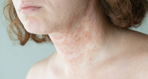
Discoid eczema on the neck showing severe skin inflammation and discolouration

Complications of Discoid Eczema

Complications of Discoid Eczema The complications of discoid eczema arise mainly from scratching, infection, and long-term skin damage. While the condition is not life-threatening, it can cause significant physical discomfort, emotional distress, and in some cases, permanent skin changes. Timely treatment and ongoing care are essential to prevent these complications from affecting quality of life. One of the most common complications of discoid eczema is secondary bacterial infection. When itchy patches are scratched, the skin’s protective barrier breaks down, allowing bacteria—especially Staphylococcus aureus—to enter. Infected eczema becomes swollen, red, warm, and may ooze yellow fluid. If left untreated, the infection can spread to other areas or lead to cellulitis, which sometimes requires hospital treatment. Long-term or repeated scratching can lead to lichenification, a thickening and hardening of the skin. These areas often become darker or lighter than the surrounding skin, and the texture can remain altered even after inflammation subsides. This scarring can cause cosmetic concerns, especially when lesions are visible on the arms or legs. Another potential outcome is post-inflammatory pigmentation. After the lesions heal, they may leave behind dark or light marks that can last for several months. This is particularly noticeable in people with darker skin tones, and although the pigmentation is not harmful, it can cause self-consciousness or distress. The complications of discoid eczema are not only physical. Many people experience emotional challenges related to the condition. Constant itching disrupts sleep, while the appearance of lesions may lead to social anxiety or embarrassment. Some individuals report depression or reduced self-esteem, particularly during prolonged flare-ups. Children with discoid eczema may struggle at school due to distraction from itching or discomfort. Adults may avoid social or professional interactions if the eczema is visible or misinterpreted as a contagious skin condition. These psychosocial effects are sometimes overlooked but are just as important to address in comprehensive care. Complications of Discoid Eczema In rare cases, long-term use of high-potency steroid creams without medical supervision can lead to skin thinning, stretch marks, or hormonal side effects. That’s why treatment should always be guided by a qualified healthcare provider who monitors for adverse effects. In South Africa, another complication is inconsistent access to care. People in rural or underserved areas may not receive a confirmed diagnosis or access to specialist treatments. This increases the risk of chronic skin damage, persistent symptoms, and poor disease control. Preventing the complications of discoid eczema begins with proper treatment, consistent skincare, and patient education. With the right support, most people can manage the condition effectively and avoid the more serious outcomes. [Next: Back to Overview →]

Complications of Discoid Eczema Read More »

3D medical illustration of diphtheria affecting the throat with highlighted inflammation

Diphtheria

Diphtheria Diphtheria is a dangerous bacterial illness that primarily targets the throat and nose but may also affect the skin and internal organs. Caused by Corynebacterium diphtheriae, this spreads from person to person through respiratory droplets, making it highly contagious in the absence of vaccination. While it’s rare in countries with established immunisation programmes, pockets of outbreaks still occur, especially where vaccination rates are low. The infection causes a thick grey membrane to form across the throat or tonsils, leading to serious breathing or swallowing difficulties. If untreated, the bacteria’s toxins can damage the heart, kidneys, and nervous system, sometimes fatally. This disease remains life-threatening without immediate treatment, particularly in vulnerable groups. Although largely eliminated in the UK and many Western countries, diphtheria still appears in areas of conflict, displacement, or poverty. In South Africa, routine immunisation through the public health system has significantly reduced the cases, but lapses in vaccine coverage have led to isolated outbreaks. These incidents highlight the need for ongoing vigilance and public education. Diphtheria Globally, vaccine hesitancy and uneven access to healthcare services are contributing to the disease’s re-emergence in some regions. Public health organisations stress the importance of the DTP (diphtheria, tetanus, pertussis) vaccine as a cornerstone of disease prevention. Despite progress, diphtheria continues to threaten communities with low immunisation uptake or declining booster compliance. Early recognition, immediate treatment, and strong immunisation efforts remain essential to control and eventually eliminate diphtheria on a global scale. [Next: Causes of Diphtheria →]

Diphtheria Read More »

Causes of Diphtheria

Causes of Diphtheria

Causes of Diphtheria The causes of diphtheria stem from infection by the bacterium Corynebacterium diphtheriae. This microbe releases toxins that damage tissues and disrupt normal bodily functions. The primary cause is airborne transmission—when an infected person coughs or sneezes, they release droplets that can easily infect others, particularly in crowded or unsanitary conditions. Direct contact with infected wounds or items such as shared utensils and towels can also spread the bacteria. These non-respiratory cases are usually linked to cutaneous diphtheria, which causes painful skin sores. Although less severe, this form remains contagious. Unvaccinated individuals are at the highest risk. In particular, young children, the elderly, and those with weakened immune systems are vulnerable. Globally, causes of outbreaks often involve lapses in public health systems, vaccine misinformation, or displacement from war and poverty. In South Africa, outbreaks have occurred in communities where immunisation services are inconsistent. While the diphtheria vaccine is part of South Africa’s public immunisation programme, barriers such as long distances to clinics, stock issues, or vaccine hesitancy sometimes prevent full coverage. This leaves some children and adults unprotected, increasing the likelihood of localised outbreaks. Causes of Diphtheria The most severe causes of diphtheria complications are linked to the bacterium’s toxin. Once in the bloodstream, the toxin can travel to the heart, nervous system, and kidneys. These systemic effects explain why diphtheria can be deadly if not treated immediately. Understanding the causes of diphtheria is key to preventing its spread. Public health education, consistent immunisation, and infection control measures remain the most effective tools to address both transmission and complications. [Next: Symptoms of Diphtheria →]

Causes of Diphtheria Read More »

Scroll to Top