Mental Matters

Author name: admin

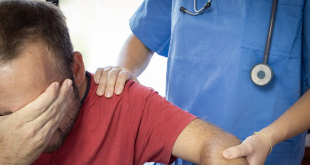
Distressed man holding his face while a healthcare professional provides support

Dislocated Shoulder

Dislocated Shoulder A dislocated shoulder occurs when the upper arm bone (humerus) pops out of the shoulder socket (glenoid). It is one of the most common joint dislocations seen in both sports injuries and everyday accidents. A dislocated shoulder can happen in many ways—from falling onto an outstretched arm to receiving a heavy blow during contact sports. It often results in sudden, severe pain and visible shoulder deformity. The shoulder is the body’s most mobile joint, which also makes it the most unstable. Its wide range of motion means that even a minor force applied in the wrong direction can push the ball of the joint out of its socket. Most injuries are anterior, meaning the bone moves forward out of the joint. Less commonly, the shoulder can dislocate backwards or downwards, depending on the type of trauma. Once dislocated, it may swell quickly and become very painful. Moving the arm becomes extremely difficult, and numbness, tingling, or weakness may spread down the arm. The dislocation often stretches or tears surrounding ligaments and tendons, increasing the risk of future instability or further injury. Dislocated Shoulder In South Africa, dislocated shoulder injuries are regularly seen in rugby, mountain biking, trail running, and mining-related accidents. Delayed treatment or repeated dislocations can lead to chronic shoulder instability, making early care vital. In areas with limited orthopaedic services, this can sometimes delay proper reduction and rehabilitation. Internationally, shoulder dislocations are common in both athletic and non-athletic populations. Older adults may experience them from simple falls, while young people more often dislocate their joints during high-speed or contact activities. Once a shoulder has dislocated, there’s a higher risk it will happen again, especially without proper rehabilitation. Understanding the nature of the injury, how to manage it, and how to prevent recurrence is essential for long-term shoulder health. [Next: Causes of Dislocated Shoulder →]

Dislocated Shoulder Read More »

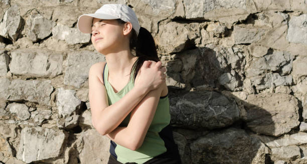
Man clutching his shoulder in pain after possible injury during outdoor activity

Causes of Dislocated Shoulder

Causes of Dislocated Shoulder The causes of dislocated shoulder injuries are varied, but most involve sudden trauma or excessive force applied to the shoulder joint. The shoulder’s mobility makes it vulnerable to being forced beyond its natural limits, especially during high-impact or awkward movements. A common cause of dislocated shoulder is falling directly onto an outstretched hand or elbow, which transmits force to the shoulder and forces the humeral head out of the socket. Contact sports, such as rugby, American football, and hockey, are leading contributors to shoulder dislocations. A heavy tackle or fall can easily dislodge the joint. Non-contact sports like gymnastics, swimming, and volleyball can also be involved, particularly when athletes land awkwardly or overstretch the shoulder. In South Africa, rugby and soccer-related shoulder injuries are widespread, particularly among school-age athletes. Vehicle accidents and workplace injuries are also well-documented causes of dislocated shoulder events. A sudden jolt, a fall from height, or being struck by a heavy object can dislocate the joint. In mining, agriculture, and construction industries—common sectors in South Africa—the physical demands and risk of falls or accidents make shoulder dislocations a regular occurrence. Some individuals are more prone to shoulder dislocation due to anatomical factors. These include naturally loose ligaments, a shallow socket, or previous injury to the shoulder joint. Recurrent dislocations are often seen in people who had their first dislocated shoulder in adolescence or early adulthood. The supporting structures never fully recover, leading to increased vulnerability. Genetics may also contribute. People with conditions such as hypermobility syndrome, Ehlers-Danlos syndrome, or connective tissue disorders are more likely to suffer joint dislocations, including at the shoulder. Their ligaments are more elastic, and joints are more prone to instability. Causes of Dislocated Shoulder Poor conditioning or muscle weakness can also play a role. Weak shoulder muscles may not offer enough support during physical activities, allowing the joint to slip out. Additionally, improper lifting techniques or sudden movements in gym environments are common risk factors. In older adults, dislocated shoulder injuries are often due to simple falls. As bone density decreases with age, the risk of fractures or dislocations increases even with low-impact trauma. In South Africa, where uneven pathways and unsafe stairwells are common in some urban and rural areas, elderly individuals may be at higher risk for shoulder injuries during everyday activities. Understanding the causes of dislocated shoulder can help prevent future injuries. Whether through proper training, workplace safety, or strengthening exercises, addressing risk factors reduces the chances of dislocation and improves joint stability. [Next: Symptoms of Dislocated Shoulder →]

Causes of Dislocated Shoulder Read More »

Middle-aged woman clutching painful shoulder with visible signs of dislocation

Symptoms of Dislocated Shoulder

Symptoms of Dislocated Shoulder The symptoms of dislocated shoulder usually begin instantly at the time of injury. Individuals often feel a sharp, intense pain in the shoulder, followed by an immediate inability to move the arm. A dislocated shoulder typically causes visible changes in the contour of the shoulder, with the joint appearing out of place or misshapen. In some cases, the shoulder may droop or look square instead of rounded. Other common symptoms of dislocated shoulder include swelling, bruising, and a feeling that the shoulder has “popped out” of position. Many people report hearing or feeling a snapping sound at the moment of injury. Attempting to move the arm may increase pain and make the shoulder feel unstable or wobbly. Tingling, numbness, or weakness in the arm, hand, or fingers may also occur if nerves are compressed or stretched during the dislocation. In more serious cases, the dislocation may damage blood vessels or the brachial plexus, a complex network of nerves supplying the arm. These symptoms should always be taken seriously and assessed by a healthcare provider. Other Symptoms A visible lump or bulge in the front or back of the shoulder is often noticeable, especially in leaner individuals. The arm is typically held close to the body in a protective position, with the elbow slightly bent. Any attempt to rotate or raise the arm tends to be extremely painful. Furthermore, it may increase the risk of further damage. In the case of recurrent dislocations, the symptoms may be less severe. The shoulder may slip in and out of place more easily, often with minimal trauma. These episodes still result in pain, swelling, and instability but may not involve as much visible deformity. In South Africa, where sports injuries are common. Symptoms of dislocated shoulder are often first treated at local clinics or sports events. Early identification and referral to a hospital or orthopaedic specialist are essential. For proper care and to prevent complications like fractures or long-term instability. Symptoms of Dislocated Shoulder It is important to differentiate shoulder dislocation from other injuries like rotator cuff tears. Additionally, fractures or shoulder impingement which may present with similar symptoms. Medical evaluation, including X-rays or an MRI, is usually required to confirm the diagnosis and determine the severity of the damage. Recognising the symptoms of dislocated shoulder and seeking prompt treatment reduces the risk of further injury and ensures a more successful recovery. Ignoring the warning signs or self-adjusting the joint can lead to long-term problems and chronic shoulder dysfunction. [Next: Diagnosis of Dislocated Shoulder →]

Symptoms of Dislocated Shoulder Read More »

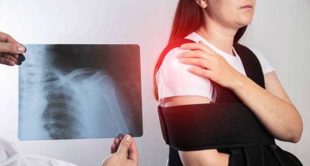
X-ray diagnosis of dislocated shoulder with woman wearing a shoulder brace

Diagnosis of Dislocated Shoulder

Diagnosis of Dislocated Shoulder The diagnosis of dislocated shoulder usually begins with a physical examination and a review of how the injury occurred. Most dislocations are easily recognisable due to the visible deformity of the shoulder, severe pain, and restricted movement. However, medical evaluation is still essential to confirm the diagnosis and assess any related damage. Such as fractures, torn ligaments, or nerve injury. A dislocated shoulder must be treated with care to avoid complications. During the examination, the healthcare provider will inspect the shoulder for signs of asymmetry, swelling, or unusual positioning. The patient is often unable to lift or rotate the arm. Any attempt to move the joint typically causes pain or resistance. In many cases, the shoulder appears “flattened” on one side. With a noticeable bulge at the front or back where the humeral head has shifted. The next step in the diagnosis of dislocated shoulder is imaging. X-rays are the standard test used to confirm whether the shoulder is dislocated and to check for associated fractures. In some cases, small bone fragments may break off during the dislocation, which can only be detected on a radiograph. An MRI may be ordered if soft tissue damage is suspected. Such as tears in the rotator cuff, labrum, or surrounding ligaments. X-Rays in SA In South Africa, X-rays are typically available in hospitals and larger clinics, though access may be limited in rural areas. Patients in these regions are often referred to regional hospitals for imaging, which can cause delays. Despite this, early and accurate diagnosis of dislocated shoulder remains critical to proper management and full recovery. In cases of recurrent dislocations, doctors may perform additional assessments to check for chronic instability. These include special physical tests to determine if the joint can be easily dislocated again. Identifying underlying anatomical abnormalities—such as a shallow shoulder socket or weak supporting muscles—can help inform the long-term treatment plan. Diagnosis of Dislocated Shoulder Doctors also check for nerve or blood vessel damage. Particularly if the patient experiences numbness, tingling, or coldness in the affected arm. The axillary nerve, which runs near the shoulder joint, is particularly at risk during dislocations and must be assessed carefully. Once the diagnosis of dislocated shoulder is confirmed, the medical team proceeds with reducing the dislocation—gently guiding the bone back into place—and developing a treatment strategy. This includes managing pain, preventing recurrence, and strengthening the joint through physiotherapy. An accurate diagnosis ensures that no complications are missed and that the patient receives the most appropriate care based on the severity and type of dislocation. [Next: Treatment of Dislocated Shoulder →]

Diagnosis of Dislocated Shoulder Read More »

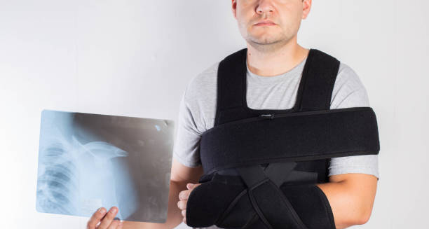
Man in shoulder brace holding X-ray after treatment for dislocated shoulder

Treatment of Dislocated Shoulder

Treatment of Dislocated Shoulder The treatment of dislocated shoulder focuses first on reducing the dislocation, followed by pain management, rehabilitation, and in some cases, surgical intervention. Reducing the dislocation means guiding the humeral head back into its socket. A trained medical professional performs this procedure, often using sedation or muscle relaxants to ease the process and reduce discomfort. Once the shoulder is back in place, doctors immobilise the arm with a sling or brace to allow the ligaments and soft tissues to heal. The immobilisation period usually lasts one to three weeks, depending on the severity of the injury and the patient’s overall health. During this time, doctors advise patients to rest the shoulder and avoid overhead movements. Doctors manage pain and inflammation with non-steroidal anti-inflammatory drugs (NSAIDs) like ibuprofen. Apply ice packs several times a day during the first 48 to 72 hours to reduce swelling. After the initial phase, the focus of treatment of dislocated shoulder shifts towards physiotherapy. Rehabilitation is crucial to restoring strength and mobility. Physiotherapists guide patients through exercises designed to strengthen the rotator cuff and shoulder stabilisers. Range-of-motion work helps prevent stiffness, while resistance training improves muscle balance. A comprehensive rehab programme can also address any muscular imbalances that contributed to the dislocation. Surgeons may recommend surgery for recurrent dislocations or associated structural damage. Surgical treatment of dislocated shoulder may involve tightening stretched ligaments, repairing torn labral tissue, or even bone grafting to deepen the shoulder socket. Surgeons often use arthroscopic procedures because they offer quicker recovery and less post-operative discomfort. Treatment of Dislocated Shoulder In South Africa, surgical options are available through both public and private healthcare systems. However, public hospitals may have longer waiting lists, especially for elective procedures like shoulder stabilisation surgery. Rural communities often face challenges accessing follow-up physiotherapy because ongoing rehab services may be limited. Doctors may recommend bracing during sports or high-risk activities, especially for individuals with a history of dislocation. Long-term treatment focuses on preventing recurrence, and athletes are often encouraged to work with trainers or physiotherapists to adjust their techniques and strengthen vulnerable areas. With the right treatment of dislocated shoulder, most patients regain full function within a few months. However, ignoring rehabilitation or returning to activity too early increases the risk of repeat injuries and long-term joint instability. [Next: Complications of Dislocated Shoulder →]

Treatment of Dislocated Shoulder Read More »

Complications of Dislocated Shoulder

Complications of Dislocated Shoulder

Complications of Dislocated Shoulder The complications of dislocated shoulder can vary depending on the severity of the injury, the speed of treatment, and whether the dislocation recurs. Although many people recover well, some may experience ongoing shoulder instability, chronic pain, or nerve damage. Proper management is vital to reduce the risk of long-term issues after a dislocated shoulder. One of the most common complications is recurrent dislocation. After the initial injury, the ligaments and tissues around the shoulder may remain stretched or torn, reducing the joint’s ability to stay in place. This is particularly common in younger individuals and athletes who return to activity too quickly. Repeated dislocations can lead to progressive damage and weaken the shoulder further. Another major complication is damage to cartilage and bone. During a dislocated shoulder, the humeral head may scrape against the socket, tearing the labrum (a ring of cartilage) or causing small fractures. Over time, this wear and tear can lead to shoulder arthritis, especially if left untreated. Cartilage injuries are difficult to heal and may require surgical repair. Nerve damage is another risk, especially to the axillary nerve, which wraps around the shoulder joint. When this nerve is stretched or compressed during dislocation, it can cause numbness, tingling, or muscle weakness in the upper arm. While most nerve injuries are temporary, some can lead to long-term weakness if not identified and treated early. Vascular injury is rare but serious. A dislocated shoulder can compress or tear nearby blood vessels, leading to reduced blood flow to the arm. Symptoms such as coldness, paleness, or absence of a pulse require urgent medical attention. Older adults are more vulnerable to vascular complications due to reduced tissue elasticity. Psychological complications are also worth noting. After a traumatic dislocation, some individuals develop a fear of re-injury or lose confidence in their shoulder’s stability. This can lead to activity avoidance, reduced physical fitness, and poor mental well-being. Support from physiotherapists and mental health professionals can help address these concerns. Complications of Dislocated Shoulder In South Africa, the complications are sometimes compounded by delayed access to specialist care or rehabilitation. Rural patients may receive emergency care for the dislocation but not the follow-up therapy needed to restore full function. Without guided rehab, the risk of long-term complications increases. Surgical complications, such as infection, stiffness, or hardware failure, may occur in patients undergoing corrective procedures. However, these risks are generally low when surgeries are performed in accredited facilities and followed by proper rehabilitation. Preventing the complications of dislocated shoulder involves early intervention, thorough rehabilitation, and attention to long-term joint health. With the right care, most individuals can return to their normal activities with minimal or no lasting effects. [Next: Back to Overview →]

Complications of Dislocated Shoulder Read More »

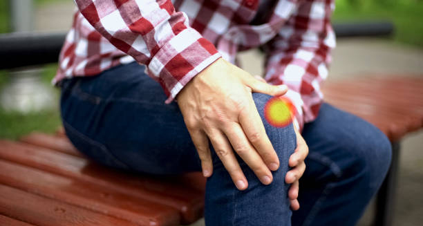
Man sitting on bench clutching knee with visible pain indicating dislocated kneecap

Dislocated Kneecap

Dislocated Kneecap A dislocated kneecap occurs when the patella. Which normally sits in a groove at the front of the knee joint, slips out of place, usually to the side. This injury often happens suddenly during sports or physical activity, particularly when the knee is twisted or experiences a direct blow. A dislocated kneecap can be both painful and disabling. Making walking difficult and sometimes causing long-term instability if not treated correctly. The kneecap plays an essential role in helping the thigh muscles straighten the leg. When the patella moves out of its groove, it can cause intense pain, swelling, and a visible deformity in the front of the knee. People may also feel the kneecap “pop” out and then back into place or find they cannot bend or straighten the knee normally. In some cases, the dislocation becomes a recurring problem, especially in individuals with loose ligaments or previous knee injuries. Although a dislocated kneecap can affect anyone. It is more common in teenagers and young adults, especially those who are active in contact sports or activities requiring rapid changes in direction. Women are also more likely to experience this injury due to differences in anatomy and muscle alignment. People with hypermobility syndromes or shallow patellar grooves are also at greater risk. In South Africa, where rugby, netball, and athletics are popular among youth, knee injuries—including patellar dislocations—are commonly seen in emergency departments. Immediate attention from medical professionals helps reduce complications and ensures proper management of the injury. Dislocated Kneecap Globally, the increasing popularity of recreational sports means that dislocated kneecap cases are becoming more frequent. Fortunately, most individuals recover well with prompt diagnosis, appropriate treatment, and rehabilitation exercises to strengthen the muscles around the knee and prevent recurrence. Understanding what causes this injury, how to identify it, and what treatment options are available can make a big difference in long-term knee health. [Next: Causes of Dislocated Kneecap →]

Dislocated Kneecap Read More »

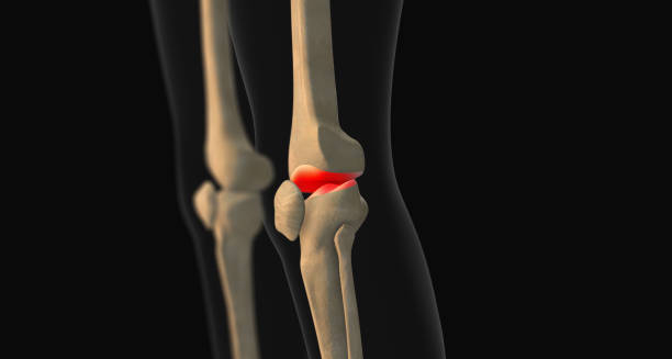
3D illustration showing dislocated kneecap in human knee anatomy

Causes of Dislocated Kneecap

Causes of Dislocated Kneecap The causes of dislocated kneecap injuries are most commonly linked to trauma or sudden movements that force the patella out of its normal groove. This can happen when a person plants their foot and twists the leg, especially during high-speed or pivoting activities. A direct blow to the knee, such as a tackle in rugby or a fall during running, can also lead to a dislocated kneecap. Sports that involve quick directional changes, jumping, or impact—such as football, basketball, and hockey—frequently see cases of this injury. Gymnasts and dancers, who require high levels of flexibility and knee rotation, may also be at increased risk. However, not all dislocations are due to high-impact incidents. Some people have underlying anatomical issues that make their kneecap more likely to dislocate. One of the most significant causes of dislocated kneecap is a shallow femoral groove—the channel in the thigh bone that guides the patella during movement. If this groove is not deep enough, the patella can easily slide out of place. Weakness in the quadriceps muscles, which help stabilise the knee, can also contribute. When these muscles aren’t strong enough to hold the kneecap in position, even minor stress can lead to dislocation. Ligament laxity is another major factor. People with naturally looser ligaments, such as those with hypermobility disorders, are more prone to joint dislocations, including the patella. Genetics can also play a role, with some families more likely to experience joint instability or abnormal knee alignment. Poor footwear, inadequate warm-up, and uneven terrain may further increase the risk, particularly in sports settings. In South Africa, where many rural or township schools lack proper playing surfaces or athletic footwear, children and adolescents may be more vulnerable to knee injuries, including dislocated kneecap episodes. Causes of Dislocated Kneecap Additionally, prior dislocations make future ones more likely. Once the ligaments and tissues around the patella have been stretched or torn, they are less capable of holding the kneecap in its proper position. Without adequate rehabilitation, this can become a chronic issue. Identifying the underlying causes of dislocated kneecap helps guide prevention strategies and treatment plans. Whether the injury stems from sports, anatomy, or repetitive strain, addressing these factors is key to reducing the chance of recurrence and maintaining knee stability over time. [Next: Symptoms of Dislocated Kneecap →]

Causes of Dislocated Kneecap Read More »

Woman holding her knee in pain, showing symptoms of a dislocated kneecap

Symptoms of Dislocated Kneecap

Symptoms of Dislocated Kneecap The symptoms of dislocated kneecap are usually immediate and unmistakable. When the patella slips out of place, individuals often feel a sharp, sudden pain in the front of the knee, followed by swelling and visible deformity. A dislocated kneecap may cause the entire knee to look misshapen or as if the kneecap is sitting off to one side. A common sign is the sensation of something “popping out” or shifting within the knee joint. Some people report hearing a snapping sound at the time of injury. The knee often gives way or buckles, making it nearly impossible to bear weight. In many cases, the kneecap remains dislocated until a healthcare professional manually puts it back into place—a process called reduction. In some cases, the patella may relocate spontaneously, but pain and swelling persist. Other typical symptoms of dislocated kneecap include limited range of motion, tenderness around the kneecap, and bruising that may develop after a few hours. The joint may feel unstable or wobbly, particularly when attempting to stand or walk. Muscle spasms around the knee are also common, as the body tries to protect the injured area. After the initial injury, the knee often becomes stiff and swollen. Pain tends to worsen with movement, and some individuals may develop a fear of bending or placing weight on the knee. If a piece of bone or cartilage breaks off during the dislocation—known as an osteochondral injury—locking or catching of the joint may occur. In younger athletes, especially those involved in high-impact sports, these symptoms of dislocated kneecap may mimic other injuries such as ligament tears or meniscus damage. That’s why a proper medical assessment is essential. Imaging tests, such as X-rays or MRIs, help confirm the dislocation and identify any associated fractures or soft tissue damage. Symptoms of Dislocated Kneecap In South Africa, access to prompt diagnosis may vary depending on whether a person visits a private or public healthcare facility. In rural areas, delays in receiving imaging or orthopaedic assessment can lead to complications if the kneecap is not properly realigned. Recognising the symptoms of dislocated kneecap quickly allows for timely treatment and reduces the risk of long-term joint instability. With appropriate care, most people make a full recovery, but ignoring symptoms can result in repeat dislocations or chronic knee problems. [Next: Diagnosis of Dislocated Kneecap →]

Symptoms of Dislocated Kneecap Read More »

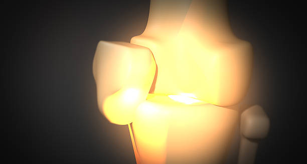
3D medical illustration showing dislocated kneecap in glowing joint anatomy

Diagnosis of Dislocated Kneecap

Diagnosis of Dislocated Kneecap The diagnosis of dislocated kneecap begins with a physical examination and a detailed account of how the injury occurred. Doctors often recognise the condition immediately based on visible signs—such as an obvious misalignment of the kneecap, swelling, and the patient’s inability to straighten or bear weight on the affected leg. However, confirming the full extent of the injury and ruling out associated damage requires further evaluation. In the early stage of diagnosis of dislocated kneecap, the medical professional may attempt gentle palpation of the knee to assess pain points and determine if the patella is still dislocated. If the kneecap has already returned to position on its own, swelling, bruising, and tenderness may still provide strong clues. Nevertheless, imaging is essential to assess the condition fully. An X-ray is often the first imaging test performed. It helps confirm the position of the kneecap and check for fractures or loose bone fragments. In some cases, dislocation can cause part of the bone or cartilage to break off—a complication that needs surgical attention. If soft tissue injuries are suspected, such as ligament tears or cartilage damage, an MRI (magnetic resonance imaging) may be ordered for a more detailed view. In South Africa, access to MRI can vary. While private medical facilities often provide immediate imaging, public hospitals may experience delays due to limited resources. Despite this, timely diagnosis of dislocated kneecap is crucial for effective treatment and to prevent repeat injuries. Doctors also assess knee stability after the kneecap has been reduced (returned to position). They examine how well the patella moves within its groove and check the strength of surrounding muscles and ligaments. In some cases, additional conditions—like patellar tracking disorder or shallow trochlear grooves—may be identified during this process, which could explain why the dislocation happened. Diagnosis of Dislocated Kneecap Children and adolescents may be referred to paediatric orthopaedic specialists, especially if the dislocation affects growth areas or if there’s concern about future instability. Athletes and active individuals might also receive sport-specific assessments to guide rehabilitation and return-to-play decisions. A complete diagnosis of dislocated kneecap is the foundation for building an effective recovery plan. It not only confirms the injury but also identifies any underlying structural problems that could cause recurrence. Early, accurate diagnosis ensures patients receive appropriate treatment, reducing long-term impact and supporting full return to daily activities or sport. [Next: Treatment of Dislocated Kneecap →]

Diagnosis of Dislocated Kneecap Read More »

Scroll to Top