Mental Matters

Author name: admin

Treatment of Diabetes

Treatment of Diabetes

Treatment of Diabetes Treatment of diabetes aims to control blood sugar levels, prevent issues, and promote overall health. Depending on the type and severity of the condition, diabetes treatment may involve lifestyle changes, medication, insulin therapy, or a combination of all three. A multidisciplinary approach—including GPs, endocrinologists, dietitians, and diabetes educators—is often required. Lifestyle Modifications Diet Emphasis on low-glycaemic index foods: whole grains, legumes, vegetables Limit sugary drinks, refined carbs, and processed snacks Carbohydrate counting helps manage meal-time glucose spikes Exercise Regular physical activity improves insulin sensitivity and lowers blood sugar Aim for at least 150 minutes per week of moderate aerobic activity Weight Management Losing 5–10% of body weight can significantly improve Type 2 diabetes control Even small reductions in weight have measurable benefits Smoking Cessation and Alcohol Moderation Smoking increases cardiovascular risks Alcohol can disrupt blood sugar balance, especially in insulin users Medications Type 1 Diabetes Requires insulin therapy from the point of diagnosis Multiple daily injections or insulin pump therapy Type 2 Diabetes First-line treatment is metformin Other oral or injectable medications include sulphonylureas, SGLT2 inhibitors, DPP-4 inhibitors, and GLP-1 agonists Insulin may be added if blood sugar remains uncontrolled Gestational Diabetes Managed through diet and exercise Insulin prescribed if lifestyle changes do not suffice Monitoring and Follow-Up Regular HbA1c tests to assess long-term control Self-monitoring of blood glucose (SMBG) helps track daily variations Continuous glucose monitors (CGMs) are increasingly used for real-time feedback Additional Support as Treatment of Diabetes Psychological support is vital to address diabetes-related distress and burnout Diabetes education empowers individuals with self-care skills In summary, treatment of diabetes involves a personalised blend of lifestyle change and medical intervention. Consistency, education, and support are key to achieving good glycaemic control and avoiding complications. [Next: Complications and Recovery from Diabetes →]

Treatment of Diabetes Read More »

Woman using a continuous glucose monitor on her arm to manage diabetes complications and recovery

Complications and Recovery from Diabetes

Complications and Recovery from Diabetes Complications and recovery from diabetes are closely tied to blood sugar management and early intervention. While diabetes is a chronic condition, many of its complications are preventable or manageable with proactive care. Recovery in this context refers not to curing diabetes, but to restoring and preserving quality of life. Acute Complications Hypoglycaemia (Low Blood Sugar) Symptoms: shakiness, confusion, sweating, blurred vision Causes: missed meals, excess insulin, intense exercise Immediate treatment: fast-acting carbohydrates (e.g., fruit juice, glucose tablets) Hyperglycaemia (High Blood Sugar) Symptoms: thirst, frequent urination, fatigue Prolonged hyperglycaemia can lead to: Diabetic ketoacidosis (DKA) in Type 1 Hyperosmolar hyperglycaemic state (HHS) in Type 2 Both conditions are medical emergencies requiring hospital care. Chronic Complications and Recovery from Diabetes Cardiovascular Disease Increased risk of heart attack and stroke Linked to hypertension and abnormal cholesterol Kidney Disease (Diabetic Nephropathy) Damaged blood vessels in the kidneys lead to protein leakage May progress to kidney failure requiring dialysis Eye Damage (Diabetic Retinopathy) High glucose damages retinal blood vessels Can result in blindness if untreated Nerve Damage (Neuropathy) Commonly affects feet and hands Leads to numbness, tingling, or pain Increases risk of foot ulcers and amputations Foot Complications Poor circulation and nerve damage lead to slow-healing wounds Annual foot checks are essential Recovery and Prevention Tight glucose control reduces the risk of complications by over 50% Early detection through screening improves long-term outcomes Regular check-ups for eyes, kidneys, feet, and heart are essential Living Well with Diabetes With structured care plans and informed self-management, most people can lead active lives Peer support groups and mental health care enhance recovery Remission is possible in some cases of Type 2 diabetes through significant lifestyle changes In conclusion, complications and recovery from diabetes hinge on early detection, education, and continuous care. With a holistic approach, individuals can avoid severe complications and maintain a high quality of life. [Next: Back to Overview →]

Complications and Recovery from Diabetes Read More »

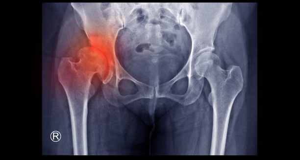
X-ray showing developmental dysplasia of the hip

Developmental Dysplasia of the Hip

Developmental Dysplasia of the Hip – Overview Developmental dysplasia of the hip (DDH) is a condition where the hip joint does not form properly in infants and young children. In this condition, the ball at the top of the thigh bone is not securely held in the socket of the pelvis. This can lead to instability, partial dislocation, or complete dislocation of the joint. Early detection and treatment are essential to avoid long-term mobility problems and joint degeneration. The hip joint is a ball-and-socket structure. In babies with DDH, the socket may be too shallow, or the ligaments holding the joint in place may be too loose. This abnormal development can begin before birth or continue to evolve after the child is born. The condition ranges in severity from mild instability to full dislocation. Additionally, it can affect one or both hips, although the left hip is more commonly involved. Developmental of this is more common in girls, first-born children, breech presentations. Additionally, in babies with a family history of hip problems. It is also more prevalent in cultures that practise tight swaddling of infants with extended legs. Which can restrict normal hip development. Developmental Dysplasia of the Hip When identified and managed early, most children with DDH develop normal hip function and avoid complications. However, if left untreated, the condition can lead to chronic pain, limping, leg length differences, and early-onset arthritis. In summary, developmental dysplasia of the hip is a condition that affects the normal development of the hip joint in infants and children. Prompt diagnosis and intervention can restore normal joint function and prevent future mobility issues. [Next: Causes of Developmental Dysplasia of the Hip →]

Developmental Dysplasia of the Hip Read More »

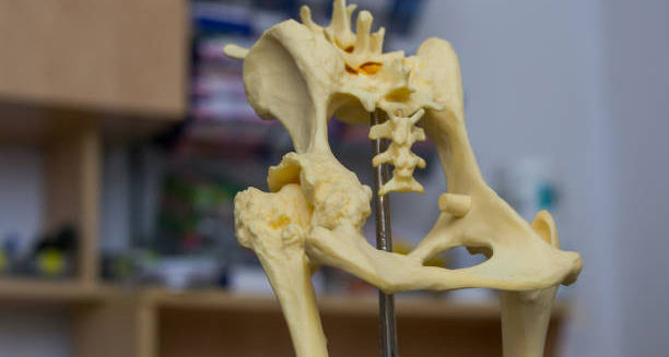
Anatomical model showing hip joint deformation

Causes of Developmental Dysplasia of the Hip

Causes of Developmental Dysplasia of the Hip Causes of developmental dysplasia of the hip are multifactorial, involving a combination of genetic, mechanical, and environmental factors. In many cases, the exact reason for developmental dysplasia of the hip is not clear. However, certain risk factors and developmental influences are strongly associated with the condition. Genetic Factors Family history plays a significant role. If a parent or sibling has had DDH, a child’s risk of developing the condition increases. Some studies suggest a genetic predisposition related to joint laxity. Where the ligaments and connective tissues are more elastic, allowing the hip to become unstable more easily. Mechanical and Positional Factors The position of the baby in the womb can influence hip development. Breech presentation (when the baby is born bottom-first rather than head-first) places additional pressure on the hips and limits their ability to move freely. This increased pressure can prevent the ball of the hip joint from settling properly in the socket. First-born babies are also at increased risk due to the tighter space in the uterus. Which may restrict movement and proper joint alignment. The left hip is more frequently affected because of the typical position of the foetus in the womb, with the left hip pressed against the mother’s spine. Postnatal Influences Certain cultural practices, particularly swaddling methods that keep a baby’s legs tightly extended and together, can contribute to the development or worsening of DDH. Swaddling that does not allow natural hip flexion and abduction can inhibit proper hip development. Hormonal Influence | Causes of Developmental Dysplasia of the Hip During pregnancy, maternal hormones like relaxin help loosen the mother’s joints in preparation for birth. These hormones may also affect the baby’s joints, especially in female infants, making their hip joints more susceptible to instability. In summary, the causes of developmental dysplasia of the hip are linked to genetics, in-utero positioning, hormonal effects, and cultural practices. Understanding these risk factors is key to prevention, early detection, and effective management. [Next: Symptoms of Developmental Dysplasia of the Hip →]

Causes of Developmental Dysplasia of the Hip Read More »

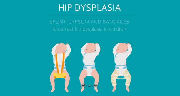
Illustration of infants with hip dysplasia braces

Symptoms of Developmental Dysplasia of the Hip

Symptoms of Developmental Dysplasia of the Hip Symptoms of developmental dysplasia of the hip can vary significantly depending on the age of the child and the severity of the condition. In many cases, particularly in newborns, developmental dysplasia of the hip does not cause any obvious signs and is only detected during routine screening. However, as a child grows, more noticeable symptoms may develop if the condition remains untreated. Symptoms in Newborns and Infants Hip instability: The hip may feel loose or dislocate with gentle movement during physical examination. Asymmetry in leg length: One leg may appear shorter than the other. Uneven skin folds: Extra or uneven folds may be noticeable on the thighs or buttocks. Limited range of motion: Difficulty in spreading the infant’s thighs apart during nappy changes. Audible ‘click’ or ‘clunk’: A sound or sensation during hip movement that may indicate dislocation. Routine screening by a healthcare provider includes the Ortolani and Barlow manoeuvres to detect any hip instability in the first few weeks of life. Symptoms in Older Infants and Toddlers Delayed walking: The child may begin walking later than expected. Limping or waddle: Walking may appear uneven or abnormal. Toe walking: The child may favour walking on their toes on the affected side. Exaggerated lumbar curve: An inward curve of the lower back (lordosis) may develop in bilateral DDH. In some cases, especially when the condition is mild, symptoms may be so subtle that they go unnoticed until a child begins walking or even later in childhood. Adolescent or Adult Symptoms | Symptoms of Developmental Dysplasia of the Hip If undiagnosed until adolescence or adulthood, individuals may experience: Hip or groin pain Stiffness and limited mobility Early onset osteoarthritis In conclusion, symptoms of developmental dysplasia of the hip are age-dependent and range from subtle instability in infants to noticeable gait problems in toddlers. Early screening and awareness of risk factors are essential for timely diagnosis and intervention. [Next: Diagnosis of Developmental Dysplasia of the Hip →]

Symptoms of Developmental Dysplasia of the Hip Read More »

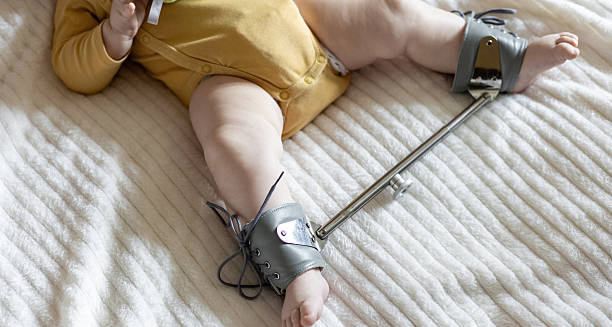
Infant wearing a Pavlik harness for hip dysplasia treatment

Diagnosis of Developmental Dysplasia of the Hip

Diagnosis of Developmental Dysplasia of the Hip Diagnosis of developmental dysplasia of the hip typically involves a combination of physical examination, imaging, and patient history. Since early detection dramatically improves outcomes, routine screening for developmental dysplasia of the hip is a standard part of newborn care in many countries. Clinical Examination In newborns and young infants, healthcare professionals perform specific manoeuvres during routine check-ups: Barlow test: The examiner attempts to gently dislocate the hip. Ortolani test: The examiner attempts to relocate a dislocated hip with a gentle ‘clunk.’ These tests are most effective within the first two to three months of life, as the infant’s muscles are still soft and flexible. As the child grows, the reliability of these tests decreases. Imaging Tests & Diagnosis of Developmental Dysplasia of the Hip When physical examination is inconclusive or the infant is at higher risk (e.g. breech birth, family history), imaging studies are used: Ultrasound: The preferred method for diagnosing DDH in infants under six months. It allows visualisation of the hip joint’s soft tissues and structure. X-ray: Used after six months of age, once the bones begin to ossify and become visible on radiographs. Screening Protocols Many countries follow national screening protocols, which may include: Newborn physical exams at birth and six-week check-ups Ultrasound screening for at-risk infants at 4–6 weeks Follow-up imaging at three to six months if abnormalities are detected Later Diagnosis In older children or adults presenting with gait issues or hip pain, diagnosis is based on clinical findings and X-rays. Delayed diagnosis often requires more invasive interventions. In summary, diagnosis of developmental dysplasia of the hip relies on early screening, clinical tests, and appropriate imaging. Timely diagnosis leads to more effective, less invasive treatment and significantly better long-term outcomes. [Next: Treatment of Developmental Dysplasia of the Hip →]

Diagnosis of Developmental Dysplasia of the Hip Read More »

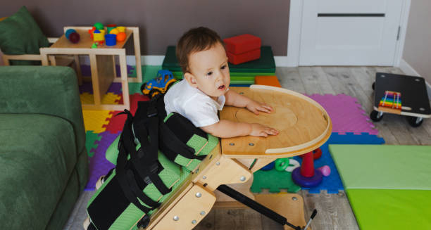
Toddler receiving physical therapy in a standing support chair for hip dysplasia

Treatment of Developmental Dysplasia of the Hip

Treatment of Developmental Dysplasia of the Hip Treatment of developmental dysplasia of the hip depends on the child’s age at diagnosis and the severity of the condition. Early intervention is highly effective and often non-surgical. When developmental dysplasia of the hip is detected in newborns, conservative methods like bracing usually suffice. Delayed cases may require surgery. Newborns to 6 Months Pavlik harness: A soft brace that holds the hips in a flexed, abducted position. It allows the joint to develop correctly while still permitting some movement. Treatment duration usually lasts 6–12 weeks, with periodic monitoring using ultrasounds. Success rates are high if the condition is detected early. 6 Months to 2 Years If bracing is ineffective or diagnosis is delayed, a closed reduction under general anaesthesia may be necessary. The hip is gently manipulated into place and held in position with a hip spica cast. Additional imaging such as an arthrogram may be used to confirm hip positioning during the procedure. Over 2 Years Open reduction surgery may be required to realign the joint if non-surgical methods fail or if the hip has become rigid. This is often followed by casting and sometimes further surgical interventions to reshape the hip socket or femur. Rehabilitation and Monitoring Children require regular follow-ups with imaging to ensure proper hip development. Physiotherapy may be recommended to restore full mobility and strength, particularly after casting or surgery. Outcomes | Treatment of Developmental Dysplasia of the Hip Most children treated early develop normal, functional hips. Delay in diagnosis can lead to increased need for surgical intervention and long-term complications. In conclusion, the treatment of developmental dysplasia of the hip is most successful when started early. With the right care, many children go on to lead active, pain-free lives. [Next: Complications and Recovery from Developmental Dysplasia of the Hip →]

Treatment of Developmental Dysplasia of the Hip Read More »

Mother carrying child in hip-healthy baby carrier during recovery from hip dysplasia

Complications and Recovery from Developmental Dysplasia of the Hip

Complications and Recovery from Developmental Dysplasia of the Hip Complications and recovery from developmental dysplasia of the hip depend heavily on the timing of diagnosis and the type of treatment used. With early intervention, developmental dysplasia of the hip often resolves completely. However, delayed or inadequate treatment can lead to persistent issues and long-term joint damage. Potential Complications Residual dysplasia: Incomplete formation of the hip socket even after treatment, requiring further monitoring or surgery. Avascular necrosis: Damage to the blood supply of the femoral head, leading to bone death and joint collapse. Re-dislocation: The hip may become unstable again after bracing or surgery. Leg length discrepancy: One leg may grow shorter than the other due to changes in hip alignment. Osteoarthritis: Poorly aligned or damaged hips are at higher risk of developing arthritis in early adulthood. Post-Treatment Recovery | | Complications and Recovery from Developmental Dysplasia of the Hip Most children recover well with proper follow-up and physical therapy. Bracing generally allows full return to mobility within weeks after treatment ends. Surgery typically requires a longer recovery, including casting and gradual reintroduction to weight-bearing activities. Psychological and Developmental Support Long-term casting or hospitalisation may affect a child’s emotional well-being or developmental milestones. Parental support, early intervention services, and occupational therapy can help children stay on track. Adult Considerations Adults who were untreated or inadequately treated as children may require hip replacement surgery due to chronic pain or severe arthritis. Regular check-ups can detect early signs of degeneration. In summary, complications and recovery from developmental dysplasia of the hip are largely preventable with early diagnosis and proper management. Ongoing follow-up ensures that the joint continues to develop correctly and minimises long-term problems. [Next: Back to Overview →]

Complications and Recovery from Developmental Dysplasia of the Hip Read More »

Cartoon illustration showing symptoms and treatment of a detached retina

Detached Retina

Detached Retina – Overview A detached retina is a serious eye condition where the retina—the thin layer of tissue at the back of the eye—pulls away from its normal position. This separation cuts off the retina’s supply of oxygen and nutrients, which can result in permanent vision loss if not treated promptly. A detached retina is considered a medical emergency and requires immediate attention from an ophthalmologist. The retina plays a vital role in vision. It captures light and converts it into electrical signals sent to the brain via the optic nerve. When the retina detaches, this process is disrupted, causing blurred vision, flashes of light, floaters, or a shadow across the field of vision. If left untreated, the condition can lead to total blindness in the affected eye. Retinal detachment most commonly occurs due to ageing, injury, or underlying eye diseases. The condition affects about 1 in 10,000 people each year, often in those aged 50 and older. However, younger individuals with high myopia (short-sightedness), a family history of retinal problems, or those who have had previous eye surgery are also at increased risk. Detached Retina There are three main types of retinal detachment: rhegmatogenous (caused by a tear or hole in the retina), tractional (where scar tissue pulls the retina away), and exudative (caused by fluid build-up beneath the retina without a tear). Each type requires different treatment approaches. In summary, a detached retina is a potentially sight-threatening condition that demands swift diagnosis and surgical intervention. Early recognition of symptoms and access to professional care significantly improve the chances of preserving vision. [Next: Causes of Detached Retina →]

Detached Retina Read More »

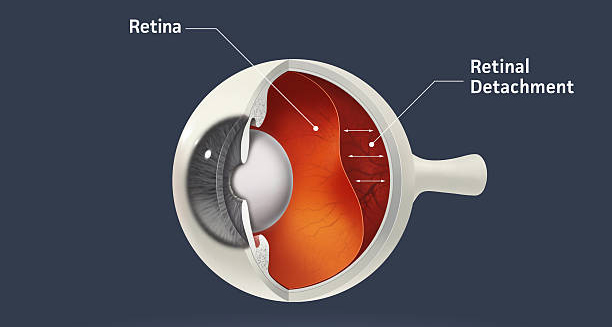
Cross-section diagram showing retinal detachment inside the eye

Causes of Detached Retina

Causes of Detached Retina Causes of detached retina are varied, but they all lead to the separation of the retina from the underlying layers that supply it with oxygen and nutrients. A detached retina can occur suddenly or gradually, and understanding the underlying causes can aid in prevention and early treatment. Rhegmatogenous Retinal Detachment This is the most common type and is caused by a tear or hole in the retina: Ageing: As people age, the vitreous gel inside the eye shrinks and may pull away from the retina, causing a tear Myopia (short-sightedness): People with severe myopia have longer eyeballs, which stretches the retina and makes it thinner and more prone to tearing Eye injury or trauma: Blunt or penetrating injuries can cause tears or breaks in the retina Previous eye surgery: Procedures like cataract removal increase the risk of retinal detachment, especially in the first year after surgery Once a tear occurs, fluid from the vitreous gel can seep through the opening and collect underneath the retina, causing it to lift off. Tractional Retinal Detachment | Causes of Detached Retina This form is often seen in people with: Diabetic retinopathy: Scar tissue from abnormal blood vessels pulls the retina away from its base Retinal vein occlusion: Blocked blood vessels may lead to fibrosis and traction on the retina Inflammatory conditions: Diseases like uveitis can result in scarring and traction This type develops slowly and is typically painless. Exudative Retinal Detachment This type occurs when fluid builds up beneath the retina without any tears or breaks: Tumours: Ocular melanoma or metastatic cancers can leak fluid under the retina Inflammation: Conditions like scleritis or Coats’ disease can cause abnormal fluid accumulation Systemic diseases: High blood pressure or kidney disorders may contribute to fluid leakage in the eye Other Risk Factors Family history of retinal detachment Lattice degeneration: Thinning of the peripheral retina, increasing vulnerability Extreme sports or sudden head movements: Though rare, can increase risk In conclusion, causes of detached retina span age-related degeneration, trauma, disease, and anatomical predisposition. Knowing the risk factors allows at-risk individuals to monitor their eye health and seek urgent care when symptoms arise. [Next: Symptoms of Detached Retina →]

Causes of Detached Retina Read More »

Scroll to Top