Mental Matters

Author name: admin

Treatment of Creutzfeldt-Jakob Disease

Treatment of Creutzfeldt-Jakob Disease

Treatment of Creutzfeldt-Jakob Disease Treatment of Creutzfeldt-Jakob disease focuses on supportive and palliative care, as there is currently no cure or disease-modifying therapy available. Because Creutzfeldt-Jakob disease progresses rapidly, the primary goals are comfort, symptom control, and maintaining dignity throughout the disease process. 1. Symptom Management Doctors use medications to alleviate distressing symptoms: 2. Nutritional Support As patients lose the ability to eat and swallow safely. A feeding tube can provide comfort and nutrition, but the decision depends on individual preferences. 3. Mobility and Safety | Treatment of Creutzfeldt-Jakob Disease Physiotherapists and occupational therapists may provide support during the early stages to reduce fall risk and encourage gentle movement. With proper nursing care, patients avoid bedsores and infections linked to immobility. 4. Psychological and Emotional Support Both patients and families require emotional support. Counselling, support groups, and hospice services are important resources to help families prepare for end-of-life decisions and cope with grief. 5. Palliative and End-of-Life Care As the disease progresses, most individuals require round-the-clock care. Hospice services focus on comfort, dignity, and emotional well-being. Clinicians and families should address artificial nutrition, hydration, and resuscitation decisions early in care planning. 6. Infection Control Although CJD is not contagious in everyday contact, special precautions are taken in hospitals during surgical procedures or post-mortem examination due to the durability of prions on instruments. Research is ongoing, exploring treatments that block prion propagation, but no therapies have proven effective to date. Ultimately, the treatment of Creutzfeldt-Jakob disease is centred on compassionate care and supporting both patient and family through a deeply challenging and rapid disease trajectory. [Next: Complications and Recovery from Creutzfeldt-Jakob Disease →]
Treatment of Creutzfeldt-Jakob Disease Read More »

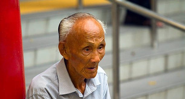
Elderly man sitting with a distant, solemn expression, symbolising cognitive and neurological decline associated with Creutzfeldt-Jakob disease.

Complications and Recovery from Creutzfeldt-Jakob Disease

Complications and Recovery from Creutzfeldt-Jakob Disease Complications and recovery from Creutzfeldt-Jakob disease must be understood in the context of a terminal illness. While Creutzfeldt-Jakob disease cannot be cured or reversed, supportive measures aim to reduce suffering and manage complications that arise during its rapid course. 1. Loss of Independence Patients quickly lose the ability to walk, talk, eat, and care for themselves. Most become bedbound within a few months, requiring full-time assistance for even the simplest tasks. 2. Communication Difficulties As cognitive and language abilities deteriorate, communication becomes limited. Speech therapists may offer early support, but in late stages, families rely on non-verbal cues and emotional presence. 3. Infections | Complications and Recovery from Creutzfeldt-Jakob Disease Immobilised patients are at high risk of chest infections, bedsores, and urinary tract infections. Pneumonia is a common cause of death. Good hygiene and positioning help reduce risk. 4. Seizures and Neurological Crises Frequent seizures, muscle rigidity, and startle responses may cause distress. These are controlled with medications but often worsen as the disease progresses. 5. Nutritional and Hydration Issues Difficulty swallowing and appetite loss lead to malnutrition and dehydration. Families must make difficult decisions about artificial feeding and end-of-life preferences. 6. Psychological and Social Impact CJD is traumatic for families, as loved ones often change dramatically within weeks. Counselling and hospice involvement are crucial for emotional support and grief management. Recovery Outlook Recovery from CJD is not possible. The disease progresses inevitably toward coma and death. However, with early diagnosis and excellent palliative care, many patients experience a peaceful and dignified end-of-life phase. In conclusion, complications and recovery from Creutzfeldt-Jakob disease must focus on maximising comfort, providing emotional support, and ensuring that patients pass with dignity. The rare and devastating nature of this disease underscores the importance of early recognition and compassionate care. [Next: Back to Overview →]

Complications and Recovery from Creutzfeldt-Jakob Disease Read More »

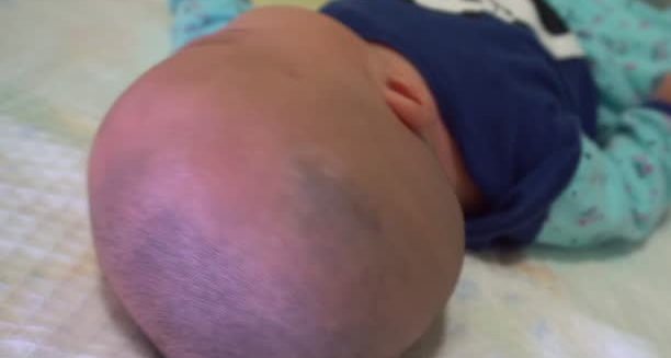
Close-up of an infant’s head from behind, showing abnormal skull shape suggestive of craniosynostosis.

Craniosynostosis

Craniosynostosis – Overview Craniosynostosis is a birth defect in which one or more of the sutures in a baby’s skull close too early. Before the brain has finished growing. This premature fusion affects the shape of the head and may increase pressure inside the skull. Craniosynostosis can vary in severity. Depending on how many sutures are involved and whether it occurs as part of a genetic syndrome or in isolation. The human skull is made up of several bones joined by flexible sutures. Furthermore, allowing the skull to expand as the brain grows. In craniosynostosis, these sutures fuse too early, forcing the brain to grow in the direction of the remaining open sutures. This results in an abnormally shaped head, and in severe cases, it can lead to developmental delays, visual impairment, and neurological issues. Craniosynostosis Craniosynostosis can be non-syndromic, which means it occurs without other abnormalities, or syndromic, where it is associated with genetic conditions such as Apert, Crouzon, or Pfeiffer syndrome. Non-syndromic cases are more common and typically involve just one suture. Early detection and treatment are crucial. In many cases, surgery is required to correct the shape of the skull and allow the brain to grow normally. When treated promptly, the long-term outlook is often positive. In summary, craniosynostosis is a condition involving premature skull suture fusion that affects head shape and brain development. With early diagnosis and appropriate intervention, most children go on to lead healthy lives. Importance of Early Intervention Early diagnosis of craniosynostosis is key to achieving the best possible outcomes. Pediatricians and specialists often detect abnormal head shapes during routine check-ups, prompting imaging tests to confirm the diagnosis. When addressed promptly — usually within the first year of life — surgical treatment can relieve pressure on the brain, correct deformities, and support normal development. Delays in treatment may increase the risk of complications, highlighting the importance of early intervention. Supporting Families and Long-Term Outlook Raising a child with craniosynostosis can be emotionally challenging for families, especially in syndromic cases requiring multiple surgeries or ongoing care. Support groups, genetic counselling, and coordinated medical care can ease the journey. Fortunately, with appropriate medical and surgical management, many children go on to reach developmental milestones and lead healthy, active lives. Advances in surgical techniques and multidisciplinary care continue to improve both cosmetic and functional outcomes. [Next: Causes of Craniosynostosis →]

Craniosynostosis Read More »

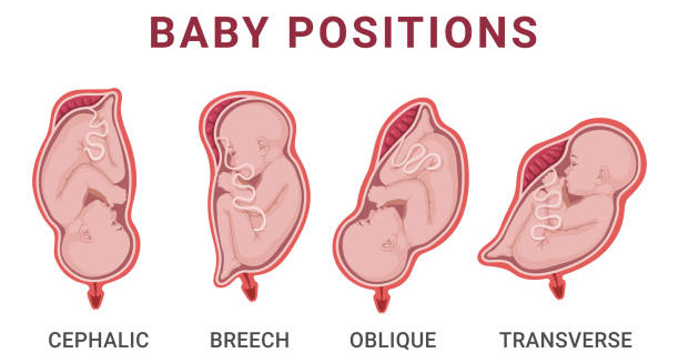
Illustration of various fetal positions in the womb, including cephalic, breech, oblique, and transverse, which may influence cranial development.

Causes of Craniosynostosis

Causes of Craniosynostosis Causes of craniosynostosis can be genetic, environmental, or a combination of both. While many cases arise without a known reason, understanding the underlying mechanisms can help parents and healthcare providers identify the condition early. Craniosynostosis occurs when one or more cranial sutures close too soon, disrupting normal skull and brain growth. Genetic Factors | Causes of Craniosynostosis Approximately 15–30% of cases are associated with known genetic syndromes, such as: Apert syndrome Crouzon syndrome Pfeiffer syndrome Muenke syndrome These syndromic forms of craniosynostosis often involve multiple sutures and come with other physical abnormalities, such as limb deformities or facial asymmetry. Mutations in genes such as FGFR2, FGFR3, and TWIST1 have been identified as common culprits. Non-Syndromic Causes In most cases, craniosynostosis occurs in isolation, without other medical conditions or inherited traits. These non-syndromic cases may result from: Spontaneous gene mutations Intrauterine constraints, such as limited space in the womb Abnormal foetal positioning Multiple pregnancies, which increase pressure on the baby’s head Environmental Factors While not as well-established, certain environmental factors may contribute to craniosynostosis, including: Maternal smoking Advanced paternal age Certain fertility treatments Use of specific medications during pregnancy More research is needed to fully understand how these factors interact with genetic predispositions. Prematurity and Metabolic Disorders Some premature infants are at greater risk due to altered bone development. Additionally, metabolic disorders affecting calcium or phosphate levels can influence early suture fusion. In conclusion, causes of craniosynostosis vary widely, ranging from genetic mutations to external pressures and unknown factors. Recognising risk factors helps guide screening, especially in newborns with abnormal head shapes or family history of related conditions. [Next: Symptoms of Craniosynostosis →]

Causes of Craniosynostosis Read More »

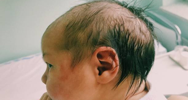
Side profile of an infant with an abnormal head shape and visible ridges on the skull, indicating symptoms of craniosynostosis.

Symptoms of Craniosynostosis

Symptoms of Craniosynostosis Symptoms of craniosynostosis typically become noticeable within the first few months of life, as the baby’s head begins to grow abnormally due to premature suture fusion. While some cases are immediately obvious, others may be subtler and detected only during routine health checks. Recognising the signs of craniosynostosis early is vital for ensuring timely treatment. Abnormal Head Shape The most prominent symptom is an unusual head shape that does not improve or changes asymmetrically over time. The shape depends on which suture is affected: Sagittal suture fusion causes a long, narrow head (scaphocephaly) Coronal suture fusion leads to a flat forehead on one side (plagiocephaly) Metopic suture fusion results in a pointed forehead (trigonocephaly) Lambdoid suture fusion causes flattening at the back of the head Unlike positional plagiocephaly (flat head syndrome), the skull in craniosynostosis feels rigid or ridged over the affected suture. Palpable Ridge | Symptoms of Craniosynostosis Parents or doctors may feel a raised, firm ridge along the fused suture line. This is caused by the overlapping of bones where the suture has prematurely closed. Facial Asymmetry Some children develop visible facial imbalance, such as uneven eye placement, misaligned ears, or a shifted jawline, particularly in more severe or syndromic cases. Slow or No Head Growth As the skull cannot expand normally, the baby’s head circumference may not increase at the expected rate. Head growth should be carefully monitored during routine paediatric check-ups. Neurological Symptoms (in advanced cases) Increased intracranial pressure Developmental delays Irritability, vomiting, or poor feeding Bulging fontanelle Seizures (rare) In summary, symptoms of craniosynostosis include an abnormal head shape, palpable ridges, and possible developmental issues. Identifying these signs early can lead to prompt intervention and better outcomes. [Next: Diagnosis of Craniosynostosis →]

Symptoms of Craniosynostosis Read More »

Paediatric doctor showing an X-ray to a mother and child during a consultation, representing craniosynostosis diagnosis.

Diagnosis of Craniosynostosis

Diagnosis of Craniosynostosis Diagnosis of craniosynostosis typically begins with a visual and physical assessment of the infant’s skull shape and growth pattern. Since craniosynostosis often presents with noticeable head asymmetry or ridges along the sutures, early detection is often possible during routine check-ups in the first months of life. Physical Examination A paediatrician will: Examine the baby’s head shape and symmetry Feel for ridges or hardened sutures Measure head circumference and compare it to age-based growth charts Assess fontanelles (soft spots) for early closure The presence of a non-growing or oddly shaped skull, combined with a visible or palpable ridge, often prompts further investigation. Imaging Tests | Diagnosis of Craniosynostosis To confirm the diagnosis and assess the extent of the condition, the following imaging tools are used: X-rays: May show early suture closure or overlapping skull bones CT scans with 3D reconstruction: The gold standard for confirming craniosynostosis, showing detailed skull structure and fused sutures MRI: Used if there are concerns about brain development or soft tissue involvement Genetic Testing In suspected syndromic cases—where craniosynostosis is accompanied by limb abnormalities, facial differences, or developmental delays—genetic testing may be recommended. This can identify mutations in genes like FGFR2, FGFR3, or TWIST1 and help guide long-term care planning. Differential Diagnosis Doctors must distinguish craniosynostosis from other causes of abnormal head shape: Positional plagiocephaly: Caused by lying in the same position, but sutures remain open Hydrocephalus: Enlarged head due to fluid build-up Other cranial malformations or syndromes Multidisciplinary Involvement Diagnosis is usually confirmed and managed through a team that may include: Paediatricians Neurosurgeons Craniofacial surgeons Geneticists In summary, diagnosis of craniosynostosis is based on clinical examination and imaging. Accurate diagnosis ensures proper treatment planning and helps prevent complications related to increased intracranial pressure or delayed brain development. [Next: Treatment of Craniosynostosis →]

Diagnosis of Craniosynostosis Read More »

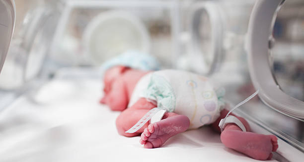
Newborn baby lying in an incubator with medical monitoring, representing early treatment for craniosynostosis.

Treatment for Syndromic Craniosynostosis

Treatment of Craniosynostosis Treatment of craniosynostosis depends on the severity of the condition, which sutures are affected, and whether it is syndromic or non-syndromic. The main goal of treating craniosynostosis is to correct skull shape, relieve any pressure on the brain, and allow for normal brain growth and development. Surgical Intervention Most moderate to severe cases require surgery, ideally performed before 12 months of age to take advantage of the skull’s natural flexibility. Types of Surgery Cranial vault remodelling: Involves removing and reshaping affected parts of the skull Endoscopic strip craniectomy: A minimally invasive option for infants under six months, involving small incisions and removal of the fused suture Spring-assisted surgery or helmet therapy: Used in some cases after endoscopic surgery to guide skull growth Surgery is typically performed by a craniofacial surgical team and may require a short hospital stay and post-operative care. Non-Surgical Management | Treatment of Craniosynostosis In mild cases or when surgery is not immediately necessary, doctors may recommend: Helmet therapy: A custom-fitted helmet gently reshapes the skull over time Regular monitoring: To track skull growth and ensure brain development is not affected Helmet therapy is usually most effective when started before 12 months of age and worn for several months under professional supervision. Treatment for Syndromic Craniosynostosis Children with syndromic forms may require multiple surgeries and ongoing medical care, including: Facial surgery for mid-face or orbital corrections Airway and hearing assessments Developmental therapy and educational support A coordinated team of specialists is essential for these cases, as multiple organ systems may be involved. Post-Surgical Care Recovery includes: Follow-up imaging to assess bone growth Monitoring for signs of raised intracranial pressure Support for speech, motor, or cognitive delays if present In summary, treatment of craniosynostosis often involves surgery to correct skull shape and relieve pressure. With timely intervention and multidisciplinary care, most children experience excellent long-term outcomes. [Next: Complications and Recovery from Craniosynostosis →]

Treatment for Syndromic Craniosynostosis Read More »

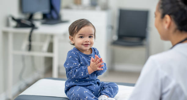
Smiling toddler in pyjamas sitting on a medical exam table, interacting with a doctor, symbolising recovery from craniosynostosis.

Complications and Recovery from Craniosynostosis

Complications and Recovery from Craniosynostosis Complications and recovery from craniosynostosis depend on the type and severity of the condition, the timing of diagnosis, and the quality of treatment. While craniosynostosis is often correctable with surgery, delayed intervention or syndromic forms may result in additional challenges requiring long-term support. Potential Complications Increased intracranial pressure: A major concern in untreated cases, this can lead to: Headaches Nausea and vomiting Vision problems Cognitive or developmental delays Facial and cranial asymmetry: If left uncorrected, the deformity may become more pronounced and lead to psychosocial concerns Neurological issues: In syndromic cases, learning difficulties, speech delays, and behavioural challenges may develop Surgical complications: While rare, possible risks include infection, bleeding, or the need for revision surgery Recovery After Surgery | Complications and Recovery from Craniosynostosis Most children recover well and return home within a few days Stitches or staples are removed after about one to two weeks Swelling and bruising are common in the first few days but subside quickly Follow-up appointments ensure the skull is growing normally and that no pressure build-up is occurring Long-Term Monitoring Ongoing care is important to: Track head circumference Monitor developmental milestones Identify any signs of recurrent suture fusion or pressure issues Support vision, hearing, and motor development Children with syndromic craniosynostosis often require continued specialist input throughout childhood. Emotional and Social Support Families may benefit from: Counselling to help manage anxiety surrounding surgery Parent support groups Early intervention programmes if developmental delays occur In conclusion, complications and recovery from craniosynostosis vary by case, but early treatment typically results in excellent outcomes. With careful monitoring and support, most children go on to lead healthy, active lives. [Next: Back to Overview →]

Complications and Recovery from Craniosynostosis Read More »

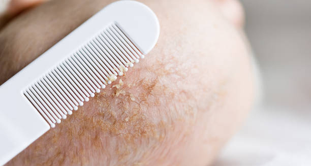
Close-up of a baby’s scalp with visible cradle cap flakes being gently removed using a fine-tooth comb.

Cradle Cap

Cradle Cap – Overview Cradle cap is a common and harmless skin condition that affects newborns and infants, characterised by greasy, yellowish, scaly patches on the scalp. Also known as infantile seborrhoeic dermatitis, this typically appears within the first few weeks of life and may last for several months. Although it can look concerning to new parents, it is not contagious, painful, or a sign of poor hygiene. The condition primarily affects the scalp but may also appear behind the ears, on the eyebrows, eyelids, or in the nappy area. The scales may flake off over time, and in some cases, hair may fall out with the crusts, though it usually grows back. The skin underneath can appear red or irritated but rarely causes discomfort to the baby. Cradle Cap Cradle cap develops when the oil-producing sebaceous glands in a baby’s skin become overactive, possibly due to residual maternal hormones in the baby’s body. This overproduction of sebum traps dead skin cells, leading to the formation of thick, crusty patches. Unlike other rashes, this does not itch or cause pain, and babies typically remain unbothered by it. The condition is self-limiting and often resolves on its own without medical treatment. However, gentle care routines can help speed up the healing process and prevent recurrence. In rare cases, if the area becomes infected or inflamed, further treatment may be required. In summary, this condition is a common, non-serious skin condition in babies marked by greasy, scaly patches on the scalp. With regular care and patience, it usually clears up on its own without complications. [Next: Causes of Cradle Cap →]

Cradle Cap Read More »

Newborn baby wearing a soft knitted bonnet, gently touched by a caregiver, illustrating care for conditions like cradle cap.

Causes of Cradle Cap

Causes of Cradle Cap Causes of cradle cap are not fully understood, but the condition is thought to result from a combination of overactive sebaceous glands and skin sensitivity. Though it appears alarming, cradle cap is not caused by poor hygiene, allergies, or infection. It is a natural and temporary skin response in many infants and requires only gentle management. Overactive Sebaceous Glands Newborns retain a small amount of maternal hormones for several weeks after birth. These hormones stimulate the oil (sebum) glands in the baby’s skin, particularly on the scalp. The excess sebum traps old skin cells and prevents them from naturally shedding, forming greasy, scaly patches. Yeast Overgrowth | Causes of Cradle Cap Another possible contributing factor is the presence of a yeast-like fungus called Malassezia, which thrives in oily skin environments. This yeast may contribute to inflammation and the build-up of scales. However, it is generally harmless and lives on most people’s skin without causing issues. Environmental Factors Although not a direct cause, warm or humid conditions can worsen cradle cap by increasing oil production and moisture retention. This can make the scales appear thicker or more widespread. Genetic Predisposition Some infants may be more prone to cradle cap due to genetic factors. Babies with a family history of skin conditions like eczema or seborrhoeic dermatitis may be more likely to develop cradle cap, although it is not classified as an allergic or autoimmune disorder. Normal Skin Development The condition is sometimes simply a part of the baby’s skin adapting to the external environment after birth. The immaturity of the skin barrier in newborns may make them more susceptible to skin conditions such as cradle cap. It is important to reassure parents that cradle cap is not contagious and cannot be transmitted between children. It also does not result from washing the baby’s hair too infrequently or too often. In conclusion, causes of cradle cap are largely linked to temporary hormonal changes and skin oil production. It is a benign condition that typically clears up with time and basic skin care practices. [Next: Symptoms of Cradle Cap →]

Causes of Cradle Cap Read More »

Scroll to Top