Mental Matters

Author name: admin

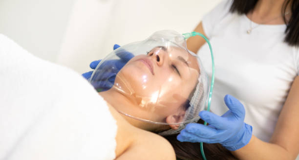
Woman receiving oxygen therapy through a face mask for cluster headache relief

Treatment of Cluster Headaches

Treatment of Cluster Headaches Treatment of cluster headaches focuses on relieving pain quickly during an attack and preventing future episodes. Because cluster headaches are so intense, immediate and effective intervention is essential. There are three main pillars of management: acute treatment, preventive treatment, and lifestyle modifications. 1. Acute Treatments (Abortive Therapy) The goal is to stop pain rapidly once an attack begins: 2. Preventive Treatments | Treatment of Cluster Headaches These are started at the onset of a cluster period to reduce frequency and severity: 3. Newer and Surgical Options For resistant cases, options include: 4. Lifestyle Changes | Treatment of Cluster Headaches Although not causes, triggers such as alcohol, strong smells, and irregular sleep can worsen symptoms. Maintaining a consistent daily routine and avoiding known triggers during active periods is essential. With the right combination of abortive and preventive therapies, the treatment of cluster headaches can significantly improve quality of life. Early intervention and specialist care are crucial for long-term success. [Next: Complications and Recovery from Cluster Headaches →]

Treatment of Cluster Headaches Read More »

Man in glasses grimacing with pain and holding his forehead during a cluster headache

Complications and Recovery from Cluster Headaches

Complications and Recovery from Cluster Headaches Complications and recovery from cluster headaches depend on the severity and duration of the condition. While cluster headaches are not life-threatening, they can cause substantial personal and social disruption, especially without adequate treatment. Many patients report that the condition affects every aspect of their lives—from relationships to employment. 1. Psychological Effects | Complications and Recovery from Cluster Headaches The intensity and unpredictability of attacks can lead to: Some sufferers feel isolated or misunderstood due to the rarity of the condition, which may impact their mental health. 2. Medication Side Effects Long-term use of preventive medications like verapamil or lithium requires regular monitoring due to potential side effects, such as cardiac issues or kidney dysfunction. 3. Work and Daily Life Disruption Frequent attacks can make it difficult to hold a job or maintain a social life. Night-time attacks may lead to chronic sleep deprivation, further affecting mood and concentration. 4. Risk of Chronic Cluster Headaches In a minority of cases, the condition becomes chronic, meaning attacks occur without significant remission periods. This form is harder to manage and may require more aggressive treatment strategies. 5. Recovery Outlook While there is no cure, most people with episodic cluster headaches experience periods of remission. With proper treatment, they can lead full and active lives. Adherence to treatment plans, regular check-ups, and support from healthcare professionals and patient groups can make a significant difference. 6. Support and Education Connecting with others through support groups and education empowers patients. Learning how to recognise early signs of a cluster period and having a treatment plan in place improves confidence and reduces the emotional burden. In conclusion, complications and recovery from cluster headaches centre on managing both the physical symptoms and emotional impact. With the right support and treatment, many patients find relief and regain control over their lives. [Next: Back to Overview →]

Complications and Recovery from Cluster Headaches Read More »

Club Foot

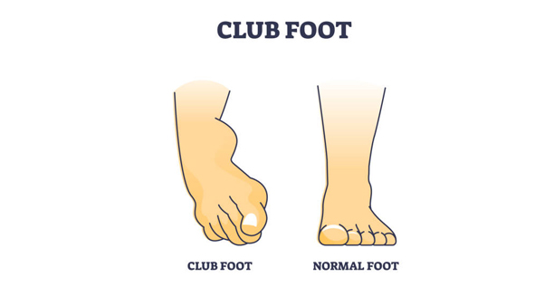
Club Foot – Overview Club foot is a congenital condition in which a baby’s foot or feet are twisted out of their normal position. The term talipes equinovarus describes a deformity where the foot points downward and inward, often resembling the shape of a golf club. It can affect one or both feet and occurs in roughly 1 in every 1,000 live births, making it one of the most common musculoskeletal birth defects. This condition develops during pregnancy and is typically detected at birth or during prenatal scans. The bones, muscles, tendons, and blood vessels of the affected foot are abnormally shaped or positioned, making it difficult or impossible for the child to walk normally without treatment. Although the condition itself is not painful, if left untreated, it can lead to long-term disability, difficulty in walking, and social or psychological issues due to visible deformity. Club Foot Fortunately, with early diagnosis and proper intervention, the outcomes for children with talipes equinovarus are excellent. Most babies respond well to non-surgical treatment methods, particularly the Ponseti method, which involves gentle manipulation, casting, and bracing. In more severe or resistant cases, surgical intervention may be required. In summary, club foot is a treatable birth defect involving the abnormal positioning of the foot or feet. Early treatment leads to good outcomes, allowing children to walk, run, and lead active lives without restrictions. Hope Through Early Treatment Early diagnosis and intervention are key to correcting it and ensuring normal mobility. With prompt and consistent treatment — particularly in infancy — most children regain full function and experience no lasting limitations. Techniques like the Ponseti method have transformed the prognosis for affected children, offering non-invasive solutions with high success rates. Looking Ahead Ongoing care, including follow-ups and bracing, plays a vital role in preventing relapse. Education and support for parents are also crucial, as managing club foot requires commitment and consistency in the early years. With the right care, children born with it can look forward to healthy, active lives without physical or social barriers. [Next: Causes of Club Foot →]

Club Foot Read More »

Medical illustration comparing a club foot to a normal foot

Causes of Club Foot

Causes of Club Foot Causes of club foot are not entirely understood, though several genetic and environmental factors appear to contribute to its development. While club foot often occurs in otherwise healthy infants, some cases are associated with other conditions or syndromes. Understanding the causes of club foot helps guide research and improve early detection strategies. 1. Genetic Factors Evidence suggests a strong genetic component. If a parent had club foot, there is a significantly increased chance their child may inherit the condition. The risk is also higher if a sibling is affected. Several genes associated with muscle and skeletal development have been implicated, though no single gene has been identified as the sole cause. 2. Position in the Womb | Causes of Club Foot Limited space in the uterus may affect foetal positioning, especially in the third trimester. Club foot can develop when the foot is compressed or restrained, particularly in cases of multiple births or low amniotic fluid levels. 3. Neuromuscular Conditions Some babies are born with club foot as part of a wider syndrome involving the nervous or muscular system. Conditions like spina bifida or arthrogryposis are examples where club foot may occur alongside other abnormalities. 4. Environmental Factors Although harder to pinpoint, maternal smoking, infections during pregnancy, or certain medications may slightly increase the risk of club foot. Poor maternal nutrition and exposure to harmful substances during early foetal development have also been suggested as contributing factors. 5. Idiopathic Cases | Causes of Club Foot In most cases, club foot appears without any associated health problems or identifiable cause. These are referred to as idiopathic club feet and account for the majority of cases. Despite the unknown cause, these cases usually respond very well to treatment. The causes are likely multifactorial, involving a combination of genetic predisposition and environmental influence. Ongoing research aims to uncover more precise mechanisms and improve prevention and early intervention strategies. [Next: Symptoms of Club Foot →]

Causes of Club Foot Read More »

Close-up of a newborn’s foot showing signs of club foot deformity

Symptoms of Club Foot

Symptoms of Club Foot Symptoms of club foot are usually visible at birth or detected on prenatal scans. The condition affects the structure and position of the foot or feet, and though club foot itself is not painful in infants, its visual and physical effects are clear and distinct. 1. Foot Positioning | Symptoms of Club Foot The most noticeable symptom is the abnormal inward and downward rotation of the foot. In classic club foot: Both feet can be affected (bilateral club foot), although one-sided cases are also common. 2. Tightness and Stiffness The Achilles tendon at the back of the heel is typically tight and shortened, which contributes to the downward positioning of the heel. The calf muscles may also be underdeveloped, giving the lower leg a thinner appearance on the affected side. 3. Limited Range of Motion | Symptoms of Club Foot The foot has restricted movement. Attempts to move the foot into a normal position are met with resistance, though the condition is not painful for the newborn. 4. Associated Changes in Leg Shape In some cases, the shin may appear slightly curved or shortened. The affected foot and leg can be smaller than the unaffected side, especially in unilateral cases. 5. Walking Difficulties (if Untreated) Without treatment, club foot leads to problems with mobility: Early identification of the symptoms of club foot allows for prompt treatment, which can begin within the first weeks of life. With appropriate care, most children achieve full mobility without pain or long-term disability. [Next: Diagnosis of Club Foot →]

Symptoms of Club Foot Read More »

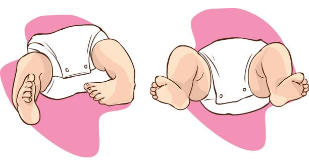
Medical illustration comparing baby with club foot and normal foot position

Diagnosis of Club Foot

Diagnosis of Club Foot Diagnosis of club foot is usually straightforward, as the condition is visible at birth. Routine prenatal ultrasounds may reveal club foot in some cases. Allowing for early planning and referral to specialists. An accurate and timely diagnosis is essential for starting treatment as early as possible. 1. Physical Examination at Birth Doctors typically identify club foot immediately after birth through visual assessment. The position and flexibility of the foot are evaluated. Clinicians use the Pirani and Dimeglio scales to grade deformity severity. These tools help assess the likelihood of treatment success and the need for surgery. 2. Prenatal Diagnosis | Diagnosis of Club Foot High-resolution ultrasound imaging can detect club foot during pregnancy, usually between 18 and 22 weeks of gestation. Once confirmed, parents meet an orthopaedic specialist to receive counseling and plan post-birth treatment. 3. Distinguishing Between Types It’s important to determine whether the club foot is: Further investigations, including genetic tests or MRI scans, help diagnose suspected abnormalities in complex cases. 4. Monitoring During Growth After initial treatment begins, regular follow-up appointments and imaging (usually X-rays) help monitor progress. To maintain proper correction, doctors regularly check foot positioning, bone growth, and joint flexibility. 5. Parental Education | Diagnosis of Club Foot Parents are often involved in early treatment, especially when bracing is required. Diagnosis includes education about the importance of compliance with home care routines, casting schedules, and potential long-term follow-up. A precise diagnosis of club foot sets the foundation for successful treatment. Early intervention, guided by an experienced orthopaedic team, leads to excellent outcomes in the vast majority of cases. [Next: Treatment of Club Foot →]

Diagnosis of Club Foot Read More »

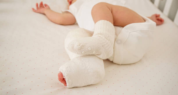
Infant in white onesie with both feet in corrective plaster casts for club foot

Treatment of Club Foot

Treatment of Club Foot Treatment of club foot aims to correct the position of the foot and ensure long-term function, mobility, and comfort. Thanks to advances in medical techniques, most children with club foot grow up to walk, run, and play without any significant restrictions. The key to success is early, consistent, and specialised intervention. 1. Ponseti Method The Ponseti method is the gold standard for treating club foot and has revolutionised outcomes worldwide. It involves: This method is over 90% effective when parents follow the bracing regimen carefully. 2. French Functional Method This approach involves daily stretching, taping, and physiotherapy. While less common than Ponseti, it may be used in specific cases or cultural contexts. 3. Surgical Treatment | Treatment of Club Foot Surgery is considered when non-surgical methods fail or if the club foot is extremely rigid. Procedures may involve: Surgery can carry risks like stiffness or overcorrection, so it’s reserved for resistant cases. 4. Parental Involvement Success relies heavily on parental commitment to the bracing phase. Education and support are vital to ensure compliance and reduce relapse. 5. Long-Term Monitoring | Treatment of Club Foot Children with corrected club foot need periodic assessments during early growth to catch any recurrence early. Footwear modifications, orthotics, or physiotherapy may be recommended to support long-term function. In short, treatment of club foot is most effective when started early—preferably within the first few weeks of life. With modern methods, children typically go on to live active, healthy lives without any visible deformity. [Next: Complications and Recovery from Club Foot →]

Treatment of Club Foot Read More »

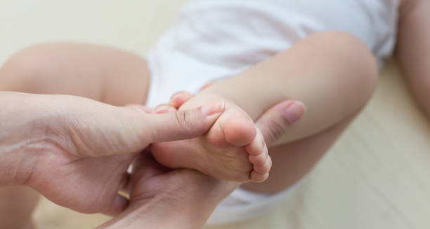
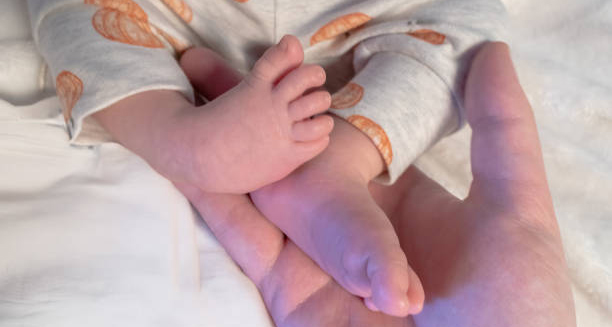
Adult hand supporting the feet of an infant with club foot

Complications and Recovery from Club Foot

Complications and Recovery from Club Foot Complications and recovery from club foot depend largely on the timing and quality of treatment. When properly managed, club foot has an excellent prognosis, but delayed or incomplete care can lead to physical limitations and social challenges. Understanding the possible complications ensures that parents and clinicians remain vigilant during recovery. 1. Risk of Recurrence The most common complication is recurrence, which typically happens due to poor adherence to bracing routines after initial casting. Without continued use of the brace as recommended, the foot can revert to its abnormal position. 2. Stiffness and Reduced Range of Motion In some cases, especially after surgery, the corrected foot may become stiff. While children often adapt well, slight limitations in movement or foot flexibility may remain, particularly during activities requiring a full range of motion. 3. Unequal Leg Size | Complications and Recovery from Club Foot Mild discrepancies in leg length or foot size are common, especially in unilateral club foot. These differences are usually minimal and do not affect walking ability. 4. Pain and Arthritis Although rare, untreated or poorly treated club foot can result in chronic pain or early-onset arthritis in the foot or ankle due to abnormal joint development. 5. Need for Additional Surgery If relapse occurs or the initial correction was incomplete, additional procedures may be required. Secondary surgeries are often minor but may include tendon transfers or revision casting. Recovery and Outlook | Complications and Recovery from Club Foot Recovery begins with correction in infancy and continues with bracing through toddlerhood. Most children: By school age, many children show no visible signs of ever having had club foot. Emotional and social development is typically unaffected when treatment is prompt and successful. Parental Support and Education Support groups, education, and regular follow-ups help families stay engaged in care. Understanding the importance of long-term bracing and appointments is crucial for avoiding setbacks. In conclusion, complications and recovery from club foot are manageable when addressed early. With dedication and proper medical guidance, children with club foot almost always lead full, active, and pain-free lives. [Next: Back to Overview →]

Complications and Recovery from Club Foot Read More »

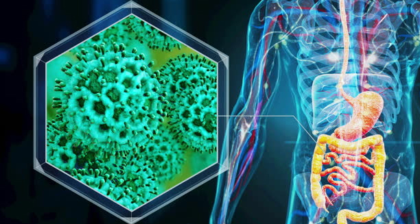
Microscopic view of Clostridium difficile bacteria and human digestive system

Clostridium difficile

Clostridium difficile – Overview Clostridium difficile, often referred to as C. difficile or C. diff. A type of bacteria that causes inflammation of the colon, a condition known as colitis. A CDI infection typically occurs after antibiotic use. Which disrupts the normal balance of bacteria in the gut and allows C. difficile to multiply and release toxins. These toxins damage the intestinal lining, leading to symptoms such as diarrhoea, abdominal pain, and fever. C. difficile is found naturally in the environment and can be present in small numbers in the intestines of healthy individuals. However, in the right conditions—particularly when protective gut bacteria are eliminated by antibiotics—it can rapidly grow and cause illness. Older adults, hospitalised patients, and those with weakened immune systems are especially vulnerable. Clostridium difficile – Overview The infection ranges from mild diarrhoea to life-threatening inflammation of the bowel. In some cases, complications such as toxic megacolon, sepsis, or even death may occur. C. difficile is highly contagious, spreading via spores that can survive for months on surfaces such as bed rails, toilet seats, and hands. Due to its association with healthcare environments, C. difficile has become a major concern in hospitals and care facilities. Rigorous hygiene protocols and antimicrobial stewardship are essential to preventing its spread. In summary, Clostridium difficile is a dangerous bacterial infection that often follows antibiotic use and poses significant risks in medical settings. Early detection, isolation, and treatment are vital to preventing serious outcomes and limiting transmission. [Next: Causes of Clostridium difficile →]

Clostridium difficile Read More »

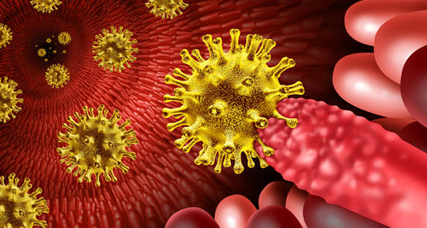
3D illustration of Clostridium difficile bacteria invading intestinal tissue

Causes of Clostridium difficile

Causes of Clostridium difficile Causes of Clostridium difficile infection are primarily linked to the disruption of the normal gut microbiome. Most commonly through antibiotic use. Loss of healthy gut bacteria allows Clostridium difficile to grow unchecked and produce toxins. Causing inflammation and damage to the intestines. 1. Antibiotic Use The most significant cause is the use of broad-spectrum antibiotics. Doctors have closely linked clindamycin, fluoroquinolones, cephalosporins, and penicillins to an increased risk of CDI. These antibiotics kill beneficial gut bacteria, reducing the natural competition that keeps C. difficile in check. 2. Hospital or Care Facility Stay C. difficile is commonly acquired in hospitals, nursing homes, and other care facilities where antibiotics are frequently used and close contact between patients facilitates spread. The bacteria are passed through spores that can contaminate hands, surfaces, and equipment. 3. Weakened Immune System | Causes of Clostridium difficile People with compromised immune systems—such as cancer patients, transplant recipients, or those on immunosuppressive drugs—are more susceptible to infection and complications due to their reduced ability to fight off bacterial overgrowth. 4. Gastrointestinal Procedures Surgical interventions involving the digestive system, as well as the use of feeding tubes or prolonged use of proton pump inhibitors (PPIs), can increase the risk of developing CDI. PPIs may alter stomach acidity, allowing more bacteria to survive and reach the intestines. 5. Age and Comorbidities Older adults are significantly more at risk. People over 65 account for a large proportion of CDI cases, especially those with other chronic conditions like diabetes or kidney disease. 6. Previous CDI | Causes of Clostridium difficile Having had a C. difficile infection in the past greatly increases the risk of recurrence, especially within 2–3 months after the initial episode. Recurrent infections are often more difficult to treat. The causes of Clostridium difficile reflect a complex interplay of antibiotic exposure, environmental contamination, and host vulnerability. Awareness of these risk factors is key to both prevention and early intervention. [Next: Symptoms of Clostridium difficile →]

Causes of Clostridium difficile Read More »

Scroll to Top