Mental Matters

Author name: admin

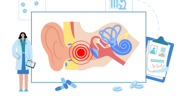
Diagram of the human ear showing cholesteatoma inside the middle ear

Cholesteatoma

Cholesteatoma – Overview Cholesteatoma is a destructive yet non-cancerous growth of skin cells that occurs in the middle ear, behind the eardrum. Although it is not a tumour in the traditional sense, a cholesteatoma can cause serious complications if left untreated. It expands slowly but progressively, damaging the delicate bones of hearing, and in severe cases, spreading into the skull base or brain. Cholesteatomas form when the skin lining of the outer ear canal invades the middle ear space. This often happens due to repeated infections or Eustachian tube dysfunction. When the Eustachian tube (which connects the middle ear to the back of the throat) does not function properly, negative pressure can develop in the middle ear. This pulls the eardrum inward, forming a pocket that traps dead skin and debris. Over time, this pocket expands into a cholesteatoma. There are two main types: congenital cholesteatoma, which is present at birth due to developmental abnormalities, and acquired cholesteatoma, which occurs later in life and is much more common. The acquired type typically affects people with a history of chronic middle ear infections, grommet insertion, or perforated eardrums. Despite being benign, the growth can erode the ossicles (the tiny bones that transmit sound), the mastoid bone, and even reach the inner ear or brain in rare cases. It can cause permanent hearing loss, facial nerve paralysis, dizziness, and life-threatening infections if not treated. For this reason, early diagnosis and surgical removal are critical. Cholesteatoma Cholesteatomas usually affect only one ear, although in rare instances, both ears may be involved. They can affect both children and adults, with slightly higher prevalence in individuals who have poor access to medical care or are immunocompromised. The condition is often identified due to persistent ear discharge, a sense of ear fullness, and progressive hearing loss. Sometimes, more severe symptoms like facial muscle weakness or vertigo may be the first clues to deeper complications. Although cholesteatoma is rare, it is considered serious due to its aggressive nature. Surgical removal is the only effective treatment, and ongoing monitoring is needed to prevent recurrence. Recurrence is possible even after successful surgery, which makes long-term follow-up essential. In summary, cholesteatoma is a dangerous but manageable ear condition that requires surgical treatment. It is not cancerous, but it behaves in a locally invasive way and must be dealt with promptly to avoid long-term damage. [Next: Causes of Cholesteatoma →]

Cholesteatoma Read More »

Woman using cotton swab in ear with discomfort, indicating poor ear hygiene

Causes of Cholesteatoma

Causes of Cholesteatoma Causes of cholesteatoma fall into two main categories: congenital and acquired. In both cases, the condition results from the build-up of squamous epithelium (skin cells) in the middle ear, which is a space normally lined with respiratory epithelium. Over time, this abnormal skin collection enlarges, becomes infected, and begins to erode surrounding structures. While congenital cases are rare, the majority of cholesteatoma diagnoses are acquired. The most common mechanism involves Eustachian tube dysfunction. When the Eustachian tube fails to equalise air pressure in the middle ear, negative pressure builds up. This can pull part of the eardrum inward, forming a retraction pocket. These pockets collect dead skin and keratin, which are normally shed and expelled in a healthy ear. In a blocked or retracted pocket, they become trapped and begin to accumulate. Chronic otitis media, or long-term middle ear infection, is another leading cause. Repeated infections weaken the structure of the eardrum and create an environment in which skin migration into the middle ear becomes more likely. This allows a cholesteatoma to form gradually over time, usually without pain in the early stages. Causes of Cholesteatoma Perforated eardrums, whether caused by trauma, infection, or surgical intervention (such as grommet insertion), may also play a role. Through these perforations, skin cells can invade the middle ear and begin forming a cholesteatoma sac. In rare cases, cholesteatoma is congenital, meaning a small nest of skin cells becomes trapped in the middle ear during foetal development. These cases are usually diagnosed in children, often when they present with hearing loss despite no history of infections or trauma. Other contributing factors include cleft palate, craniofacial abnormalities, and immunosuppression, all of which are linked to higher risks of Eustachian tube malfunction and chronic infections. Even severe allergies and sinus problems may indirectly contribute by creating nasal congestion and affecting tube drainage. Causes of Cholesteatoma Some researchers also suggest that a family history of chronic ear disease may slightly increase susceptibility, though cholesteatoma is not considered a genetic condition. Understanding the causes of cholesteatoma is important because early intervention in chronic ear problems can prevent it from forming. Treating underlying infections promptly, monitoring children with ear abnormalities, and educating high-risk groups about symptoms can reduce incidence and recurrence. [Next: Symptoms of Cholesteatoma →]

Causes of Cholesteatoma Read More »

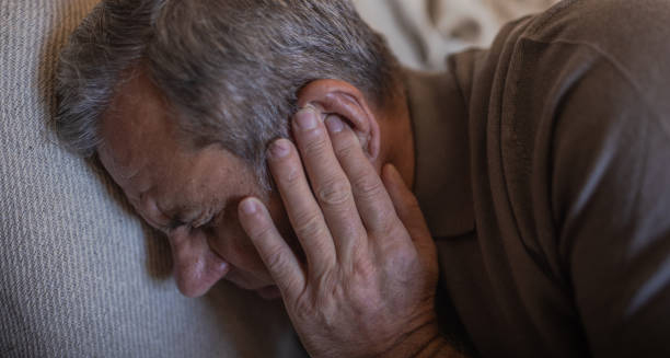
Elderly man holding his ear in pain, indicating symptoms of cholesteatoma

Symptoms of Cholesteatoma

Symptoms of Cholesteatoma Symptoms of cholesteatoma may be subtle at first but worsen as the growth expands. Since cholesteatoma can erode bone and surrounding structures, its symptoms range from simple hearing issues to more serious neurological signs. Recognising these early signs allows for quicker diagnosis and reduces the risk of permanent complications. The most common symptom is chronic ear discharge, often foul-smelling and continuous. Unlike occasional fluid drainage from an ear infection, cholesteatoma discharge tends to persist for weeks or months and doesn’t respond to ordinary ear drops. This is due to the trapped, infected keratin debris constantly draining through a hole or retraction in the eardrum. Another hallmark symptom is hearing loss in the affected ear. Initially, this hearing loss is conductive in nature, caused by the blockage of sound transmission through the ossicles. As the cholesteatoma erodes these small bones, the hearing loss becomes more severe and may also involve sensorineural components if the inner ear is affected. Symptoms of Cholesteatoma Many patients report a feeling of fullness or pressure in the ear. This may be accompanied by mild discomfort or the sense that something is stuck. While pain is not a typical early feature, some individuals do report dull aching or sharp episodes, especially if infection is present. Tinnitus, or ringing in the ear, is another frequent complaint. This may present as a high-pitched whistle or low-frequency humming and can be constant or intermittent. The presence of tinnitus often indicates irritation of the auditory nerve or inner ear. In advanced cases, the cholesteatoma may extend into the balance organs, leading to dizziness or vertigo. This is particularly concerning as it implies erosion beyond the middle ear into the labyrinth, which may affect spatial awareness and movement coordination. More severe symptoms include facial muscle weakness or twitching. The facial nerve runs through the middle ear, and cholesteatomas that invade this area may compress or damage it, resulting in drooping or paralysis of facial muscles. Symptoms of Cholesteatoma Other signs can include recurrent ear infections, frequent need for antibiotics, and a visible white mass behind the eardrum during an ear exam. In children, delayed speech or poor academic performance may be linked to undiagnosed hearing loss from a growing cholesteatoma. Not all symptoms occur at once, and some patients experience only one or two signs. Regardless, persistent discharge or progressive hearing changes should always trigger evaluation by an ENT specialist. [Next: Diagnosis of Cholesteatoma →]

Symptoms of Cholesteatoma Read More »

ENT specialist examining a diagram of the inner and middle ear for cholesteatoma diagnosis

Diagnosis of Cholesteatoma

Diagnosis of Cholesteatoma Diagnosis of cholesteatoma relies on careful clinical evaluation, supported by hearing tests and imaging. Because a cholesteatoma can silently erode bone and spread into surrounding structures, early detection is crucial to avoid permanent damage. Many patients present with long-standing symptoms like ear discharge or hearing loss, which can be overlooked or misdiagnosed without proper assessment. The process begins with a full medical history. The ENT specialist will ask about chronic ear infections, surgeries like grommet insertion, childhood ear disease, or symptoms like ear discharge, hearing changes, dizziness, or facial weakness. The history helps differentiate cholesteatoma from common middle ear infections or conditions like otitis media with effusion. The next step is a detailed otoscopic examination. Using a handheld otoscope or, preferably, a microscope or endoscope, the clinician inspects the ear canal and eardrum. Signs suggestive of cholesteatoma include: Diagnosis of Cholesteatoma Advanced examination with otoendoscopy provides a clearer and more magnified view of hidden recesses, making it easier to detect small or early cholesteatomas. An audiogram or hearing test is essential to assess the extent of hearing loss. Most patients show conductive hearing loss, where sound cannot pass effectively through the middle ear. In some cases, particularly if the inner ear is involved, there may also be sensorineural hearing loss. Speech discrimination scores may also be reduced. Tympanometry is a valuable test that measures eardrum compliance and middle ear pressure. It can confirm eustachian tube dysfunction, fluid build-up, or abnormal movement of the tympanic membrane. High-resolution CT scanning of the temporal bones is the gold standard for evaluating the extent of the disease. It provides detailed images of: In specific cases, an MRI may be requested to identify soft tissue extension or complications like brain abscess, meningitis, or sigmoid sinus thrombosis. Post-operatively, MRI with diffusion-weighted imaging is also used to monitor recurrence. Diagnosis of Cholesteatoma In infected cases, a microbial culture may be taken to guide antibiotic therapy. However, bacterial identification does not confirm or exclude the presence of a cholesteatoma. A confirmed diagnosis of cholesteatoma warrants surgical referral. Early identification allows for conservative and less extensive surgery, while delayed diagnosis may result in more complex and high-risk procedures. [Next: Treatment of Cholesteatoma →]

Diagnosis of Cholesteatoma Read More »

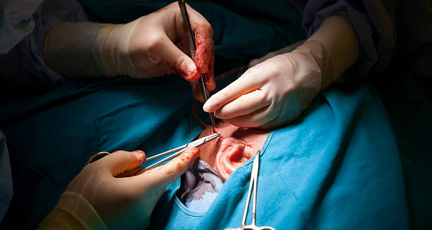
Surgeon performing cholesteatoma removal surgery on a patient’s ear

Treatment of Cholesteatoma

Treatment of Cholesteatoma Treatment of cholesteatoma involves surgical removal, as medication alone cannot eliminate the condition. Although non-cancerous, a cholesteatoma behaves aggressively — expanding and destroying delicate middle ear structures. Prompt surgery is therefore critical to remove the lesion, prevent complications, and restore or preserve hearing. The most common procedure is a mastoidectomy, performed under general anaesthesia. This involves removing the cholesteatoma and any infected or eroded tissue from the mastoid bone and middle ear. The surgery may be done in one of two approaches: Often, the surgery is combined with tympanoplasty, where the eardrum is reconstructed using graft material, usually taken from the patient’s own tissue (e.g., temporalis fascia or cartilage). If the ossicles have been damaged, ossiculoplasty may also be performed, using prosthetic implants or reshaped natural bone to reconnect the hearing chain. Treatment of Cholesteatoma In severe cases or when total removal is uncertain, a second-look procedure is scheduled six to twelve months later. This allows the surgeon to inspect the ear for any residual cholesteatoma and perform further reconstruction if necessary. Pre-surgical preparation may involve treating infections with antibiotics or ear drops to reduce inflammation. Patients are also advised to avoid getting water into the ear and to cease smoking, which can impair healing and Eustachian tube function. Post-operatively, the ear is packed and bandaged. Recovery is typically smooth, with most patients discharged within 24–48 hours. Regular follow-up is essential to monitor healing, ensure no recurrence, and assess hearing improvement. Hearing outcomes vary. Some patients experience full restoration, especially when ossicles are preserved or reconstructed. Others may need a hearing aid post-surgery. If the inner ear was affected, partial or complete hearing loss may be permanent. Treatment of Cholesteatoma Rehabilitation includes keeping the ear dry, avoiding flights or diving during recovery, and attending all follow-up appointments. In canal wall-down procedures, patients may require periodic ear cleaning to maintain hygiene in the surgically created cavity. Because cholesteatomas can recur — even years later — long-term surveillance with otoendoscopy or MRI is important, especially in children or high-risk adults. In summary, treatment of cholesteatoma is a surgical priority. With skilled care and regular monitoring, most patients recover well, regain hearing, and avoid long-term complications. [Next: Complications and Recovery from Cholesteatoma →]

Treatment of Cholesteatoma Read More »

Surgical treatment of cholesteatoma being performed on a patient’s ear

Complications and Recovery from Cholesteatoma

Complications and Recovery from Cholesteatoma Complications and recovery from cholesteatoma depend on how soon the condition is diagnosed and how extensively it has spread. Although cholesteatoma is not cancerous, its destructive nature can lead to irreversible damage if treatment is delayed. Early surgery significantly improves prognosis and reduces the likelihood of long-term issues. Potential Complications | Complications and Recovery from Cholesteatoma Recovery Process | Complications and Recovery from Cholesteatoma Recovery after surgery is generally smooth, provided the ear remains dry and clean. Most patients are advised to: Hearing often improves within weeks or months. In cases with severe ossicle damage, further surgery may be planned after the ear has healed. Children and patients who had canal wall-down procedures will need more regular inspections. Complications are rare with early treatment, but delays can lead to irreversible damage. Therefore, educating patients about signs of recurrence — such as discharge or hearing decline — is vital. In summary, complications and recovery from cholesteatoma are determined by the timing and completeness of treatment. With appropriate surgery and long-term care, most patients enjoy excellent outcomes and restored hearing function. [Next: Back to Overview →]

Complications and Recovery from Cholesteatoma Read More »

Illustration of bacteria causing cholera and a person clutching their stomach in pain

Cholera

Cholera – Overview The Cholera disease is a severe bacterial infection of the small intestine caused by ingesting water or food contaminated with Vibrio cholerae. This condition can cause rapid and life-threatening dehydration due to profuse diarrhoea and vomiting. Without prompt treatment, it can lead to shock, organ failure, and death within hours — but with timely care, most people recover fully. The disease spreads through contaminated water or food, particularly in areas with poor sanitation, overcrowding, or lack of access to clean drinking water. Cholera is especially dangerous during humanitarian crises, natural disasters, or in regions without proper sewage infrastructure. Although this condition is rare in many developed countries, it remains a significant public health concern in parts of Africa, South Asia, and Latin America. Globally, it causes an estimated 1.3 to 4 million cases and up to 143,000 deaths annually, according to the World Health Organization. Cholera Symptoms can range from mild to severe, but the hallmark sign is a sudden onset of watery diarrhoea that resembles rice water. Rapid fluid loss can result in electrolyte imbalances, muscle cramps, and circulatory collapse. This can be fatal, especially in children, the elderly, and immunocompromised individuals. Prevention focuses on access to safe drinking water, proper sanitation, hygiene practices, and, in some areas, oral cholera vaccines. With fast rehydration therapy, antibiotics (when needed), and supportive care, most patients make a full recovery. [Next: Causes of Cholera →]

Cholera Read More »

Hand in glove collecting water sample from contaminated natural source

Causes of Cholera

Causes of Cholera Causes of cholera centre around the ingestion of food or water contaminated with the bacterium Vibrio cholerae. Once inside the body, the bacteria multiply and release a toxin that causes severe diarrhoea. Understanding the causes of cholera is essential for controlling outbreaks and preventing transmission. 1. Contaminated Water The most common source is drinking or using water that contains Vibrio cholerae. This contamination often results from: In regions with inadequate sanitation, the risk of contamination is especially high during floods or after natural disasters. 2. Contaminated Food | Causes of Cholera Food can also become contaminated if: Improper food hygiene contributes significantly to cholera transmission in both households and street food settings. 3. Person-to-Person Spread (Rare) Direct person-to-person transmission of cholera is uncommon, but sharing contaminated food or surfaces can still spread the bacteria. 4. Poor Sanitation and Hygiene Environments without proper toilet facilities, hand-washing infrastructure, or waste disposal systems are ideal for cholera to spread. Refugee camps, crowded urban slums, and disaster zones are high-risk areas. 5. Travel to Affected Areas Travellers to regions experiencing cholera outbreaks are at risk if they consume unboiled water, raw fruits or vegetables, or uncooked seafood. 6. Asymptomatic Carriers | Causes of Cholera Some people infected with cholera may show no symptoms but still shed the bacteria in their stool, silently contributing to its spread. Recognising the causes of cholera helps public health workers implement effective containment strategies, including clean water provision, hygiene promotion, and food safety education. [Next: Symptoms of Cholera →]

Causes of Cholera Read More »

Woman holding stomach in discomfort during outdoor hike near waterfall

Symptoms of Cholera

Symptoms of Cholera Symptoms of cholera can develop within hours or days of exposure, ranging from mild diarrhoea to severe dehydration and death. Recognising cholera symptoms early is crucial, especially in outbreak settings where rapid response can save lives. 1. Profuse Watery Diarrhoea The most distinctive symptom is sudden, painless, watery diarrhoea, often described as: This diarrhoea is caused by the cholera toxin triggering the release of water and electrolytes from intestinal cells. 2. Vomiting | Symptoms of Cholera Severe vomiting may accompany diarrhoea, contributing further to fluid and electrolyte loss. It usually appears early in the illness and can be forceful. 3. Rapid Dehydration Due to the extreme fluid loss, patients may quickly show signs of dehydration, including: In children, dehydration may present as lethargy, sunken fontanelle, and crying without tears. 4. Muscle Cramps The loss of sodium, potassium, and other electrolytes can cause painful muscle cramps, particularly in the legs and abdomen. 5. Shock and Collapse | Symptoms of Cholera Without immediate treatment, the body may go into hypovolaemic shock, where blood volume drops so low that organs can’t function. Symptoms include: This is a life-threatening emergency that requires urgent care. 6. Mild or Asymptomatic Cases Some people infected with this condition may only have mild diarrhoea or no symptoms at all. However, they can still spread the bacteria through contaminated stool. Recognising the symptoms of cholera early — particularly in children or vulnerable populations — allows for lifesaving rehydration and reduces transmission. [Next: Diagnosis of Cholera →]

Symptoms of Cholera Read More »

Cholera-labelled syringe held by gloved hands next to a transparent globe

Diagnosis of Cholera

Diagnosis of Cholera Diagnosis of cholera relies on clinical assessment during an outbreak, supported by laboratory confirmation. In high-risk settings, early recognition of cholera ensures rapid intervention and limits further spread. 1. Clinical Diagnosis In outbreak situations or endemic regions, a suspected case is defined as: Doctors may make a provisional diagnosis based on these signs, especially if multiple cases emerge in a short time in the same area. 2. Stool Sample Analysis | Diagnosis of Cholera To confirm cholera, a stool culture or rectal swab is tested in a laboratory for the presence of Vibrio cholerae. Techniques include: While confirmation helps track outbreaks, treatment usually starts based on symptoms before lab results are available. 3. Testing for Dehydration Doctors also assess the severity of dehydration using: This assessment guides the type and speed of rehydration therapy. 4. Public Health Reporting | Diagnosis of Cholera Suspected or confirmed cholera cases must be reported to health authorities. Early identification of cases can prevent larger outbreaks and direct emergency resources to the area. In resource-limited or crisis environments, the diagnosis of cholera often hinges on recognising symptoms quickly and starting treatment without delay. Confirmation helps contain the outbreak but is secondary to saving lives. [Next: Treatment of Cholera →]

Diagnosis of Cholera Read More »

Scroll to Top