Mental Matters

Author name: admin

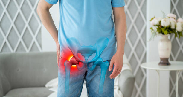
Person experiencing hip pain with medical overlay highlighting inflammation

Symptoms of Bursitis

Symptoms of Bursitis Symptoms of bursitis vary depending on the location and severity of the inflammation, but the most common symptoms of bursitis include pain, swelling, tenderness, and reduced range of motion in the affected joint. Pain is often the first and most noticeable symptom. It may begin gradually or suddenly and usually worsens with movement or pressure on the area. For example, people with shoulder bursitis may find it difficult to lift their arm or sleep on one side. Swelling is usually visible and felt around the joint. In more superficial bursae, such as those near the elbow or knee, the affected area may look red, warm, and swollen. This can cause joint stiffness or tightness during movement. Tenderness over the inflamed bursa is typical. Pressing the area lightly often triggers discomfort. Some individuals report a dull ache or sharp pain, depending on whether the bursa is infected or purely inflamed. Symptoms of Bursitis Limited mobility is another common issue. As the swelling increases, joint movement becomes restricted, making it hard to perform daily tasks like walking, dressing, or lifting. In chronic bursitis, stiffness may remain even after the pain improves. In cases of septic (infected) bursitis, symptoms may include fever, chills, redness, warmth, and severe pain. The skin over the bursa may feel hot, and the joint may be very sensitive to touch. This requires urgent medical attention to prevent the infection from spreading. Common sites and their symptoms include: Recognising symptoms of bursitis early helps reduce flare-up duration and improves treatment outcomes. If symptoms last longer than a few days, worsen, or involve signs of infection, medical advice should be sought. [Next: Diagnosis of Bursitis →]
Symptoms of Bursitis Read More »

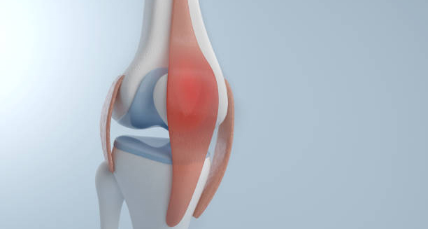
Medical illustration of knee joint highlighting bursitis inflammatio

Diagnosis of Bursitis

Diagnosis of Bursitis Diagnosis of bursitis involves evaluating a person’s symptoms, physical condition, and sometimes using imaging or lab tests to rule out other conditions. A proper diagnosis of bursitis allows for effective treatment and helps prevent complications like chronic pain or joint damage. Doctors begin by taking a full medical history, asking about the onset, location, duration, and triggers of the pain. They also consider occupational or recreational activities that may contribute to joint overuse. During the physical exam, the doctor will inspect the affected joint for swelling, redness, warmth, and tenderness. They may also check your range of motion and identify specific movements or postures that trigger discomfort. In most straightforward cases, diagnosis is clinical and no further testing is needed. However, when symptoms are severe, persistent, or involve signs of infection, additional tools may be used: Diagnosis of Bursitis Blood tests may also be ordered to check for infection markers, autoimmune diseases, or gout, particularly in recurrent or unexplained cases of bursitis. In chronic cases or where structural abnormalities are involved, referral to a rheumatologist, orthopaedic specialist, or physiotherapist may be needed for further evaluation. A correct diagnosis of bursitis ensures that treatment is tailored appropriately — whether rest and ice will suffice or if medical procedures, antibiotics, or rehabilitation are required. [Next: Treatment of Bursitis →]

Diagnosis of Bursitis Read More »

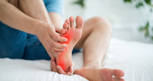
Man holding foot with red inflamed area indicating bursitis

Treatment of Bursitis

Treatment of Bursitis Treatment of bursitis focuses on reducing inflammation, relieving pain, and restoring joint function. In most cases, treatment of bursitis begins with conservative measures such as rest, ice, and over-the-counter medication. More persistent or severe cases may require injections or physical therapy. For acute, non-infectious bursitis: If symptoms persist for several weeks or the condition becomes chronic, doctors may explore more advanced treatment options: For septic bursitis: Treatment of Bursitis For bursitis caused by an underlying condition such as gout or rheumatoid arthritis, treatment involves managing the primary disease alongside the bursitis. Lifestyle adjustments — such as warming up before exercise, wearing proper footwear, and using ergonomic tools at work — are crucial in preventing recurrence. With prompt care, most people recover fully within a few weeks. Successful treatment of bursitis reduces the chance of long-term complications and restores mobility and comfort. [Next: Complications and Recovery from Bursitis →]

Treatment of Bursitis Read More »

Elderly man holding shoulder in pain due to bursitis

Complications and Recovery from Bursitis

Complications and Recovery from Bursitis Complications and recovery from bursitis depend on the severity, cause, and how early treatment begins. While most people recover quickly, delayed or incorrect management can lead to complications from bursitis that affect mobility and quality of life. Common complications include: Recovery from bursitis usually takes a few days to several weeks, depending on the type and treatment approach. With proper rest, ice, and medication, mild cases resolve quickly. Physical therapy often accelerates recovery and helps prevent recurrence by improving joint mechanics and posture. For individuals who require steroid injections or have had septic bursitis, follow-up care is essential. This may include repeated imaging, lab tests, or physiotherapy sessions. Long-term strategies to aid recovery include: Complications and Recovery from Bursitis Emotional well-being is also important. Chronic bursitis can impact daily life, leading to frustration or inactivity. Support from healthcare professionals and loved ones helps ensure a more complete recovery. With early care and preventive habits, complications and recovery from bursitis are manageable, and most individuals return to full function without long-term limitations. [Next > Back to Overview]

Complications and Recovery from Bursitis Read More »

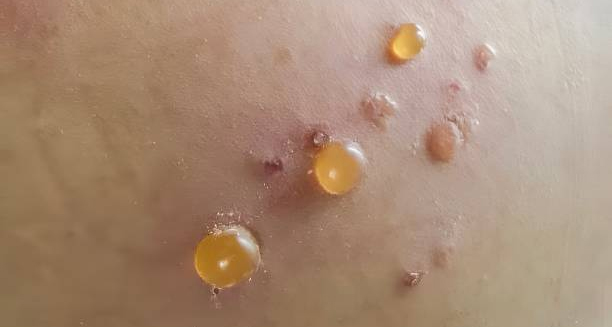
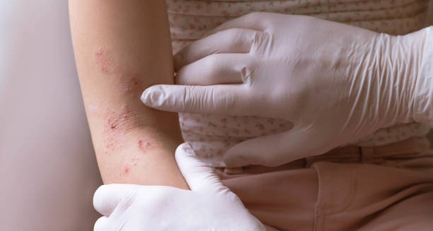
Cluster of fluid-filled blisters on inflamed skin caused by bullous pemphigoid

Bullous Pemphigoid

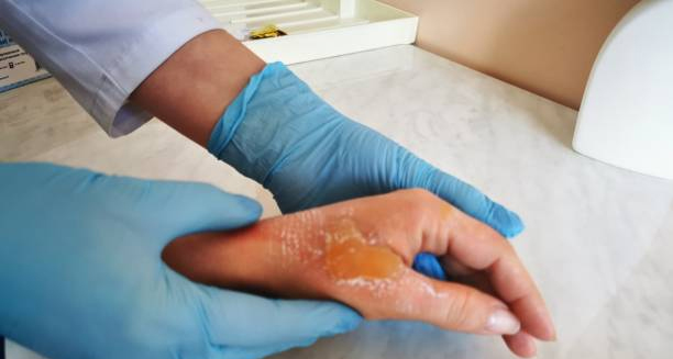
Bullous Pemphigoid – Overview Bullous pemphigoid is a rare autoimmune skin condition that causes large, fluid-filled blisters to form on areas of the skin that often flex — such as the abdomen, arms, legs, and groin. This is most commonly affects older adults and can range from mild to severely disabling depending on the extent and location of the blisters. This chronic condition occurs when the body’s immune system mistakenly attacks the basement membrane — the thin layer of tissue that anchors the outer layer of skin (epidermis) to the underlying dermis. As a result, inflammation develops and the skin layers separate, forming blisters. The blisters are usually firm, dome-shaped, and filled with clear fluid. They do not rupture easily and are often very itchy. In some cases, redness and welts appear before blistering begins. The condition typically lasts for months or even years, with periods of flare-ups and remission. Bullous pemphigoid While not contagious or cancerous, this condition can significantly affect a person’s quality of life. Scratching and broken blisters may lead to infections, scarring, or skin thickening. Treatment usually involves corticosteroids and immunosuppressants to calm the immune response, along with topical ointments to manage itching and inflammation. With proper care, most individuals can control the disease and reduce symptoms over time. [Next: Causes of Bullous Pemphigoid →]

Bullous Pemphigoid Read More »

Doctor examining a large blister on a patient's hand wearing blue gloves

Causes of Bullous Pemphigoid

Causes of Bullous Pemphigoid Causes of bullous pemphigoid stem from an autoimmune malfunction, though the exact trigger is not always clear. The most well-established causes of bullous pemphigoid include age-related immune system changes, certain medications, underlying illnesses, and skin trauma. In this condition, the immune system produces antibodies against proteins in the basement membrane zone of the skin. Specifically, it targets two proteins — BP180 and BP230 — which help bind the epidermis to the dermis. When these proteins are attacked, it causes inflammation and fluid accumulation between the skin layers, leading to blister formation. Age is the most significant risk factor. Bullous pemphigoid mainly affects individuals over the age of 60 and becomes more common with advancing age. This suggests that immune dysregulation in older adults plays a key role. Certain medications have been linked to triggering bullous pemphigoid in some people. These include diuretics (like furosemide), antibiotics (like penicillin), and drugs used to treat diabetes (such as DPP-4 inhibitors). Reactions can occur weeks or even months after starting the medication. Causes of bullous pemphigoid Skin trauma — such as surgery, burns, radiation therapy, or chronic wounds — can also precede the onset of bullous pemphigoid in affected individuals. Physical stress to the skin may provoke an immune response that targets the skin’s structural proteins. There is also evidence that some neurological diseases like Parkinson’s disease, multiple sclerosis, and dementia are associated with a higher risk of developing bullous pemphigoid. The exact link is not fully understood but may relate to overlapping immune system dysfunction. Genetic predisposition appears to play a minor role, and bullous pemphigoid is generally not inherited. Environmental triggers, infections, and seasonal changes have also been proposed, but more research is needed to confirm their influence. Understanding the causes of bullous pemphigoid helps guide prevention strategies, especially in high-risk individuals — such as avoiding known drug triggers and monitoring for early symptoms. [Next: Symptoms of Bullous Pemphigoid →]

Causes of Bullous Pemphigoid Read More »

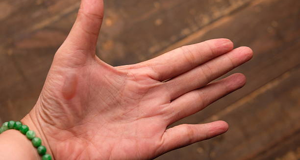
Close-up of a hand with a large blister on the palm

Symptoms of Bullous Pemphigoid

Symptoms of Bullous Pemphigoid Symptoms of bullous pemphigoid often begin subtly before progressing to widespread blistering. The most common symptoms of bullous pemphigoid include intense itching, red or raised skin, and large, fluid-filled blisters that typically do not rupture easily. In many patients, the disease starts with itching and redness, sometimes mistaken for eczema or hives. This phase may last for days or even weeks before any visible blisters appear. The skin may feel sore or tight, and small urticarial (hive-like) patches may develop. Blisters then form on the affected areas. These are usually 1–5 cm in diameter, tense (due to being deep within the skin), and filled with clear or sometimes blood-tinged fluid. Unlike blisters in other skin conditions, bullous pemphigoid blisters tend to remain intact for longer and rupture less easily. The most common sites include the lower abdomen, groin, inner thighs, armpits, and flexural surfaces like the elbows and knees. In some cases, the blisters may be widespread, covering the trunk and limbs. The palms and soles can also be involved, and the affected skin may become thickened or crusted over time. Itching is often intense and persistent, causing considerable discomfort. In people with fragile skin, scratching may lead to skin tears or secondary bacterial infections. Symptoms of Bullous Pemphigoid In rarer cases, the mucous membranes (inside the mouth, nose, or genitals) may be affected, leading to painful ulcers or erosions. However, this is less common in bullous pemphigoid than in similar disorders such as mucous membrane pemphigoid. People with severe or prolonged disease may also experience fatigue, weight loss, or low mood due to chronic discomfort and disrupted sleep. Because the appearance of symptoms of bullous pemphigoid can vary, especially in the early stages, a skin biopsy and blood tests are often required to confirm the diagnosis and differentiate it from other blistering diseases. [Next: Diagnosis of Bullous Pemphigoid →]

Symptoms of Bullous Pemphigoid Read More »

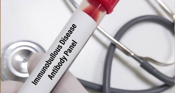
Blood sample labelled "Immunobullous Disease Antibody Panel" with stethoscope in background

Diagnosis of Bullous Pemphigoid

Diagnosis of Bullous Pemphigoid Diagnosis of bullous pemphigoid involves a combination of clinical examination, skin biopsy, and laboratory testing. A confirmed diagnosis of bullous pemphigoid ensures appropriate treatment. Additionally, helps rule out other similar skin conditions, such as pemphigus vulgaris or dermatitis herpetiformis. The diagnostic process begins with a full medical history and visual inspection of the skin. A dermatologist will look for the pattern, size, and location of the blisters. As well as the condition of the surrounding skin. The presence of firm, fluid-filled blisters on red, itchy skin — particularly in older adults — raises suspicion. To confirm the diagnosis, a skin biopsy is performed. A small sample of blistered or inflamed skin is removed and examined under a microscope. This helps determine the level at which the skin layers are separating and whether immune cells are present. Direct immunofluorescence testing is also done on skin tissue. This test detects deposits of antibodies (IgG) and complement proteins (C3) at the junction between the epidermis and dermis. The presence of a linear pattern of these immune proteins is highly characteristic of bullous pemphigoid. Diagnosis of Bullous Pemphigoid Blood tests can also be helpful. An ELISA test may be used to detect circulating autoantibodies to BP180 and BP230 — the proteins targeted by the immune system. Elevated levels of these antibodies support the diagnosis and can also be used to monitor disease activity over time. In cases where the diagnosis remains unclear. Indirect immunofluorescence or salt-split skin testing may be used to distinguish bullous pemphigoid from other autoimmune blistering disorders. Early and accurate diagnosis of bullous pemphigoid is crucial because starting the correct treatment early can significantly reduce symptoms, prevent complications, and improve quality of life. [Next: Treatment of Bullous Pemphigoid →]

Diagnosis of Bullous Pemphigoid Read More »

Doctor examining a patient's arm with blistering skin condition

Treatment of Bullous Pemphigoid

Treatment of Bullous Pemphigoid Treatment of bullous pemphigoid aims to control blister formation, reduce itching, and suppress the immune system’s attack on the skin. Effective treatment of bullous pemphigoid depends on the severity of the condition and the overall health of the patient. The cornerstone of treatment is corticosteroids, which help reduce inflammation and stop blistering. For mild, localised cases, a potent topical steroid cream such as clobetasol may be sufficient. For more widespread disease, oral steroids like prednisolone are commonly prescribed. While corticosteroids are effective, they can cause significant side effects — particularly in elderly patients — including high blood pressure, osteoporosis, and increased risk of infections. Therefore, doctors often try to minimise the steroid dose or use steroid-sparing alternatives. Immunosuppressant medications such as azathioprine, methotrexate, or mycophenolate mofetil may be added to reduce steroid use and maintain long-term control. These drugs work by dampening the overactive immune response responsible for blister formation. Tetracycline antibiotics like doxycycline, sometimes combined with nicotinamide (a form of vitamin B3), have shown effectiveness in controlling inflammation in milder cases, offering a less risky option for some patients. Biologic therapies such as rituximab may be considered in severe or treatment-resistant cases. These target specific immune cells involved in antibody production and have shown promise in reducing flare-ups and steroid dependence. Treatment of Bullous Pemphigoid Additional treatments include antihistamines to manage itching, skin hygiene practices to prevent infection, and wound care for broken blisters. Maintaining good nutrition and hydration is also vital during flare-ups. Close monitoring is essential to adjust medications, check for side effects, and assess healing progress. Treatment may be needed for months or even years, depending on disease activity. Successful treatment of bullous pemphigoid requires an individualised approach, balancing symptom control with long-term safety — especially in older adults who may have other chronic health conditions. [Next: Complications and Recovery from Bullous Pemphigoid →]

Treatment of Bullous Pemphigoid Read More »

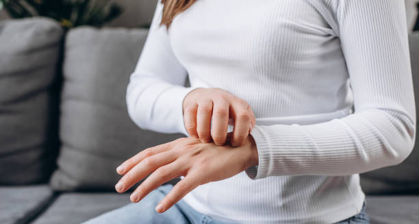
Woman scratching her irritated hand while sitting on a couch

Complications and Recovery from Bullous Pemphigoid

Complications and Recovery from Bullous Pemphigoid Complications and recovery from bullous pemphigoid are shaped by disease severity, treatment response, and overall patient health. While most people recover well with treatment, complications of bullous pemphigoid can arise from both the disease and its long-term therapy. One of the main complications is secondary skin infection. Open blisters can become entry points for bacteria, leading to cellulitis or more serious infections. Proper wound care, hygiene, and sometimes antibiotics are necessary to reduce this risk. Scarring and skin thickening may occur, especially in areas with repeated blistering. Pigment changes, both lighter and darker patches, can develop during the healing process. In some cases, movement may become restricted if scarring affects areas near joints. Steroid side effects are a significant concern in long-term treatment. These include weight gain, mood swings, osteoporosis, high blood pressure, blood sugar issues, and increased susceptibility to infection. Immunosuppressant drugs also carry a risk of liver toxicity, bone marrow suppression, or reactivation of latent infections. Emotional effects such as anxiety, frustration, or depression are common due to chronic pain, visible skin damage, and limitations on daily life. Mental health support, counselling, or support groups can play a critical role in recovery. Complications and Recovery from Bullous Pemphigoid Recovery times vary. Some people respond quickly and enter remission within months, while others may require years of intermittent treatment. Flare-ups can still occur, especially during times of stress or after reducing medication, but they usually become less frequent and easier to manage over time. Lifestyle measures to support recovery include: With early diagnosis, consistent management, and holistic support, most individuals with bullous pemphigoid can control the condition and enjoy a good quality of life — even if long-term treatment is needed. Back to Overview

Complications and Recovery from Bullous Pemphigoid Read More »

Scroll to Top