Mental Matters

Author name: admin

Causes of Macular Hole

Causes of Macular Hole

The causes of macular hole mainly stem from age-related changes in the eye. But other medical and mechanical factors can also lead to this condition. A macular hole is a full-thickness break in the macula — the central part of the retina responsible for clear, detailed vision. Knowing what causes this damage helps with early detection and prevention. Most macular holes happen spontaneously. They result from natural changes in the eye’s internal gel, called the vitreous. But trauma, severe nearsightedness, retinal diseases, and some eye surgeries can also cause holes. Whether the hole develops slowly or suddenly, understanding its cause helps eye doctors choose the best treatment and predict outcomes. Age-Related Vitreous Changes The most common cause of macular hole relates to ageing, especially changes in the vitreous humour. This jelly-like substance fills the inside of the eye. As we get older, the vitreous slowly liquefies and shrinks. Eventually, it can pull away from the retina in a process called posterior vitreous detachment (PVD). PVD usually is harmless, but sometimes the vitreous stays stuck to the macula. This pulling can tear the macula, creating a hole. This type of hole is called an idiopathic macular hole, meaning it forms without a clear cause. It accounts for most cases, especially in people over 60. Eye Trauma Blunt trauma to the eye also causes macular holes. A sudden hit sends shockwaves through the eye’s structures, damaging the retina. This can cause a hole right away or over time. Traumatic macular holes are more common in younger people who get injuries from sports, accidents, or fights. These holes often come with other eye damage, like bruises or bleeding. High Myopia (Severe Nearsightedness) People with high myopia face a higher risk of macular holes. In very nearsighted eyes, the eyeball stretches and thins the retina. This stretching weakens the macula, making it prone to breaks. High myopia can also cause conditions like macular schisis (splitting of retinal layers). If untreated, this can lead to full-thickness holes. Retinal Diseases Some retinal diseases also cause macular holes by damaging the macula’s structure: In these cases, holes develop slowly due to ongoing stress on the retina. Eye Surgery Certain eye surgeries, especially cataract removal, may increase the chance of macular holes later. Cataract surgery itself doesn’t cause holes directly. But changes in the vitreous or retina after surgery can pull on the macula. Inflammation after surgery can also lead to hole formation. People who already have early macular changes or vitreomacular adhesion before surgery face a higher risk. Inflammatory Eye Conditions Chronic inflammation inside the eye, like uveitis, can damage the macula. Persistent inflammation may cause swelling, scar tissue, or traction — all potential causes of macular holes. Systemic inflammatory diseases such as sarcoidosis or Behçet’s disease can also affect the retina and increase risk. Genetic and Congenital Factors Though rare, some macular holes relate to inherited or congenital eye problems. Juvenile retinoschisis is one example. These cases often occur in younger patients with retinal instability. Some families may have a genetic predisposition to macular diseases, though no single gene explains idiopathic holes. Vitreomacular Traction (VMT) Syndrome Vitreomacular traction syndrome happens when the vitreous partly detaches but stays stuck to the macula. This creates pulling forces that can lead to macular holes, especially in early stages. If caught early, treatments like ocriplasmin injections may release the traction and prevent full-thickness holes. Summary The causes of macular hole are varied but usually involve age-related vitreous changes, trauma, or chronic stress on the retina. Idiopathic holes are most common, especially after age 60. However, high myopia, eye surgery, inflammatory diseases, and genetic factors also contribute. Knowing the cause guides treatment and helps predict vision outcomes. Regular eye exams and early detection are key to preventing progression into a full-thickness macular hole. [Next: Symptoms of Macular Hole →]


Causes of Macular Hole Read More »

Symptoms of Macular Hole

Symptoms of Macular Hole

The symptoms of macular hole can develop gradually or appear suddenly, depending on the stage and underlying cause of the condition. The macula controls our sharp central vision. Any damage here leads to serious changes in how we see. While side vision usually stays normal, central vision problems affect daily life. Many people struggle with reading, driving, or recognising faces. At first, the signs may be mild. They can go unnoticed, especially if only one eye is affected. But as the condition gets worse, the visual problems become more obvious and harder to ignore. Spotting these changes early helps people get treatment in time and keep more of their vision. Blurry or Wavy Central Vision One of the first and most common symptoms of macular hole is blurry central vision. People often think they just need new glasses. It usually starts in one eye, and they might only notice it when they cover the other. As the hole grows, straight lines—like tiles or door frames—start to look bent or twisted. This visual change is called metamorphopsia. It becomes especially frustrating while reading. Words may seem broken, or entire parts of the page might go missing. Blind Spot in the Centre A dark or blank area in the middle of what you see is another key sign. This blind spot, called a scotoma, gets more obvious as the hole gets bigger. You might find it hard to see someone’s face clearly or read small print. Tasks like sewing or texting also become difficult. Some people describe this spot as a “shadow,” “smudge,” or a piece missing from the center of their vision. Central Vision Loss and Difficulty with Detail As the hole grows deeper, vision becomes worse. At first, this may not seem like a big problem. But over time, sharpness in the affected eye may drop a lot. In severe cases, vision may fall to the point where that eye meets the legal definition of blindness—though side vision remains. When only one eye is affected, the brain can mask the issue. That’s why many people miss the early signs. It’s important to check each eye separately to catch problems sooner. Trouble with Everyday Tasks Another group of symptoms of macular hole involves tasks that need sharp central vision. These challenges may include: Losing this type of vision affects your ability to live independently. It can also lower your confidence in daily life. Amsler Grid Distortion and Testing Doctors often use a test called the Amsler Grid to spot problems in the macula. It’s a simple grid of straight lines with a dot in the middle. People with macular holes often see wavy or missing lines near the center. Some might see a small dark spot instead of the dot. This test not only helps detect the condition but also tracks changes after treatment. Early Signs Often Show in One Eye Macular holes usually start in one eye. Because of this, the healthy eye takes over, and the brain fills in the missing parts. This makes it easy to miss the early signs. To catch the symptoms of macular hole early, test your vision one eye at a time. This is especially important for people over 60 or those with risk factors like severe nearsightedness or past eye problems. Comparing with Macular Degeneration Some signs of macular hole look like those from age-related macular degeneration (AMD). However, there are key differences: Knowing these differences helps doctors make the right diagnosis and choose the right treatment plan. Mental and Emotional Impact Vision loss doesn’t just affect your eyes. It can also take a toll on your emotions. Many people feel frustrated when they can’t see clearly or do the things they enjoy. You may feel anxious about losing more vision. Losing independence can lead to sadness or even depression. This is especially true if both eyes eventually become affected. Eye doctors and counselors can offer helpful support. Emotional care is just as important as physical treatment during this time. When to See an Eye Doctor You should see a specialist right away if you notice: Early treatment, especially for full-thickness holes, can make a big difference. Acting quickly can help save your central vision. Summary The symptoms of macular hole include blurry vision, distorted lines, and a growing blind spot in the center of your sight. These changes may start slowly, but they often get worse over time. Without treatment, full-thickness holes can lead to serious vision loss. Spotting the signs early and getting help from an eye doctor can protect your sight and improve your quality of life. [Next: Diagnosis of Macular Hole →]

Symptoms of Macular Hole Read More »

Diagnosis of Macular Hole

Diagnosis of Macular Hole

An accurate and timely diagnosis of macular hole is essential to prevent permanent central vision loss. Many people with macular holes experience blurry vision or central blind spots. These symptoms are similar to other eye diseases like macular degeneration. That’s why a full eye exam is needed to tell the difference. Eye doctors use a mix of patient history, vision tests, and detailed scans to confirm the condition and its stage. In the early stages, this condition can go unnoticed, especially if it affects just one eye. So, regular check-ups and early awareness are very important for a successful outcome. Common Symptoms and Patient History The diagnosis of macular hole often starts by listening to the patient. The doctor will ask about symptoms and past eye health. Common signs that raise concern include: People over 60, those with severe nearsightedness, or anyone who recently had cataract surgery are more at risk. If you notice a slow change in central vision, the eye doctor will likely suggest further tests. Vision Tests: Amsler Grid and Acuity Checks During the exam, the doctor checks how well each eye can see. People with a macular hole usually have poor central vision in one eye, but side vision stays normal. The Amsler grid is a quick test to spot problems in the macula. You focus on the center of a grid. If some lines look wavy or vanish, it may mean there’s a macular hole. This helps doctors find the exact part of the eye that is damaged. Direct Eye Exam with a Slit Lamp Next, the eye specialist looks closely at the retina using a slit-lamp microscope. This tool shows fine details at the back of the eye. In early stages, the retina might look a bit uneven. In later stages, the doctor may notice: These visible signs offer helpful clues and guide the next steps. High-Detail Imaging with OCT The most accurate way to confirm the diagnosis of macular hole is with Optical Coherence Tomography (OCT). This scan gives high-resolution, cross-section images of the retina without touching the eye. With OCT, doctors can: OCT also helps grade the stage of the hole: This staging helps doctors decide when and how to treat the hole. Extra Tools: Fundus Photography and More Doctors also use fundus photography to take color pictures of the retina. These images help track changes over time. Though not as exact as OCT, it’s useful for spotting other problems like scar tissue. It also helps explain the condition to patients visually. When Fluorescein Angiography Is Needed Sometimes, the signs are unclear or mixed with other eye issues. In such cases, doctors may use fluorescein angiography. This test involves a dye injection into your arm. As the dye travels through your eye’s blood vessels, a special camera takes pictures. Though it’s not routine for diagnosis of macular hole, it helps rule out conditions such as: Differentiating from Other Eye Conditions Not all central vision loss means a macular hole. That’s why proper diagnosis is key. Other possible causes include: Only the right diagnosis leads to the correct treatment. Macular holes often need surgery, while other issues may not. When to Refer to a Specialist Doctors may watch Stage I holes if vision is still good. But when a full-thickness hole shows up, referral to a retinal surgeon is urgent. Waiting too long can lead to scar tissue and vision loss that surgery can’t fix. Eye doctors often repeat OCT scans over time. This helps spot changes early, especially in Stage I or II, where the hole might even close on its own — though this is rare. Summary: Early Diagnosis of Macular Hole Saves Sight The diagnosis of macular hole involves history-taking, vision tests, and detailed scanning, especially with OCT. Correct staging of the hole is vital for choosing the best treatment. The sooner the problem is found, the better the chance of saving clear vision. People over 60 or with other eye conditions should have regular check-ups to catch issues early. [Next: Treatment of Macular Hole →]

Diagnosis of Macular Hole Read More »

Treatment of Macular Hole

Treatment of Macular Hole

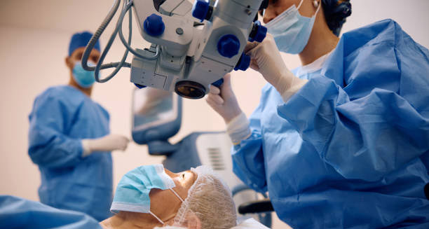
Treatment of macular hole aims to restore as much central vision as possible and prevent the condition from worsening. Modern eye care now offers very effective surgeries. These treatments work best when done early, with success rates over 90%. Because macular holes are physical tears in the central retina, eye drops or other non-surgical options rarely help in later stages. The best treatment depends on how far the macular hole has developed, its size, and the health of the eye overall. In early stages, small holes may just need regular checks. But once a full-thickness hole forms, surgery becomes the main way to avoid lasting vision loss. Understanding the Stages Before Treatment Eye doctors use a test called Optical Coherence Tomography (OCT) to see how far the macular hole has progressed: Acting early gives the best chance of saving sharp central vision. Waiting too long raises the risk of damage that may not be fixable later. Vitrectomy Surgery: The Main Treatment of Macular Hole The most common treatment of macular hole is a type of surgery called a vitrectomy. Here’s what happens during the procedure: After surgery, patients usually need to stay in a face-down position for several days. This step helps the bubble press on the macula properly. It’s one of the most important parts of the healing process and boosts the chances of success. Gas vs. Silicone Oil Tamponade in Treatment of Macular Hole Doctors mostly use gas to support healing after surgery. In tricky or repeat cases, they may use silicone oil instead. Silicone stays in the eye longer but requires another surgery to remove it later. Gas naturally fades away after a few weeks. However, during this time, patients cannot fly or go to high places because the bubble can expand and cause damage. What to Expect: Visual Recovery Timeline After a successful surgery, vision often starts to get better in about 4 to 6 weeks. Full healing can take up to 3 to 6 months. While most people don’t get perfect 20/20 vision back, many see a big improvement in what they can do. The sooner the surgery is done, the more likely the eye will recover well. If the hole stays open too long, light-sensing cells can stop working. That limits how much vision comes back. Important Postoperative Care Good care after surgery helps the treatment of macular hole work better. Doctors will give several instructions: If you notice new pain, vision loss, or floaters, contact your eye doctor right away. These signs could mean problems that need fast care. Other Treatment Options Surgery is still the top choice, but doctors sometimes explore other methods in early cases: Is Surgery Always Needed? Not always. Some very early holes don’t cause vision trouble and may not grow worse. In these cases, doctors may suggest just keeping an eye on it. However, once a full-thickness macular hole forms or vision gets worse, the treatment of macular hole should begin right away. For older people with other health issues or low chance of improvement, doctors may suggest skipping surgery after careful discussion. Possible Risks from Surgery Vitrectomy is safe for most people. But like any surgery, it has a few risks: Your surgeon will talk you through these risks and work to avoid them as much as possible. Coping with Face-Down Recovery Recovery after macular hole treatment can be hard, especially because of the face-down position. This can feel both tiring and lonely. Support from loved ones helps a lot. Special chairs, cushions, or massage tables can make the position easier. Planning ahead and asking for help can make the healing process go more smoothly. Summary: Successful Treatment of Macular Hole The treatment of macular hole works well in most cases, especially if done early. Vitrectomy surgery, combined with a gas bubble and careful recovery, gives the best chance to close the hole and bring back central vision. Acting quickly, following recovery steps, and keeping regular appointments all help to get the best result. Though other treatments may help in special cases, surgery remains the top choice today. [Next: Complications of Macular Hole →]

Treatment of Macular Hole Read More »

Complications of Macular Hole

Complications of Macular Hole

The complications of macular hole can arise from the condition itself or from surgical treatment. Although a macular hole is not usually painful, it can have a major impact on central vision. This can affect daily activities, emotional wellbeing, and long-term vision outcomes. If left untreated, a macular hole may worsen and lead to permanent central vision loss. Even after surgery, some patients experience lasting visual changes or side effects. Understanding these complications can help patients and caregivers manage expectations and plan ahead. Progressive Vision Loss One of the most serious complications of macular hole is progressive loss of central vision. When not treated in time: Peripheral vision usually remains intact, but the central vision loss can become permanent, especially if the condition progresses for months without treatment. In advanced cases, scarring may prevent full visual recovery — even after successful surgery. Macular Holes in Both Eyes Although uncommon, macular holes can develop in both eyes. This happens in about 10–15% of cases. When both eyes are affected, it can: If you’ve had a macular hole in one eye, regular monitoring of the other eye is important. Early detection can help prevent complications. Complications from Macular Hole Surgery Vitrectomy is a common and effective surgery to close a macular hole. But like any surgery, it carries some risks. 1. Cataract Formation The most common complication after surgery. 2. Retinal Detachment A rare but serious risk, affecting around 1–2% of patients. 3. Persistent or Recurrent Hole Sometimes the hole doesn’t close completely, or it reopens. 4. High Eye Pressure (IOP) Temporary rise in intraocular pressure can occur: 5. Infection (Endophthalmitis) A rare but vision-threatening infection. Visual Changes After Surgery Even if the surgery is successful, some patients continue to experience: These symptoms may improve slowly over time. However, some distortions may remain. Vision therapy and low-vision aids can help patients adjust. Emotional and Lifestyle Impacts The complications of macular hole don’t just affect vision. They can also lead to emotional and psychological challenges, including: Support from mental health professionals, occupational therapists, and low-vision support groups can make a big difference in quality of life. Missed or Delayed Diagnosis Missing the early signs of a macular hole can lead to worse outcomes: Routine eye exams are essential, especially for people with risk factors like myopia or vitreomacular traction. Tools like the Amsler grid can help catch changes early. Risk in the Fellow Eye Patients who’ve had a macular hole in one eye are at greater risk of developing one in the other. Monitoring both eyes regularly is important. Warning signs include: Early treatment in the second eye can help prevent similar complications. Summary The complications of macular hole include progressive vision loss, surgical risks like cataracts or retinal detachment, and emotional challenges. Fortunately, modern surgical treatment is often very successful — especially when started early. By understanding the potential complications and engaging in regular monitoring, patients can reduce their risk and protect their vision for the future. [Next: Back to Overview →]

Complications of Macular Hole Read More »

Overview of Macular Degeneration

Macular Degeneration

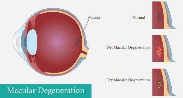
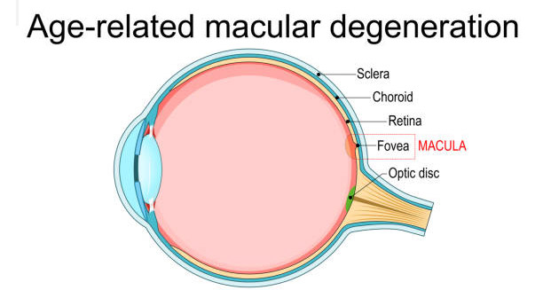
Overview of Macular Degeneration Macular degeneration — also known as age-related macular degeneration (AMD) — is a common eye condition that affects central vision. It damages the macula, which is the part of the retina responsible for sharp, straight-ahead vision. People with macular degeneration may struggle with reading, recognising faces, or driving. While peripheral (side) vision usually stays intact, the loss of central vision can be life-changing. Macular degeneration is one of the leading causes of vision loss in people over 50, especially in developed countries. As the global population ages, the number of people affected by AMD continues to rise. That’s why early detection and treatment are more important than ever. Understanding the Retina and the Macula The retina is a thin, light-sensitive layer at the back of the eye. It turns light into signals that the brain uses to create visual images. At the very centre of the retina is the macula — a small area that gives us clear, detailed vision. When the macula becomes damaged, people lose the ability to see fine details. While they won’t go completely blind, they may no longer be able to read, drive, or recognise faces clearly. Types of Macular Degeneration There are two main types of macular degeneration: dry and wet. Dry Macular Degeneration This is the most common type. It makes up about 85–90% of AMD cases. Dry AMD happens when the macula gets thinner with age. Yellow deposits called drusen also build up under the retina. Wet Macular Degeneration Wet AMD is less common but more serious. It occurs when abnormal blood vessels grow under the retina and leak blood or fluid. This damages the macula quickly. Wet AMD is a medical emergency. Fast treatment can help preserve vision and sometimes even improve it. Early Signs and How It Progresses In the early stages, macular degeneration may not cause any symptoms. Many people don’t notice anything until their vision starts to change. As the disease progresses, symptoms can include: Dry AMD progresses slowly, while wet AMD can get worse very quickly. Who Is at Risk for Macular Degeneration? Some people are more likely to develop AMD than others. The biggest risk factor is age, but many other factors can also play a role. Common risk factors include: The good news is that many of these risks can be reduced with healthy lifestyle choices. Why Early Detection Is So Important Catching macular degeneration early makes a big difference. While there is no cure for dry AMD, taking the right vitamins can slow its progression. Wet AMD can be treated with injections that help stop vision loss — and sometimes even improve it — if given early. Everyone over 50, or anyone at risk, should have a dilated eye exam once a year. Using tools like the Amsler grid at home can also help track changes in vision between appointments. Living with Macular Degeneration Losing central vision can affect every part of daily life. It may lead to: Fortunately, there are many ways to adapt. Vision aids, counselling, and support groups can help people stay independent and improve their quality of life. Summary Macular degeneration is a serious eye condition that affects the part of the retina responsible for sharp vision. It mostly affects older adults and can lead to the loss of central vision over time. While it doesn’t cause complete blindness, it can make everyday tasks difficult. Early diagnosis and treatment are key. With proper care, many people with AMD can maintain their independence and enjoy a good quality of life. [Next: Causes of Macular Degeneration →]

Macular Degeneration Read More »

Causes of Macular Degeneration

Causes of Macular Degeneration

The causes of macular degeneration are multifactorial, involving a complex interaction between genetics, environmental factors, ageing, and underlying health conditions. While the exact cause is not fully understood, extensive research has identified a number of biological and lifestyle-related contributors that increase the risk of developing this degenerative eye condition. Understanding the causes of macular degeneration is crucial for identifying those at risk and implementing early preventative strategies. Ageing and Natural Eye Changes Age is the biggest risk factor for macular degeneration. As people age, parts of the eye naturally begin to break down. The retina, especially the macula, becomes more vulnerable to damage. Here’s how ageing contributes: These changes reduce how well the macula works and can lead to vision loss. Genetic Links and Family History Genetics also play a strong role in the causes of macular degeneration. If someone in your immediate family has AMD, your risk goes up. Researchers have found that certain genes make people more likely to develop AMD. These include: These genes affect how the body handles inflammation and damage from free radicals. People with these gene types may not protect their retina as well from harm. Smoking and Oxidative Stress The retina uses a lot of oxygen, making it very sensitive to oxidative stress. Free radicals — unstable molecules — can damage cells in the eye. Over time, this damage: Smoking makes this problem worse. It’s one of the most dangerous causes of macular degeneration. Smokers have a much higher risk of AMD because: Even people who quit smoking remain at higher risk for years after. Causes of Macular Degeneration: Diet and Health Conditions What you eat also affects your eyes. A poor diet — low in antioxidants and high in unhealthy fats — increases your risk. Not getting enough nutrients like vitamins C, E, zinc, lutein, and zeaxanthin can: Medical studies like AREDS and AREDS2 showed that supplements with these nutrients help slow AMD, especially in the middle stages. In addition to diet, these health problems may raise your risk: Staying on top of your general health is one way to protect your vision. Environmental and Lifestyle Factors Many other lifestyle choices affect your chances of developing AMD. Sunlight and blue light:Too much UV light or high-energy blue light can stress the retina. Being outdoors without sunglasses or spending long hours on screens may add to the risk. Lack of exercise and obesity:Being overweight increases inflammation and can reduce blood flow to the retina. It also affects how well your body uses nutrients. Regular physical activity may lower your risk. Gender and ethnicity:Women are slightly more likely to develop AMD, possibly because they tend to live longer. Caucasians are also at higher risk than other groups, likely due to having lighter eye pigmentation. Medication Risks and Other Triggers Some drugs have been linked to higher AMD risk. These include: If you take any of these medicines, it’s important to have regular eye exams and report any vision changes quickly. Summary of the Causes of Macular Degeneration The causes of macular degeneration include a mix of age, genetics, poor lifestyle choices, and other health problems. Some risk factors, like ageing, can’t be avoided. But many others — such as smoking, poor diet, and lack of exercise — can be managed. By knowing what increases your risk and making healthy choices early, you can take steps to protect your eyesight. [Next: Symptoms of Macular Degeneration →]

Causes of Macular Degeneration Read More »

Symptoms of Macular Degeneration

Symptoms of Macular Degeneration

The symptoms of macular degeneration can vary depending on the stage and type of the condition — whether it is dry or wet age-related macular degeneration (AMD). While early macular degeneration may be asymptomatic, the disease often progresses slowly, eventually affecting central vision and interfering with essential daily activities. Identifying the symptoms of macular degeneration at an early stage can help initiate treatment strategies and reduce the risk of severe visual impairment. Early Symptoms: Often Subtle and Overlooked One of the most difficult things about AMD is that it often starts without warning. In the early stages, especially with dry AMD, most people don’t notice any changes in vision. The damage happens slowly, and the brain uses the other eye to make up for it. This often delays the discovery of the disease. Still, early signs may start to show, such as: These signs may seem small, but they should not be ignored. They are good reasons to schedule a full eye exam. Worsening Symptoms in the Intermediate Stage As the disease moves forward, symptoms of macular degeneration become clearer. Central vision gets worse, making daily tasks harder. Common symptoms at this stage include: These problems might only affect one eye at first. The other eye often fills in the gaps, which may delay noticing the problem. Symptoms of Macular Degeneration in Wet AMD Wet AMD develops faster and causes more dramatic vision changes. It happens when abnormal blood vessels grow and leak fluid under the retina. These leaks cause the macula to swell and vision to get worse quickly. Symptoms of wet AMD include: Wet AMD is serious and needs fast medical care. Quick treatment may stop the vision from getting worse and might even help improve it slightly. Bilateral Vision Loss and Its Effects In many cases, macular degeneration affects both eyes, though not always at the same time. When both eyes are affected, daily life becomes much harder. People often experience: Unlike full blindness, macular degeneration doesn’t take away side vision. Most people can still move around and see what’s to the side. But the loss of clear, direct vision affects things that give life meaning — reading, recognising faces, and enjoying hobbies. Differentiating from Other Eye Conditions Many eye diseases share similar signs, so it’s important to know when it’s really AMD. For example: If central vision suddenly changes — especially with distortion or a dark spot — it’s essential to see an eye doctor right away. Tests like the Amsler grid or OCT can confirm the symptoms of macular degeneration. Emotional and Mental Effects Living with the symptoms of macular degeneration can take a mental toll. Vision loss changes how people live, work, and connect with others. It’s common to feel: Help is available. Vision rehab, talking to a therapist, using assistive tools, and joining support groups can make a big difference. Summary of Symptoms of Macular Degeneration The symptoms of macular degeneration often start slowly, with slight blurring or distortion in central vision. Over time, it may become harder to read, drive, or recognise people’s faces. While dry AMD moves slowly, wet AMD can cause sudden and serious vision loss. Noticing symptoms early gives people a better chance to get treatment and protect the vision they still have. [Next: Diagnosis of Macular Degeneration →]

Symptoms of Macular Degeneration Read More »

Diagnosis of Macular Degeneration

Diagnosis of Macular Degeneration

The diagnosis of macular degeneration is a vital step in preserving vision and managing the progression of the disease. Because age-related macular degeneration (AMD) often begins without noticeable symptoms, many cases are only detected during routine eye exams. Early detection is crucial, especially for the wet form of AMD, which can cause rapid and severe vision loss. Timely diagnosis allows for prompt treatment that may prevent further deterioration and help preserve existing vision. Why Early Detection Matters Catching AMD early plays a huge role in treatment success, especially with the wet type. For dry AMD, while there’s still no cure, simple lifestyle changes and vitamin supplements can slow things down. For wet AMD, starting treatment early can often help maintain or even improve vision. The challenge is that AMD often starts without clear warning signs. People may not notice small vision changes until it gets worse. That’s why regular eye exams are so important, particularly for those over 50 or with known risk factors. Medical and Family History Assessment Doctors begin the diagnosis of macular degeneration by reviewing the patient’s full background. This includes: These details help the eye doctor figure out the risk level and choose the right tests. Key Eye Exam Techniques A full eye check-up with dilated pupils is essential for spotting AMD. It allows the doctor to clearly see the retina and macula. The main parts of this check include: Advanced Tools for Diagnosis of Macular Degeneration Special tests and imaging tools give a closer look at the retina and help confirm the condition. OCT is especially useful for wet AMD because it can catch small fluid changes and track damage. Staging and Identifying the Type After making the diagnosis of macular degeneration, the eye doctor will place the condition into one of these stages: These stages help guide treatment and follow-up plans. Ruling Out Other Conditions Other eye problems can look like AMD. Doctors must make sure the diagnosis is correct. Similar conditions include: Clear imaging and expert review help confirm AMD and avoid mistakes in care. Keeping an Eye on Progress Once diagnosed, patients need regular check-ups to track any changes. These may include: Sudden vision changes—like blurring, wavy lines, or central blind spots—should be reported right away. This is especially true for people with dry AMD, who might shift into the wet form. Summary of Diagnosis of Macular Degeneration The diagnosis of macular degeneration combines health history, eye exams, and advanced imaging. Spotting it early is vital, especially for wet AMD, where fast action can save vision. Regular screening and close monitoring help catch problems before they get worse. With early diagnosis, patients can begin the right treatments to protect their sight and live more independently. [Next: Treatment of Macular Degeneration →]

Diagnosis of Macular Degeneration Read More »

Treatment of Macular Degeneration

Treatment of Macular Degeneration

The treatment of macular degeneration varies a lot depending on the type and stage of the disease. While there is no cure yet, several treatment options can slow the disease, save vision, and sometimes even improve sight—especially for wet (neovascular) macular degeneration. Knowing the difference between treatments for dry and wet AMD is very important for patients, families, and doctors. Treatment of Dry Macular Degeneration Dry AMD is the more common and usually less aggressive form. Although no medicine can reverse its effects, many ways exist to manage the condition and delay vision loss. These antioxidants and minerals help lower oxidative stress and may slow the move from moderate to advanced AMD. Remember, these supplements don’t cure the disease and are mainly for people with moderate to severe dry AMD, not early stages or prevention. Besides diet, keeping a healthy weight, controlling blood pressure, and quitting smoking are key steps for long-term care. Treatment of Wet Macular Degeneration Wet AMD grows faster but responds better to treatment when caught early. It happens when abnormal blood vessels grow and leak under the retina, causing quick central vision loss. Early and ongoing care can greatly improve or keep vision stable in many patients. Common anti-VEGF drugs include: Doctors inject these medicines directly into the eye, usually once a month at first, then less often depending on results. These drugs changed AMD treatment and help many patients keep or improve vision. PDT is less common now because anti-VEGF drugs work better, but it may help patients who don’t improve with injections. Vision Rehabilitation and Assistive Technologies Even with treatment, some patients lose vision permanently. Low-vision rehab can greatly improve life quality. Helpful tools include: These tools help patients stay independent despite central vision loss. Clinical Trials and Emerging Treatments Research continues to find new ways to treat macular degeneration. Some exciting areas are: Patients interested in trials should talk to a retina specialist about options. Lifestyle Support and Mental Health Vision loss from AMD can cause strong emotions. Many patients feel frustrated, anxious, or depressed. It is important to get emotional support through counseling, support groups, or occupational therapy. Treating AMD means caring for eyesight and mental strength and independence. Summary of Treatment of Macular Degeneration The treatment of macular degeneration depends on its type—dry or wet—and how far it has progressed. Dry AMD care focuses on nutrition, watching closely, and lifestyle changes. Wet AMD needs medical care, mainly anti-VEGF injections. Vision rehab, helpful devices, and new treatments bring hope for long-term care. With early diagnosis and steady care, many people with AMD can keep useful vision and a good life. [Next: Complications of Macular Degeneration →]

Treatment of Macular Degeneration Read More »

Scroll to Top