Mental Matters

Health & Wellness

Overview of Kyphosis

Overview of Kyphosis

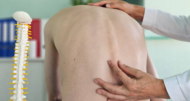
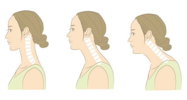
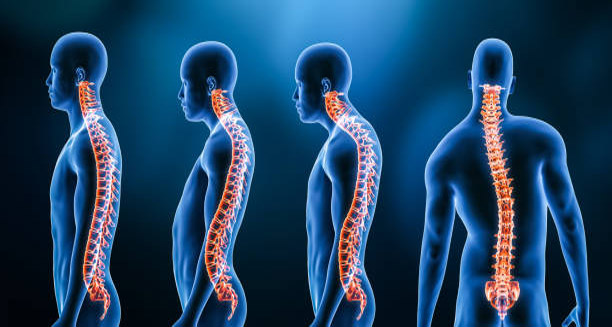
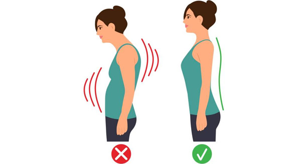
The overview of kyphosis begins with an understanding of this postural and structural condition that affects the natural curvature of the spine. Kyphosis often shows up as a forward rounding of the upper back. The overview of kyphosis includes people of all ages, but it happens more often in teens and older adults. Mild cases usually do not cause any symptoms. However, more serious cases can bring pain, stiffness, and daily limits in movement. This condition causes a strong outward bend in the upper spine, making the back look hunched. This curve might be flexible or stiff. That depends on what caused it. It’s important to tell the difference between normal posture and harmful kyphosis that needs medical care. Some people develop it slowly over time. Others get it suddenly because of an injury, illness, or their genes. Types and Causes in the Overview of Kyphosis Many types of kyphosis exist, and each type has its own cause and path. The most common one is postural kyphosis. It often shows up in teenagers because of slouching or sitting badly. This kind usually doesn’t involve bone problems. People can often fix it with exercises and better posture. In contrast, Scheuermann’s kyphosis shows up during the teen years too, but the spine bones develop in an uneven way. This type often needs more treatment. Another type, congenital kyphosis, is already present at birth. It happens when the spine doesn’t form correctly in the womb. Sometimes, surgery is needed early in life. For older people, kyphosis often happens because of bone thinning, or osteoporosis. Weak bones can break or collapse, causing the spine to round. This version, called osteoporotic kyphosis, can raise the chance of spine fractures and even affect breathing. Other conditions, such as arthritis, infections, or spine tumours, can also cause the spine to curve more. Symptoms and Emotional Impact in the Overview of Kyphosis People usually first notice kyphosis when they see a hump or curve in the upper back. Some feel very tired or stiff. Others find it hard to stand straight for a long time. Even when there’s no pain, bad posture can cause muscle strain, pinched nerves, or breathing trouble in worse cases. In more serious cases, kyphosis can lower someone’s ability to move or live fully. It might also lower their self-esteem because of how they look. This is especially true for kids and teens. A visible spine curve can lead to worry, low confidence, or sadness. Sadly, doctors might miss these emotional signs. That’s why it’s important to treat both the body and the mind. Diagnosis and Treatment in the Overview of Kyphosis Doctors find out if someone has kyphosis by doing a physical check, asking about health history, and taking scans. X-rays show the spine curve clearly. MRI or CT scans can look deeper into soft tissues or nerve problems. Getting the right diagnosis helps doctors decide if the kyphosis comes from posture, bone shape, or another illness. Treatment depends on what kind of kyphosis someone has, how serious it is, and their age. For postural kyphosis, changes in daily habits, special exercises, and good furniture may help. But if the curve is more serious or caused by misshapen bones, people might need a back brace. In some cases, surgery is the best choice. Doctors might do spinal fusion to fix and support the spine, but healing from that takes time. Prevention and Long-Term Outlook in the Overview of Kyphosis People can lower their risk of kyphosis by keeping their spine healthy. That means sitting and standing straight, eating foods rich in calcium and vitamin D, and doing exercises that strengthen bones. Older adults should get regular bone checks. Catching osteoporosis early can stop spine breaks that lead to a hunched back. Ongoing care also matters a lot. Physical therapy can make the back and stomach muscles stronger. It also helps the spine stay flexible. Sometimes, back supports can help. Learning how to lift things safely and setting up work areas to help posture also protects the spine. The overview of kyphosis should also cover what the future may look like. Some mild cases stay the same for years. But others may slowly get worse. If no one treats serious spinal curves, it can hurt breathing, press on the stomach, or even affect the nerves. These effects make daily life much harder. Full Management in the Overview of Kyphosis To manage kyphosis well, doctors must find it early and use a full care plan. Many types of health workers may be involved—general doctors, bone specialists, physical therapists, and even mental health experts. Loved ones can help too. Patients who understand their condition are more likely to follow treatment and get better results. In the end, the overview of kyphosis shows how key it is to understand all the causes, types, and effects of this spine problem. With early care and the right support, many people with kyphosis can live full, active lives. The main goal is to find the issue soon and treat both the physical and emotional parts of it. [Next: Causes of Kyphosis →]

Overview of Kyphosis Read More »

Causes of Kyphosis

Causes of Kyphosis

Understanding the causes of kyphosis is essential for accurate diagnosis and effective treatment planning. The causes of kyphosis vary depending on when it starts, a person’s health, and daily habits. Some people are born with spinal problems, while others develop them later. Poor posture, injuries, or age-related changes often play a role. Since kyphosis is a symptom rather than a single disease, its causes need to be looked at from both a medical and mechanical angle. A common cause is postural kyphosis, especially in teens and young adults. It usually results from poor posture or slouching. This type is flexible, meaning the back curve improves when the person stands up straight. It does not involve structural damage to the spine. In today’s digital world, longer screen time and more sitting have made postural kyphosis more common, particularly among students and office workers. A more serious form is Scheuermann’s kyphosis, which usually begins during teenage years. This condition comes from wedge-shaped bones in the spine. Unlike postural kyphosis, it causes a rigid curve that does not go away with better posture. Experts think Scheuermann’s disease may be caused by genetic or growth problems. It affects more males than females and often causes back pain. Congenital kyphosis is another cause. This form develops before birth when the spine does not form or connect properly. The result is a stiff and increasing curve that gets worse as the child grows. Without treatment, it may press on the spinal cord and cause nerve problems. Surgery is often needed, and early detection helps with better planning. Structural and Medical Causes of Kyphosis Degenerative changes are another major factor, especially in older adults. As people age, bones and discs in the spine break down. This leads to degenerative kyphosis, often seen with osteoarthritis or disc thinning. These changes make the spine lean forward. Weakened ligaments and stiff joints make the problem worse. Osteoporosis can also cause osteoporotic kyphosis. In this case, the spine weakens and collapses due to tiny fractures. These vertebrae turn wedge-shaped and create the hunched appearance. It often goes unnoticed until someone has back pain, shrinks in height, or the curve becomes obvious. Injuries can cause post-traumatic kyphosis. Accidents, falls, or sports injuries may fracture the spine. When these fractures heal poorly, they lead to a bent or curved back. Some cases also include nerve damage. Surgery may be needed if the curve is severe or causes other problems. Infections, such as spinal tuberculosis (Pott’s disease), can damage spinal bones and discs. This causes the spine to collapse, leading to kyphosis. It mostly affects people in developing areas or with weak immune systems. Quick treatment with antibiotics and sometimes surgery is important to stop the curve from worsening. Spinal Tumours Spinal tumours, either cancerous or not, can also lead to kyphosis. Tumours may press on the spine, weaken the bones, or cause the spine to collapse. In such cases, treating both the tumour and the spinal curve is necessary. Neuromuscular diseases like cerebral palsy, muscular dystrophy, and spinal muscular atrophy also contribute. These conditions weaken the muscles that support the spine. Over time, this leads to poor posture and spinal deformities. Treatment may include braces, therapy, or surgery. Sometimes, kyphosis results from medical treatments. For example, past spine surgery, radiation therapy, or poor surgical techniques can cause the spine to curve. These are called iatrogenic causes. They often need revision surgery. Lifestyle habits also play a role in the causes of kyphosis. Lack of exercise, poor sitting posture, and being overweight weaken the muscles that hold the spine upright. Over time, these habits encourage a curved back. Simple steps like moving more, sitting properly, and strengthening the back can help. In summary, the causes of kyphosis include many factors such as age, genetics, bone strength, posture, injuries, and other health conditions. Knowing the cause helps doctors create a better treatment plan. Acting early can stop the curve from getting worse and protect spinal health. [Next: Symptoms of Kyphosis →]

Causes of Kyphosis Read More »

Symptoms of Kyphosis

Symptoms of Kyphosis

Recognising the symptoms of kyphosis is crucial for early intervention and successful treatment. The symptoms of kyphosis can vary widely depending on the type, severity, and underlying cause of the condition. In some cases, the spinal curvature may be mild and cause no symptoms. In others, it can lead to physical discomfort, limited movement, and emotional challenges. That’s why understanding the full range of symptoms is important for healthcare providers, caregivers, and patients alike. Visible Signs and Spinal Curvature in Kyphosis The most noticeable symptom is an abnormal spinal curve, often described as a rounded or hunched back. This curve usually affects the upper (thoracic) spine and may cause someone to look stooped or slouched. In mild postural kyphosis, the change in appearance might only show after long periods of standing or sitting. In more severe forms, like Scheuermann’s disease or congenital kyphosis, the curve becomes stiff and permanent. Children or teens with these conditions may develop a visible hump, especially during growth spurts. Back pain is another common symptom, especially in adults. This pain often comes from extra strain on the muscles, ligaments, and discs caused by the spine’s poor alignment. Postural kyphosis may lead to dull, on-and-off pain, which worsens after sitting or standing too long. In contrast, Scheuermann’s kyphosis causes more constant pain due to its rigid curve and disc issues. Degenerative or osteoporotic kyphosis in older adults also often causes ongoing pain in the lower or mid-back. Fatigue, Height Loss, and Physical Limitations Fatigue is another symptom that may go unnoticed but plays a big role. The unusual spinal curve forces the muscles of the back and neck to work harder to keep the body upright. This added effort leads to tiredness, especially after physical activity. Some people also feel a sense of heaviness in the back, making it hard to complete everyday tasks or stay active. Loss of height is a frequent sign, particularly in older adults with osteoporotic kyphosis. As the spine curves and vertebrae compress, height gradually decreases. This often comes with a forward-leaning posture and trouble standing up straight. These visible changes may affect self-esteem, especially in teens and those concerned with their appearance. In severe or advanced cases, the symptoms of kyphosis go beyond discomfort. Movement in the upper back may become limited. Bending, twisting, or lifting can become harder. When the spine becomes very stiff, even simple tasks like dressing, tying shoes, or reaching overhead may be difficult. Neurological and Respiratory Symptoms of Kyphosis More serious symptoms may involve the nerves. If the spinal curve worsens or narrows the spinal canal, it can press on the spinal cord or nerve roots. This may cause tingling, numbness, or weakness in the arms or legs. In rare but serious cases, such as congenital or trauma-related kyphosis, pressure on the spinal cord can affect coordination or cause bladder and bowel issues. Kyphosis can also affect breathing. Severe upper back curves may pull the ribcage forward and down, limiting lung expansion. This can lead to short, shallow breaths or breathing problems during activity. In extreme cases, especially congenital kyphosis, respiratory failure can occur. Kyphosis Symptoms in Children and Emotional Impact Children with kyphosis may show signs like stiffness in the back, clumsiness, or avoiding physical play due to pain or self-consciousness. For teenagers, mental and emotional symptoms are important to watch. Those with visible spinal curves may be bullied or feel isolated. This can lead to anxiety, low self-esteem, or depression. Support from family, teachers, and mental health professionals is key in these situations. Sometimes, the symptoms of kyphosis develop slowly and are easy to overlook. This is often the case with postural kyphosis. People may think it’s just poor posture rather than a medical problem. That’s why regular check-ups—especially for teens and older adults—are important for spotting the curve early and stopping it from getting worse. The Importance of Proper Diagnosis Many symptoms of kyphosis overlap with other spine conditions like scoliosis, lordosis, or vertebral fractures. A proper physical exam and imaging tests are needed to make the correct diagnosis. Once kyphosis is diagnosed, tracking changes in symptoms over time helps show if it’s staying the same or getting worse. In conclusion, the symptoms of kyphosis range from posture changes to major physical and emotional challenges. Spotting symptoms early and treating the whole person—both physically and emotionally—can lead to better outcomes. Whether symptoms are mild or severe, early diagnosis and care make a big difference in quality of life for people living with kyphosis. [Next: Diagnosis of Kyphosis →]

Symptoms of Kyphosis Read More »

Diagnosis of Kyphosis

Diagnosis of Kyphosis

A thorough and timely diagnosis of kyphosis is essential in determining the severity of spinal curvature and identifying any underlying causes or complications. The diagnosis of kyphosis usually starts with a detailed medical history and physical exam, followed by imaging and functional tests. Accurate diagnosis helps distinguish between types—postural, congenital, Scheuermann’s, or degenerative—and guides treatment and monitoring. Doctors begin by asking about symptoms, family history, and related problems such as back pain, tiredness, or breathing trouble. Parents may notice changes in posture or height in children and teens. Knowing when symptoms began and if they are worsening helps decide if kyphosis is flexible (postural) or fixed (structural). Physical Exam and Imaging in Diagnosis of Kyphosis During the physical exam, the doctor observes the spine’s curve while the patient stands, bends forward, or lies down. In postural kyphosis, the curve often disappears when lying flat or standing straight. In structural kyphosis like Scheuermann’s or congenital types, the curve stays visible and does not correct with posture changes. The doctor also checks for uneven shoulder height, a prominent upper back, or an exaggerated spine curve. To confirm the diagnosis of kyphosis, imaging tests are needed. A side-view X-ray is the first step. It measures the Cobb angle, which shows the degree of curvature. A Cobb angle over 40 degrees usually means kyphosis. In teens, a sharp curve with wedge-shaped vertebrae may indicate Scheuermann’s disease, while a smooth curve suggests postural causes. Advanced imaging, such as MRI, may be done if doctors worry about spinal cord or nerve issues. MRI offers detailed pictures of the spinal cord, discs, and muscles, helping rule out tumors, cysts, or infections. CT scans are less common but useful for assessing congenital kyphosis or trauma-related problems. Neurological and Bone Health Evaluation Neurological exams are important if patients report numbness, tingling, or weakness. Reflexes, sensation, and muscle strength tests check for nerve or spinal cord compression. If signs of nerve damage appear, urgent imaging and referral to a spine specialist may follow. In older adults with osteoporosis or fractures, bone density tests like DEXA scans help assess fracture risk. Early detection of bone loss can guide treatment to prevent worsening kyphosis and new fractures. Pulmonary function tests may also be needed. Severe thoracic kyphosis can limit lung expansion. Spirometry or full lung function tests evaluate breathing capacity, especially in patients with shortness of breath or fatigue. Special Cases and Monitoring For children with suspected congenital kyphosis, early diagnosis is critical. These cases often progress fast and may need surgery to avoid deformity or nerve damage. Pediatric orthopaedic specialists manage diagnosis using X-rays, MRIs, and clinical checks. After confirming kyphosis, classifying the type guides treatment. Postural kyphosis tends to be mild and reversible with exercise and posture correction. Scheuermann’s kyphosis is rigid and may require bracing or surgery. Degenerative kyphosis relates to aging spine changes and needs therapy, pain relief, and bone health support. Proper classification ensures treatment matches the patient’s age, symptoms, and needs. Monitoring is key. Even after diagnosis, patients—especially growing kids—need regular follow-ups. X-rays every 6 to 12 months track changes. Adults with symptoms also require imaging to assess progression or complications. It is vital to distinguish kyphosis from other spine conditions like scoliosis (sideways curve) or lordosis (inward curve). Sometimes patients have kyphoscoliosis, combining curves. Accurate diagnosis avoids wrong treatment and unnecessary discomfort, stressing the need for specialist care in complex cases. Summary of Diagnosis of Kyphosis In summary, the diagnosis of kyphosis involves a stepwise approach with clinical exams, imaging, and functional tests. Whether postural or structural, early and accurate diagnosis allows timely treatment, prevents complications, and supports long-term spine health. Modern tools and awareness give patients a better chance at maintaining quality of life through early care and ongoing management. [Next: Treatment of Kyphosis →]

Diagnosis of Kyphosis Read More »

Treatment of Kyphosis

The treatment of kyphosis is highly dependent on the cause, degree of spinal curve, patient’s age, and whether pain or functional problems occur. Early treatment can greatly improve posture, reduce discomfort, and stop progression. This is especially true in teens during growth spurts and older adults with degenerative kyphosis. A range of treatments exist—from conservative to surgical—to manage this spinal condition. Conservative Treatment of Kyphosis Postural kyphosis, the most common and mildest form, usually starts with non-invasive care. Physical therapy focuses on strengthening the spinal extensor muscles, improving core stability, and increasing flexibility. Stretching the hamstrings, chest, and hip flexors helps correct muscle imbalances that worsen poor posture. Over time, a personalised exercise plan can reduce the curve and ease pain, particularly if started early. Postural education plays a key role too. Many patients, especially children and teens, don’t notice their poor posture habits. Physiotherapists teach proper sitting, standing, and moving techniques to keep the spine aligned. Simple changes—like adjusting desk or screen height—help prevent worsening and reduce discomfort. In rigid kyphosis forms such as Scheuermann’s disease, bracing may be necessary during adolescence. A thoracolumbosacral orthosis (TLSO) brace gently guides the spine toward a more natural curve. Patients usually wear the brace for many hours daily, sometimes up to 20 hours. Bracing works best before bones fully mature. Sticking to the schedule is vital, and doctors regularly check progress. While bracing won’t completely fix the curve, it can stop it from worsening and may avoid surgery. Pain Management and Lifestyle Changes Adults with degenerative kyphosis or Scheuermann’s disease often need pain relief. Over-the-counter medicines like paracetamol or NSAIDs help mild to moderate pain. Stronger drugs or steroid injections may ease nerve pain or inflammation, especially if nerve compression occurs. Lifestyle changes support treatment. Keeping a healthy weight lessens spinal pressure. Avoiding heavy lifting or high-impact sports prevents worsening. For elderly patients with osteoporosis-related kyphosis, getting enough calcium and vitamin D, plus medications like bisphosphonates, strengthens bones and lowers fracture risk. Fall prevention is critical since minor trauma can worsen spinal curves in these patients. Surgical Treatment of Kyphosis When conservative care fails or the curve passes 75 degrees, surgery may be needed. The most common operation is spinal fusion, which joins vertebrae using rods, screws, and bone grafts. The aim is to realign the spine, stabilise the curve, and relieve pressure on nerves or the spinal cord. Surgery suits patients with worsening deformity, severe pain, or neurological problems like weakness, numbness, or bladder issues. Before surgery, patients undergo thorough evaluations, including imaging and heart and lung tests. Recovery usually involves several hospital days and months of rehab. Risks include infection, bleeding, hardware problems, and damage to nearby spinal segments. Still, many patients see major improvements in posture, pain, and quality of life after surgery. Psychological and Follow-Up Care Kyphosis also affects mental health. Teens and young adults may suffer from low self-esteem due to their visible spine shape. Counselling or support groups can help them cope. Treating both body and mind leads to better outcomes, especially for younger people. Regular monitoring is key in the treatment of kyphosis. Whether conservative or surgical, doctors track the spinal curve and symptoms over time. Children and teens may need X-rays every six to twelve months, especially during growth spurts. Adults with pain or degeneration also require periodic scans and check-ups. Emerging Treatments and Special Cases New therapies include minimally invasive surgeries and biologics that help bone healing or fusion. These are still experimental but show promise. Also, posture-correcting devices and smart sensors are growing as tools to aid posture training. However, they do not replace medical care. Congenital kyphosis, where spinal defects exist from birth, needs early detection and often surgery in the first years of life. Specialist care by paediatric orthopaedic surgeons is vital to prevent neurological damage and manage curve progression. Summary of Treatment of Kyphosis In summary, the treatment of kyphosis depends on the individual’s age, symptoms, and spinal curve type. Many cases respond well to physiotherapy, bracing, and lifestyle changes. Others require surgery to restore function and avoid complications. Early diagnosis, education, and close follow-up form the foundation of effective care. A personalised treatment plan can improve posture, reduce pain, and enhance quality of life. [Next: Complications of Kyphosis →]

Treatment of Kyphosis Read More »

Complications of Kyphosis

Complications of Kyphosis

The complications of kyphosis can vary widely depending on the severity, type, age of onset, and whether the condition is treated early or allowed to progress. While some individuals experience only mild symptoms or cosmetic concerns, others face significant physical, psychological, and neurological issues. Understanding the complications of kyphosis is essential for early intervention, risk reduction, and long-term health management. One of the most immediate and noticeable complications is chronic back pain. The altered spinal alignment places extra stress on muscles, ligaments, and joints, causing persistent discomfort. This mechanical pain is especially common in adults with Scheuermann’s disease or degenerative kyphosis. Over time, continuous strain leads to muscle fatigue, spasms, and reduced mobility. These problems greatly impact daily activities and quality of life. Respiratory and Digestive Complications of Kyphosis Respiratory issues are another common complication. Severe kyphotic curves—especially those over 70 to 80 degrees—can compress the chest cavity. This compression limits lung expansion, causing shortness of breath, reduced exercise tolerance, and a higher chance of lung infections. Older adults with osteoporotic kyphosis may see worsening of pre-existing lung conditions like COPD, making their care more challenging. Digestive problems also arise when spinal curvature presses on the abdominal area. Pronounced kyphosis can push organs such as the stomach and intestines into unusual positions. This may cause acid reflux, feeling full quickly, and abdominal discomfort. Although less common than breathing issues, these gastrointestinal problems should not be overlooked or misdiagnosed. Neurological and Psychosocial Complications Neurological problems rank among the more serious complications of kyphosis. Extreme spinal curves can compress the spinal cord or nerve roots. This compression leads to tingling, numbness, weakness, or sharp pain in the arms or legs. In severe cases, kyphosis causes myelopathy—spinal cord compression that can result in poor coordination, trouble walking, or bladder and bowel issues. Early diagnosis and surgery often prevent permanent nerve damage. The psychosocial impact of kyphosis is frequently underestimated. Visible spinal curves can cause embarrassment, low self-esteem, and social withdrawal, especially in teenagers and young adults. Bullying and self-consciousness in social settings raise the risk of anxiety and depression. Adults may also struggle with body image and confidence, which affect relationships and emotional health. Functional Limitations and Fracture Risks Functional limitations are a major concern. As kyphosis worsens, standing for long periods, walking far, or lifting objects becomes difficult. In elderly patients, reduced mobility contributes to frailty, increased fall risk, and dependence on assistive devices or caregivers. These challenges reduce independence and lower quality of life. In older adults, kyphosis often links to a higher risk of vertebral fractures, especially when caused by osteoporosis. Vertebral wedging weakens the spine, making it prone to collapse even after minor injuries. Each fracture worsens the curvature, creating a vicious cycle of instability and complications. This risk is especially high in postmenopausal women and men with undiagnosed low bone density. Cardiovascular Effects and Progression In rare cases, kyphosis affects cardiovascular function. Severe thoracic spine curvature can restrict space for the heart, reducing its efficiency. This may worsen conditions like congestive heart failure, particularly in patients with existing heart problems. While not common, this complication matters in those with combined heart and lung disease. Kyphosis can also progress over time if untreated. Mild spinal curves often worsen during childhood or teenage growth spurts. In adults, degenerative disc disease, arthritis, and osteoporosis speed up curvature. A progressing deformity raises the risk of complications and reduces the success of treatments like bracing or physical therapy. Ultimately, surgery may become necessary. Psychological and Post-Surgical Considerations Living with a chronic spinal condition takes a psychological toll. Health anxiety, frustration with physical limits, and fear of worsening disability cause emotional distress for many. Counselling, therapy, or support groups often help build coping skills and improve mental health. Post-surgical complications should be acknowledged. Spinal fusion surgery can correct curvature and ease symptoms but carries risks like infection, bleeding, hardware failure, and damage to nearby spinal segments. Recovery is usually long, with months of rehabilitation, making surgery a major decision. Congenital Kyphosis and Economic Burden Children with congenital kyphosis—spinal defects present at birth—face early and serious complications. Rapid curve progression, neurological issues, and multiple surgeries are common. Close care from paediatric orthopaedic specialists is vital to reduce future problems. Finally, kyphosis carries a heavy economic burden. Ongoing treatments, lost income due to disability, and costs for mobility aids or home changes strain individuals and families. Insurance limits and poor access to specialists make managing kyphosis harder, especially in low-resource areas. Summary of Complications of Kyphosis In summary, the complications of kyphosis affect nearly every part of life—physical, emotional, and social. From chronic pain and lung problems to mental health issues and mobility loss, untreated or severe kyphosis has far-reaching consequences. Recognising these complications highlights why early diagnosis, regular monitoring, and personalised care are vital to reduce long-term effects and improve overall quality of life. [Next: Back to Overview →]

Complications of Kyphosis Read More »

Overview of Krabbe Disease

Overview of Krabbe Disease

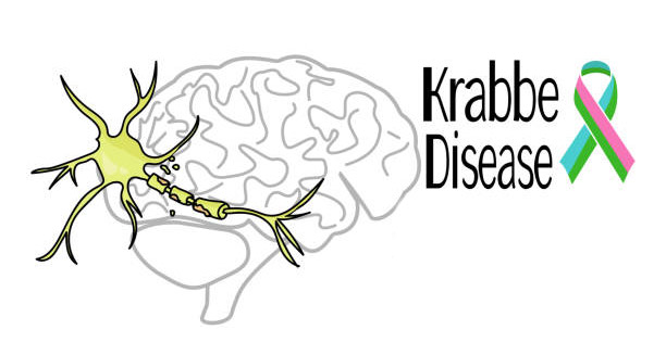
Overview of Krabbe disease is crucial for raising awareness about this rare but severe neurological disorder. Krabbe disease, also called globoid cell leukodystrophy, is a genetic condition affecting the central and peripheral nervous systems. A deficiency of the enzyme galactocerebrosidase (GALC) causes the disease. This deficiency leads to harmful substances building up in the brain and spinal cord. The result is progressive damage to the myelin sheath that protects nerve cells. Understanding the overview of Krabbe disease helps with recognising early signs, getting timely diagnosis, and exploring treatments, especially where newborn screening is not common. Krabbe disease belongs to leukodystrophies—a group of disorders with abnormal development or destruction of brain white matter (myelin). The disease usually appears in infancy, often within the first six months. However, rarer late-onset forms can show up in adolescence or adulthood. In the most common infantile form, the disease progresses fast. Affected babies lose motor skills, become extremely irritable, and have feeding problems. This infantile form is the most aggressive, with life expectancy often limited to a few years if untreated. Genetics and Pathophysiology Krabbe disease follows an autosomal recessive inheritance pattern. This means a child must inherit two faulty copies of the GALC gene—one from each parent—to develop the disease. Carriers with one defective copy usually show no symptoms but can pass the gene to children. The disease affects about 1 in 100,000 live births worldwide, but rates vary by region and group. For example, some communities in Israel and people of Scandinavian descent have higher rates due to genetic clustering. From a biological viewpoint, Krabbe disease occurs because the body cannot produce enough GALC enzyme. This enzyme breaks down fatty substances called galactolipids. When these fats, especially psychosine, build up, they become toxic to oligodendrocytes—the cells that form myelin. Widespread myelin loss disrupts nerve function throughout the body. This damage slows or blocks electrical signals in the nervous system, causing the severe neurological decline seen in Krabbe disease. Symptoms and Clinical Course The overview of Krabbe disease is incomplete without symptoms and progression. In infants, early signs include excessive crying, muscle stiffness or low muscle tone (hypotonia), irritability, and sensitivity to touch. Symptoms worsen over time, leading to seizures, blindness, deafness, and eventually a vegetative state. In contrast, late-onset forms progress slowly and may cause muscle weakness, ataxia (poor coordination), and cognitive decline. While infantile Krabbe disease is usually fatal in early childhood, those with later-onset forms can live into adolescence or adulthood with serious disability. Early diagnosis is critical given the disease’s severity and rapid infant progression. Where newborn screening tests GALC enzyme activity, Krabbe disease can sometimes be found before symptoms start. Early diagnosis allows treatments like haematopoietic stem cell transplantation (HSCT) before irreversible brain damage. Unfortunately, in countries without screening, diagnosis often comes too late for effective treatment, highlighting the need for global awareness. Current Treatments and Support The overview of Krabbe disease also includes treatment options. No cure exists yet, but HSCT shows promise when done before symptoms appear. This transplant introduces healthy donor cells to produce GALC enzyme and preserve neurological function. However, HSCT carries risks and only benefits patients treated very early. For symptomatic individuals, treatment focuses on palliative care—managing pain, nutrition, seizures, and breathing support to improve quality of life. Research continues on new therapies like gene therapy and enzyme replacement. These aim to fix the genetic defect or deliver GALC enzymes directly to the nervous system. Though still experimental, these advances offer hope for future treatments. The disease also places an emotional and social burden on families. Caring for a child with progressive neurological decline demands much energy and resources. Families often need medical, psychological, and financial support. Support groups, genetic counselling, and specialised care help families cope with the challenges. Summary of Overview of Krabbe Disease In conclusion, the overview of Krabbe disease shows a complex and tragic illness marked by rapid neurological decline due to GALC enzyme deficiency. Early diagnosis, especially through newborn screening, remains the most crucial factor in improving outcomes. Early interventions like stem cell transplantation can help. As medical science advances, hope grows for better therapies. Meanwhile, raising awareness and expanding access to screening and support are vital to improving life for those affected by Krabbe disease. [Next: Causes of Krabbe Disease →]

Overview of Krabbe Disease Read More »

Causes of Krabbe Disease

Causes of Krabbe Disease

Causes of Krabbe disease are rooted in genetics, specifically mutations in a gene critical to the nervous system’s function. Krabbe disease is an inherited autosomal recessive disorder caused by mutations in the GALC gene. This gene encodes the enzyme galactocerebrosidase. Understanding the causes of Krabbe disease is vital for diagnosis, genetic counselling, identifying at-risk groups, and developing future treatments. At its core, Krabbe disease is a lysosomal storage disorder. Lysosomes are parts of cells that break down waste materials, including complex fats. The GALC gene gives instructions to make galactocerebrosidase, an enzyme that helps break down certain fats—especially galactosylceramide and psychosine. When the enzyme is missing or deficient, these fats build up, particularly psychosine. Psychosine is toxic to oligodendrocytes, the cells that produce and maintain myelin. Myelin protects nerve fibers and helps electrical signals move efficiently through the nervous system. Damage to myelin causes the severe neurological symptoms of Krabbe disease. Genetic Inheritance and Mutation Types The genetic mutation causing Krabbe disease must come from both parents. Each parent carries one defective copy of the GALC gene but usually shows no symptoms. When two carriers have a child, there is a 25% chance the child will inherit both defective copies and develop Krabbe disease. There is a 50% chance the child will be a carrier like the parents and a 25% chance the child will inherit two healthy genes. This autosomal recessive pattern explains why Krabbe disease may appear unexpectedly in families without a history of the disorder. Researchers have identified over 130 mutations in the GALC gene that can cause Krabbe disease. Some mutations cause a total loss of enzyme activity, leading to the most severe infantile form. Others result in partial enzyme deficiency and cause milder or later-onset forms in adolescence or adulthood. The severity and progression often link directly to the remaining enzyme activity level. Certain populations show higher carrier rates due to founder effects. For example, people of Scandinavian descent and some Druze and Arab groups in Israel have higher incidences. These clusters help researchers identify common mutations and improve genetic screening in these communities. Compound Heterozygosity and Lipid Toxicity In some cases, compound heterozygosity occurs, meaning a child inherits two different GALC mutations—one from each parent. This also results in low enzyme activity but makes diagnosis and prognosis more complex since different mutations interact differently. The buildup of psychosine is especially harmful. Even small amounts are highly toxic to oligodendrocytes. Psychosine disrupts normal cell function and kills these cells, leading to myelin loss and the appearance of globoid cells—large cells filled with storage material seen in brain tissue. These globoid cells are a hallmark of Krabbe disease. Although genetics drive the disease, other factors might influence symptom onset and severity. Some research suggests that epigenetic or environmental factors, such as infections or stress, could accelerate progression in people with low enzyme activity. However, evidence is not conclusive, and these are not primary causes. Importance of Early Detection and Genetic Counselling Newborn screening that includes GALC activity testing allows early identification of affected infants. Early detection leads to prompt confirmatory genetic tests. This timing is crucial because intervention before symptoms appear offers the best chance to improve outcomes. Without screening, many infantile cases go undiagnosed until irreversible damage happens. Understanding the causes of Krabbe disease supports genetic counselling for families with known histories or high-risk populations. Carrier screening during preconception or pregnancy planning helps prospective parents make informed decisions. This knowledge also guides research into treatments like gene therapy and enzyme replacement, aiming to restore GALC function or deliver the enzyme to the nervous system. Summary of Causes of Krabbe Disease In summary, the causes of Krabbe disease lie in genetic mutations of the GALC gene. These mutations cause a critical enzyme deficiency that destroys myelin in the nervous system. The autosomal recessive inheritance requires both parents to be carriers. A wide range of mutations, often found in specific populations, affect disease severity and onset. Understanding these causes aids diagnosis, management, genetic counselling, and newborn screening. As research progresses, unraveling the genetic complexities remains key to better outcomes for those affected. [Next: Symptoms of Krabbe Disease →]

Causes of Krabbe Disease Read More »

Symptoms of Krabbe Disease

Symptoms of Krabbe Disease

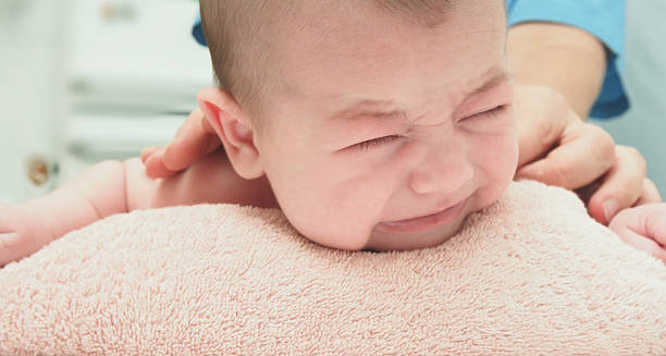
Symptoms of Krabbe disease vary widely in presentation and severity. They depend mainly on the age when symptoms start and the level of enzyme deficiency caused by GALC gene mutations. Infantile Krabbe disease is the most common and severe form. However, late-onset cases in adolescence or adulthood also occur. Knowing the symptoms of Krabbe disease helps with early detection, accurate diagnosis, and timely treatment. This is crucial due to the disease’s rapid progression and permanent damage to the nervous system. In infantile Krabbe disease, symptoms usually appear within the first six months. Early signs can be subtle and mistaken for mild developmental delays or fussiness. Babies may show extreme irritability, strong sensitivity to loud sounds or touch, and frequent crying that is hard to soothe. Parents might also notice their child becoming unusually quiet or unresponsive. As the disease progresses, clearer neurological symptoms emerge quickly. Early Neurological and Motor Symptoms Muscle stiffness and spasticity are key symptoms in infants with Krabbe disease. Affected babies often have increased muscle tone, clenched fists, or rigid limbs. This stiffening contrasts with the usual relaxed muscles of healthy infants. Along with this, seizures can occur—these are sudden bursts of abnormal brain activity that cause twitching, jerking, or full-body convulsions. Seizures often increase in number and severity as the disease worsens. Feeding difficulties are also common early symptoms. Infants may struggle to latch, suck, or swallow, leading to poor nutrition and weight gain. Feeding issues often come with vomiting or acid reflux. As neurological damage increases, many babies need feeding tubes to stay nourished and hydrated. Vision and hearing loss are notable symptoms of Krabbe disease. Babies often stop following objects with their eyes or responding to familiar sounds. As nerve damage spreads, blindness and deafness become common in late stages. These sensory losses add to developmental delays and social isolation. Progressive Motor Decline and Late-Onset Symptoms Motor skills deteriorate rapidly. Infants who reached milestones like holding up their head or rolling over may lose these abilities. Many never progress beyond basic movement and become bedridden. Muscle weakness and paralysis spread from limbs to the torso and breathing muscles. Breathing becomes hard, and many children require ventilators. Lung infections, such as pneumonia, are leading causes of death. Later-onset Krabbe disease appears in childhood, adolescence, or adulthood. Symptoms develop more slowly and may go unnoticed or be mistaken for other neurological diseases. Juvenile cases often show muscle weakness, walking difficulties, poor coordination (ataxia), and falling school or social performance. Children may seem clumsy or tired, and fine motor skills like handwriting decline. Adult-onset Krabbe disease is very rare. Symptoms may include stiff muscles, vision problems, chronic pain, and mental health issues like depression or anxiety. Adults may have gait problems, tremors, or numbness. Cognitive decline and memory loss can occur later. Although slower, adult-onset still causes significant disability. Behavioral Changes and Diagnostic Challenges A common feature across all types is symptom progression. Once symptoms start, they worsen steadily. Infantile cases often result in death before age two. This makes early symptom recognition vital for diagnosis and intervention. Families with affected children sometimes notice early signs sooner in later siblings. This can lead to faster diagnosis and early treatments like stem cell transplants, which may slow progression if done before symptoms begin. Behavioral changes happen too. Affected individuals may withdraw, respond less to stimuli, or have more crying and frustration. These behaviors are often misread as mood issues before diagnosis. Symptoms of Krabbe disease overlap with other conditions like cerebral palsy or leukodystrophies, making diagnosis tricky. Any child showing developmental regression, increased muscle tone, feeding problems, or unexplained neurological signs should get prompt testing for Krabbe disease, including enzyme assays and genetic tests. In conclusion, symptoms of Krabbe disease range widely by onset age and enzyme deficiency level. From early irritability and muscle stiffness in infants to coordination and cognitive problems in older patients, the disease progresses relentlessly. Early symptom recognition is key to prompt diagnosis, treatment, and improving quality of life. Understanding these symptoms remains essential to managing this rare but serious disease. [Next: Diagnosis of Krabbe Disease →]

Symptoms of Krabbe Disease Read More »

Diagnosis of Krabbe Disease

Diagnosis of Krabbe Disease

Diagnosis of Krabbe disease is a complex and multi-step process that often begins with observing neurological symptoms in infants or children. Sometimes, diagnosis happens through genetic screening or family history. Because Krabbe disease is rare and its early symptoms can mimic other common disorders, doctors must keep a high level of suspicion. Timely evaluation and specialised laboratory tests are essential for an accurate diagnosis of Krabbe disease. Early in the diagnostic process, clinicians observe clinical signs. Infants with Krabbe disease often show irritability, muscle stiffness, feeding difficulties, and loss of developmental skills. These signs suggest neurological problems but are not specific. They may appear in cerebral palsy or other leukodystrophies. Therefore, doctors take a full medical history and perform thorough physical and neurological exams. Often, parents report a steady decline that prompts further testing. Enzyme Testing and Genetic Confirmation A key part of the diagnosis of Krabbe disease is enzyme testing. Krabbe disease results from a lack of the enzyme galactocerebrosidase (GALC). This enzyme breaks down fats that build up in the brain and nervous system. Measuring GALC activity in blood, often using dried blood spots, serves as a reliable first test. Very low GALC activity strongly suggests Krabbe disease, especially when paired with symptoms. However, enzyme tests alone do not confirm the diagnosis. Some people have low GALC levels without symptoms. Others with borderline results may still develop the disease. Therefore, genetic testing is needed for confirmation. DNA analysis, usually from blood, finds mutations in the GALC gene that cause Krabbe disease. Finding two disease-causing mutations confirms the diagnosis. Genetic tests also help differentiate infantile, juvenile, and adult forms. Neuroimaging and Additional Tests Neuroimaging supports the diagnosis of Krabbe disease. Brain MRI scans often show characteristic damage to the myelin sheath around nerves. In infantile cases, MRI reveals widespread white matter changes, especially in the parietal and occipital lobes. In late-onset cases, damage may be more limited. Advanced imaging like magnetic resonance spectroscopy (MRS) detects chemical changes in brain tissue, adding further evidence. Doctors may also use nerve conduction studies or electromyography (EMG). These tests measure electrical signals in nerves and muscles. Slower signals and abnormal EMG results reflect nerve damage in Krabbe disease. Newborn Screening and Challenges An emerging tool in the diagnosis of Krabbe disease is newborn screening. Several countries include Krabbe disease in their mandatory newborn screening panels. These tests use dried blood spots taken shortly after birth to measure GALC enzyme levels. If results are low, follow-up testing begins quickly, often within days. The aim is to find affected infants before symptoms appear, since early stem cell transplantation can change the disease course. Newborn screening has challenges. Some infants have genetic mutations that do not cause illness, a condition called “pseudodeficiency.” This makes confirmatory testing crucial. Doctors must carefully interpret results and counsel families about what the findings mean. Additional Diagnostic Tools and Family Counseling In some cases, doctors perform cerebrospinal fluid (CSF) analysis. A lumbar puncture checks protein levels, which often rise in Krabbe disease. However, this test is less common now due to better genetic and enzyme testing. Once diagnosed, families benefit from genetic counseling. Krabbe disease follows an autosomal recessive pattern, so parents usually carry one faulty GALC gene copy each. Counseling helps families understand the risk in future pregnancies and consider prenatal or preimplantation genetic tests if they plan more children. Diagnosing Late-Onset Krabbe Disease Diagnosing older children and adults with suspected late-onset Krabbe disease is more difficult. Symptoms like muscle weakness, balance problems, and behavioral changes develop slowly and can mimic other neurological conditions, such as multiple sclerosis or motor neuron disease. Doctors rely on enzyme tests, genetic sequencing, and MRI scans to reach an accurate diagnosis. Importance of Early Diagnosis Early and accurate diagnosis of Krabbe disease greatly affects treatment options and outcomes. In infantile cases, treatment before symptoms begin—ideally within weeks after birth—offers the best chances. For symptomatic patients, care is mostly supportive but diagnosis still guides therapy and long-term planning. In conclusion, the diagnosis of Krabbe disease is detailed and involves clinical observation, enzyme tests, genetic analysis, and imaging. Although challenges remain in distinguishing symptomatic from asymptomatic cases, advances in newborn screening and molecular tests have greatly improved early detection. Confirming the diagnosis helps doctors guide care and supports families with better information and resources. [Next: Complications of Krabbe Disease →]

Diagnosis of Krabbe Disease Read More »

Scroll to Top