Mental Matters

Author name: admin

Causes of Mastoiditis

Causes of Mastoiditis

Causes of Mastoiditis Causes of Mastoiditis primarily stem from untreated or poorly managed ear infections, especially otitis media. Mastoiditis is a serious bacterial infection of the mastoid bone behind the ear. The mastoid bone contains air cells that help balance middle ear pressure and drain fluid. When these air cells get infected or filled with fluid, mastoiditis can develop. This often leads to pain and may cause life-threatening complications. The causes of mastoiditis involve multiple factors, including infection pathways, underlying conditions, anatomical issues, and risky behaviours. Understanding these factors helps doctors diagnose early and plan effective treatment. Bacterial Middle Ear Infections (Otitis Media) The most common direct cause of mastoiditis is acute otitis media, a bacterial infection of the middle ear. If untreated, the infection can spread to the mastoid bone through the aditus ad antrum, a small connection between the middle ear and mastoid air cells. This spread causes swelling, inflammation, and bone damage. Common bacteria causing otitis media and mastoiditis include: These bacteria often follow a viral upper respiratory infection. They thrive in fluid behind the eardrum and can move into the mastoid if left untreated. Inadequate or Delayed Treatment of Ear Infections Delayed or incomplete treatment of ear infections is another major cause. Some infections resolve with antibiotics, but stopping early or resistance allows bacteria to persist. This can let the infection reach the mastoid. Limited access to healthcare or underestimating the infection severity increases risk, especially in children. Chronic Otitis Media Chronic otitis media, especially with cholesteatoma (skin growth in the middle ear), can also cause mastoiditis. These infections linger for weeks or months, slowly eroding bone. Cholesteatomas damage the mastoid through infection and pressure, making them high-risk factors. Congenital or Structural Abnormalities Some people are born with ear canal or Eustachian tube abnormalities, which cause fluid buildup and repeated infections. Children’s shorter, horizontal Eustachian tubes make them more prone to mastoiditis. Other anatomical contributors include: Upper Respiratory Tract Infections Colds or flu can indirectly cause mastoiditis. URTIs often affect the Eustachian tube, causing poor middle ear ventilation and fluid buildup. This creates a breeding ground for bacteria. Though URTIs don’t directly cause mastoiditis, they can start a chain of events leading to it. Immunocompromised Conditions People with weakened immune systems—HIV/AIDS, cancer treatment, diabetes, or long-term steroids—have higher mastoiditis risk. Their bodies may not fight simple ear infections, allowing the infection to spread. Chemotherapy or radiation can damage tissues and drainage, worsening the risk. Trauma or Surgical Intervention Ear trauma or previous ear surgery, like tympanostomy tube placement, may lead to mastoiditis. Surgical procedures or injuries can introduce bacteria into the middle ear or mastoid. Even minor trauma, such as aggressive ear cleaning, can allow bacteria to spread. Eardrum perforations create direct bacterial entry points. Nosocomial (Hospital-Acquired) Infections Mastoiditis may develop as a hospital-acquired infection in patients already in hospital. ICU stays or ventilator-associated pneumonia may expose patients to bacteria resistant to standard antibiotics, increasing the risk of mastoiditis. Environmental and Behavioural Factors Lifestyle and environmental factors can influence mastoiditis risk: Age as a Risk Factor Children under two are most prone to middle ear infections due to immature immune systems and ear anatomy. Because mastoiditis often follows these infections, age is an indirect risk factor. Adults can develop mastoiditis too, but severity increases with age due to delayed recognition and other health problems. In summary, the causes of mastoiditis involve many interconnected factors. Untreated or poorly managed ear infections are the main cause, but anatomical differences, immune problems, environmental exposures, and prior ear trauma or surgery also contribute. Early recognition and management of these causes reduce the incidence and severity of mastoiditis. [Next: Symptoms of Mastoiditis →]

Causes of Mastoiditis Read More »

Symptoms of Mastoiditis

Symptoms of Mastoiditis

Symptoms of Mastoiditis Symptoms of Mastoiditis can vary depending on the severity of the infection and how long it has been present. This condition usually develops after untreated or severe middle ear infections. Early signs may be subtle but can progress into more noticeable and serious symptoms. Recognising symptoms of mastoiditis early helps prevent long-term damage and complications. In children, early signs are often mistaken for recurring ear infections, which can delay care. Understanding these symptoms aids timely medical attention. Pain and Swelling Behind the Ear One of the earliest signs of mastoiditis is pain behind the affected ear. This pain is usually persistent, throbbing, and worsens over time. The mastoid bone becomes inflamed and tender, causing noticeable swelling. The area may feel warm, firm, or look red and flushed. Severe swelling can push the ear outward or downward. Children often show pain by tugging at their ear, becoming irritable, or refusing to lie on one side. The skin over the mastoid may appear shiny and tight due to inflammation. Fever and General Malaise High fever often accompanies mastoiditis. Temperatures can rise above 38.5°C (101.3°F), with chills and sweating. This shows the body is fighting the infection. Fatigue, weakness, and malaise are also common. In severe cases, fever may persist despite standard fever-reducing medicine. Fever lasting more than a day or two with ear pain should be checked urgently. Ear Discharge (Otorrhoea) Ear discharge is another key symptom of mastoiditis. The fluid may be thick, foul-smelling, or blood-tinged. It usually drains from the ear canal and results from pus building up in the middle ear or mastoid. Sometimes, pus pressure ruptures the eardrum, releasing fluid. While this may relieve pain temporarily, it indicates a serious stage requiring immediate care. Chronic discharge lasting over two weeks, especially with swelling behind the ear, strongly suggests mastoiditis. Hearing Loss Temporary hearing loss is common in mastoiditis. Fluid buildup or middle ear damage often causes this. Sounds may seem muffled or distant. People may need to raise the TV or radio volume. If untreated, bone erosion or severe damage can cause permanent hearing loss. Children with hearing loss are at risk for delays in speech and communication if not treated promptly. Irritability, Restlessness, and Poor Feeding in Children Infants and young children may show behavioural changes rather than verbal complaints. Irritability, excessive crying, refusal to eat, and poor sleep may indicate mastoiditis. Poor feeding can lead to dehydration and weaken immunity, worsening the infection. Parents should watch for such changes after an ear infection. Headache and Neck Stiffness As infection spreads, headaches or neck stiffness may develop. This may result from inflammation of surrounding tissue or, rarely, the brain lining. Any headache, neck stiffness, and fever combination is a medical emergency due to possible meningitis. Pain may radiate to the side of the head or upper neck, making head movement uncomfortable. These symptoms, along with ear discharge or swelling, should never be ignored. Tinnitus and Vertigo Sometimes, mastoiditis affects the inner ear. This can cause tinnitus (ringing) or vertigo (spinning sensation). People may feel dizzy, unsteady, or nauseous. These symptoms suggest the infection is reaching the vestibular system, which controls balance. Though less common, they indicate a more severe disease and require prompt treatment. Swollen Lymph Nodes and Facial Swelling Nearby lymph nodes, especially around the neck and jaw, may swell and feel tender. Facial swelling on the side of the affected ear may also occur. In rare cases, abscesses form, creating a visible bulge near the ear or neck. Surgery is often needed, and such signs may indicate infection spreading systemically. Changes in Mental Status (in Severe Cases) In very severe or neglected mastoiditis, brain involvement may cause confusion, lethargy, disorientation, or seizures. These neurological changes, especially with ear pain or discharge, are medical emergencies. Early detection and treatment prevent these advanced complications. While uncommon early, these symptoms highlight the importance of vigilance and timely care. [Next: Diagnosis of Mastoiditis →]

Symptoms of Mastoiditis Read More »

Diagnosis of Mastoiditis

Diagnosis of Mastoiditis

Diagnosis of Mastoiditis Diagnosis of Mastoiditis begins with a detailed clinical assessment and a careful review of the patient’s symptoms and medical history. Mastoiditis often occurs after a severe or unresolved middle ear infection. Doctors start with a thorough ear examination. Accurate diagnosis is key to determine how serious the infection is, detect complications, and plan the best treatment. Because mastoiditis can look like other ear or head infections, quick and correct diagnosis prevents delays and serious problems. Clinical Evaluation The first step in diagnosing mastoiditis is a complete history and physical exam. Doctors ask about recent or repeated ear infections, discharge, fever, or pain behind the ear. They also check for hearing loss, balance problems, fatigue, or feeling unwell. During the exam, the doctor uses an otoscope to inspect the ear canal and eardrum. Swelling, redness, bulging eardrum, or discharge may be visible. They check behind the ear for tenderness, warmth, or swelling. If the ear sticks out, it may suggest mastoid bone swelling. For children, signs like irritability, poor feeding, or restlessness can help doctors notice the problem. These early clues often lead to more tests. Otoscopic Examination Otoscopic exams are important because mastoiditis often follows or occurs with middle ear infections. Using an otoscope, doctors look for a red, bulging, or perforated eardrum. Pus leaking from the eardrum may show the infection has spread beyond the middle ear. Doctors may see fluid behind the eardrum or limited eardrum movement. These signs suggest a complicated ear infection. In advanced cases, swelling may make the canal hard to see. This calls for imaging tests. Imaging Studies If physical exams and symptoms suggest mastoiditis, imaging helps confirm the diagnosis and shows how far the infection has spread. Common imaging includes: 1. Computed Tomography (CT) Scan of the Temporal BoneCT scans give clear images of mastoid air cells and middle ear structures. They detect fluid, swelling, bone damage, or abscesses. CT is especially helpful to find early complications, such as infection spreading to nearby tissues or skull base. Bone damage seen on CT strongly suggests mastoiditis. CT scans are often done for patients who do not improve with antibiotics, have systemic symptoms, or show signs of brain complications. 2. Magnetic Resonance Imaging (MRI)MRI is useful in complex or chronic cases. It helps when doctors suspect brain abscess, sinus thrombosis, or meningitis. MRI shows soft tissues and nearby nerves clearly. It is used when neurological symptoms appear, like confusion, seizures, or severe headaches. MRI can also rule out other deep infections or tumors. Laboratory Tests Blood and fluid tests support the diagnosis and check the body’s response. Common tests include: Finding the bacteria early allows doctors to give targeted antibiotics, reducing resistance and improving outcomes. Differential Diagnosis Doctors must distinguish mastoiditis from other ear or skull conditions: Accurate diagnosis requires combining symptoms, exams, imaging, and lab results. Mistakes may delay treatment and allow serious complications. Specialist Referral Patients suspected of mastoiditis, especially with complications, should see an ENT specialist promptly. ENT teams may do further exams like nasopharyngoscopy or hearing tests and decide if surgery is needed. In hospitals with ENT services, referrals are urgent for children, immunocompromised patients, or those not improving after 48–72 hours of medical care. [Next: Treatment of Mastoiditis →]

Diagnosis of Mastoiditis Read More »

Treatment of Mastoiditis

Treatment of Mastoiditis

Treatment of Mastoiditis The treatment of Mastoiditis typically involves a combination of aggressive medical and surgical interventions due to the potentially serious nature of this middle ear infection. Treatment of Mastoiditis depends on the severity, underlying cause, and whether complications have developed, but it is always considered a condition requiring prompt and comprehensive care. Early and effective treatment is vital to prevent the spread of infection to nearby structures like the brain or facial nerves. This section outlines medical therapies, surgical procedures, hospitalisation protocols, and the ongoing management of mastoiditis, providing an in-depth look into the clinical approach used to resolve the infection and preserve hearing and overall health. Medical Management The first step in treatment of mastoiditis is usually medical. Doctors start with strong intravenous (IV) antibiotics in hospital. This ensures enough medicine reaches the blood and infected tissue. Common antibiotics include ceftriaxone, cefotaxime, or vancomycin. Doctors adjust the choice based on culture results from ear fluid or mastoid samples. In simple acute cases, patients usually receive IV antibiotics for 7–14 days. After that, they may take oral antibiotics for another 1–2 weeks. If the patient does not improve in 24–48 hours, or symptoms get worse, doctors move to surgery. Even so, in many early cases, antibiotics alone control the infection and stop the spread. Myringotomy and Tympanostomy A myringotomy is a small cut made in the eardrum. It drains pus or fluid, reduces pressure, eases pain, and allows accurate cultures. Sometimes, doctors place a tympanostomy tube in the ear to keep it ventilated and stop more fluid build-up. Children usually need general anaesthesia for these procedures. Adults may have them with local anaesthesia if they can stay calm. These methods work best when mastoiditis occurs together with a middle ear infection. Mastoidectomy in Treatment of Mastoiditis If infection spreads into the mastoid bone and does not improve with medicine, surgery is needed. A mastoidectomy removes infected air cells from the bone. Surgeons drill into the mastoid to clear infection and support healing. There are several types: This surgery is vital in the treatment of mastoiditis when abscesses or bone erosion appear. It prevents dangerous problems like meningitis or brain abscess. Hospitalisation and Monitoring Most people with acute mastoiditis need hospital care, especially children. Doctors check for: If problems such as subperiosteal abscess, facial nerve weakness, or brain spread appear, doctors speed up surgery. Hospital stays may last from a few days to several weeks depending on recovery. Outpatient care is only safe in very mild cases with close specialist follow-up. Follow-Up and Long-Term Care After acute treatment of mastoiditis, follow-up is important. Doctors check hearing recovery and look for leftover infection. Hearing tests are repeated after a few weeks. In severe cases, scans confirm full healing. If a tube was inserted, it may stay in place for 6–12 months. Patients then need regular ENT visits. In some, repeated infections or chronic mastoiditis develop, needing further surgery or long-term antibiotics. Supportive Therapies Supportive treatments add to medicine and surgery. These include: Parents and caregivers should watch for warning signs of infection coming back. Completing all prescribed antibiotics is key. Patients should also avoid water in the ear during healing. Special Considerations in Children Children get mastoiditis more often. For them, treatment of mastoiditis must suit their age, immune health, and ability to explain symptoms. ENT surgeons usually get involved early. A team approach works best when problems arise. Doctors often choose surgery faster for children, since their narrow ear spaces make infection spread quicker. In chronic mastoiditis or cholesteatoma, children may need larger surgeries. Sometimes, staged procedures are used to clear infection first and then restore hearing later. [Next: Complications of Mastoiditis →]

Treatment of Mastoiditis Read More »

Complications of Mastoiditis

Complications of Mastoiditis

Complications of Mastoiditis The complications of mastoiditis can be serious and potentially life-threatening if the infection is not treated promptly and effectively. Complications of mastoiditis arise when the infection spreads beyond the mastoid bone, impacting surrounding tissues, structures of the ear, and even other parts of the skull and brain. While many cases of mastoiditis respond well to antibiotics and drainage, failure to diagnose or manage the condition quickly can lead to an array of medical consequences that may affect a person’s hearing, neurological function, and overall health. Hearing Loss One of the most common complications of mastoiditis is hearing loss. The mastoid bone connects closely to the middle ear. When it becomes inflamed or infected, the delicate hearing parts may suffer damage. Fluid buildup, damage to the tiny ear bones, or scarring of the eardrum can block sound transmission. At first, this hearing loss may be temporary if caused by fluid or pressure. Yet, permanent damage to the cochlea or the hearing nerve may cause sensorineural hearing loss, which usually does not improve. Sometimes, surgery like mastoidectomy (removing infected mastoid cells) or tympanoplasty (repairing the eardrum) can restore some hearing. Even so, some patients may still need hearing aids or other devices if the loss remains severe or permanent. Facial Nerve Paralysis Another alarming complication is facial nerve paralysis. The facial nerve passes through the temporal bone, which also contains the middle ear and mastoid air cells. If mastoiditis spreads or forms an abscess inside this bone, the nerve may become compressed or damaged. This can lead to muscle weakness or paralysis on one side of the face. The person may struggle to close the eye, show expressions, or control mouth movement. Doctors usually order urgent scans and may need to perform surgery to release pressure. With fast treatment, the nerve can recover slowly. But when treatment is delayed, some permanent weakness may remain. Labyrinthitis and Vertigo Infections spreading into the inner ear can cause labyrinthitis. This condition inflames the hearing and balance system. It often brings intense vertigo, dizziness, nausea, vomiting, and poor balance. Labyrinthitis disrupts daily life and can also leave lasting balance issues or hearing loss. While medicines can calm symptoms for a short time, patients also need antibiotics and sometimes drainage surgery to clear the infection. If vertigo lingers, vestibular physiotherapy can help with recovery. Intracranial Complications of Mastoiditis Some of the most dangerous complications of mastoiditis occur when infection spreads inside the skull. These include brain abscess, meningitis, or lateral sinus thrombosis. All of these conditions require emergency care. Brain Abscess A brain abscess forms when bacteria move from the infected mastoid bone into the brain. The thin temporal bone wall makes this possible. Symptoms often include a severe headache, confusion, weakness in certain body parts, or seizures. Doctors confirm diagnosis through scans like CT or MRI. Treatment includes intravenous antibiotics and, in many cases, surgical drainage of the pus. Meningitis Meningitis develops when the membranes around the brain and spinal cord become infected. The signs include very high fever, neck stiffness, sensitivity to light, vomiting, and confusion. Meningitis linked to mastoiditis can progress very quickly. Without rapid treatment with antibiotics and hospital care, it may become fatal. Lateral Sinus Thrombosis Lateral sinus thrombosis occurs when a clot forms in the brain’s venous sinuses. This happens if infection spreads from the mastoid to the sigmoid or transverse sinuses. Common symptoms are headache, fever, swelling of the optic disc, and sometimes seizures or vision changes. Doctors use MRI or MRV scans to confirm the clot. Treatment requires antibiotics and blood thinners to prevent stroke and protect the brain. Subperiosteal Abscess Another complication of mastoiditis is a subperiosteal abscess. This happens when pus collects between the mastoid bone and its covering layer. Symptoms often include swelling behind the ear, redness, and the ear lobe being pushed outward. This abscess usually needs surgical drainage along with antibiotics. If left untreated, the pus may erode bone, spread into neck muscles, or create a tunnel from the mastoid to the skin, leading to chronic drainage and infection. Chronic Suppurative Otitis Media In some patients, mastoiditis does not heal fully and turns into chronic suppurative otitis media (CSOM). This is a long-lasting middle ear infection where pus drains through a hole in the eardrum. CSOM is difficult to cure and may require months of care. Doctors may prescribe ear drops, perform surgery to repair the eardrum, or remove infected tissue. Ongoing infection can also cause a cholesteatoma, a harmful skin growth in the ear and mastoid. This growth can damage bone, reduce hearing, and spread infection if not removed by surgery. Delayed Development in Children In children, complications of mastoiditis affect more than just health. Chronic ear infections and hearing loss can delay speech, language learning, and school progress. Children who struggle to hear may also face social or behavioural issues. With early diagnosis and regular ENT follow-ups, these risks can be reduced. Proper hearing support ensures children develop normally. Systemic Infection (Sepsis) In rare but severe cases, mastoiditis can spread into the whole body and cause sepsis. Sepsis creates a strong inflammatory reaction that harms many organs. Signs of sepsis include very high fever, fast breathing, low blood pressure, confusion, and even organ failure. This condition needs urgent hospital care with fluids, antibiotics, and support for the heart and circulation. Without quick action, sepsis can be fatal. [Next: Back to Overview →]

Complications of Mastoiditis Read More »

Overview of Mastocytosis

Mastocytosis

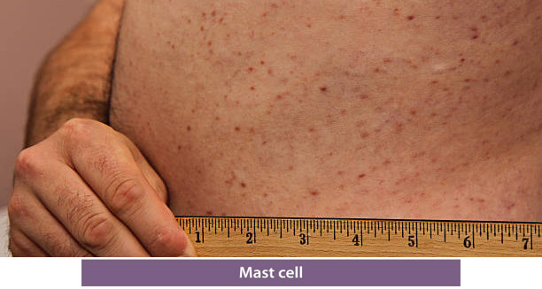
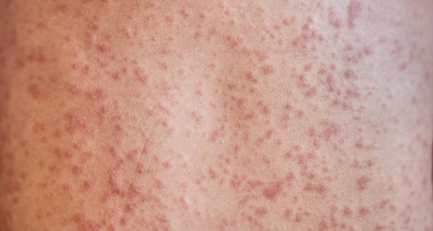
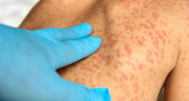
Overview of Mastocytosis Mastocytosis is a rare disorder marked by the abnormal accumulation of mast cells in the body’s tissues and organs. Mast cells are a type of white blood cell that plays a vital role in immune responses, particularly in allergic reactions and the body’s defence against pathogens. Under normal conditions, mast cells reside in the skin, respiratory tract, gastrointestinal tract, and around blood vessels and nerves. However, in mastocytosis, these cells proliferate excessively and infiltrate areas they typically don’t inhabit in such large numbers, leading to a wide range of symptoms and complications. Types and Forms of Mastocytosis This condition belongs to a group of illnesses known as mast cell disorders. Doctors divide mastocytosis into two main types. First is cutaneous mastocytosis, which affects only the skin. Second is systemic mastocytosis, which involves internal organs like the liver, spleen, bone marrow, and digestive system. Systemic mastocytosis has different levels of severity. It can be mild (called indolent), or much more serious. One rare and life-threatening type is mast cell leukemia. Cutaneous mastocytosis is more common in children. It often gets better or goes away as they grow older. The most usual form is urticaria pigmentosa. It causes reddish-brown spots or patches on the skin. These spots can itch or swell when touched, which is called Darier’s sign. In contrast, adults more often get systemic mastocytosis. This type tends to last a long time and may slowly get worse. Overview of Mastocytosis Genetics and Causes Doctors don’t fully understand what causes mastocytosis. But many patients, especially adults with systemic forms, have a specific gene change. This mutation happens in the KIT gene, which helps control mast cell growth. The change keeps the KIT receptor constantly active. This leads to out-of-control mast cell growth and buildup. Most cases happen randomly, without a family link. However, rare familial cases have been reported. This suggests that, in some people, genes may play a role in raising the risk. Common Symptoms and How They Vary Symptoms vary a lot depending on the type and severity. Most symptoms happen when mast cells get triggered and release chemicals. Common signs include: Triggers that can cause these reactions include certain foods, heat or cold, stress, medications, or insect stings. Mast cells release chemicals like histamine, heparin, and prostaglandins, which affect many systems in the body. Systemic mastocytosis can also damage organs. For example, when mast cells build up in the bone marrow, patients may have anemia, low platelets, or low white blood cells. This increases the risk of bleeding or infection. If mast cells affect the liver or spleen, the organs may become larger and work poorly. In the gut, too much acid or inflammation can lead to ulcers, diarrhoea, and trouble absorbing nutrients. Diagnosis and Testing in Mastocytosis Getting a diagnosis can be hard because symptoms often look like other illnesses. These may include allergies, autoimmune conditions, or stomach problems. That’s why doctors need several tests and a full health history to make a diagnosis. For skin forms, doctors may take a skin biopsy. To confirm systemic mastocytosis, they check serum tryptase levels, do bone marrow biopsies, and look for KIT mutations with genetic tests. Scans like ultrasound or CT can show whether organs like the liver or spleen are affected. Treatment Options and Lifestyle Management Treatment does not usually cure mastocytosis, but it helps control symptoms. Most care focuses on lowering mast cell activity. Common treatments include: In more severe systemic cases, doctors may use chemotherapy, targeted treatments like midostaurin, or stem cell transplants. Avoiding known triggers is one of the most important ways patients can manage flare-ups. Living with Mastocytosis: Long-Term Care Life with mastocytosis can be very tough, both physically and mentally. Many patients fear unexpected symptoms, especially the risk of anaphylaxis. This can limit daily activities and affect emotional health. That’s why ongoing support is so important. Education helps patients avoid triggers and manage their condition. Doctors usually advise people to carry emergency epinephrine and wear medical alert jewellery in case of serious reactions. Regular checkups help monitor organ function and track changes in symptoms over time. Research and Hope for the Future Ongoing research into mast cell behaviour and KIT gene mutations offers hope for better treatments. Scientists are learning more about how mastocytosis starts and spreads. This knowledge helps them create new drugs that may target the disease more directly and with fewer side effects. The growing medical awareness of mast cell disorders means patients are more likely to get an early diagnosis and the right care. Even though mastocytosis is complex, many people can lead a good life with proper treatment and support. Final Thoughts on the Overview of Mastocytosis To sum up, mastocytosis is a rare but serious disease that causes too many mast cells to build up in the skin or other parts of the body. The symptoms can look like allergies or more severe health issues, depending on how the disease affects each person. While there’s no cure yet, many people manage symptoms well with the right care. However, due to the chance of life-threatening problems like anaphylaxis and organ failure, patients need close follow-up and strong medical support. Thanks to ongoing research and better awareness, new treatments and better outcomes are becoming more likely. [Next: Causes of Mastocytosis →]

Mastocytosis Read More »

Causes of Mastocytosis

Causes of Mastocytosis

Causes of Mastocytosis Understanding the Causes of Mastocytosis is critical for clinicians, researchers, and patients alike, as it offers insight into how and why this rare disorder develops. The Causes of Mastocytosis are primarily rooted in genetic mutations that affect the growth and behaviour of mast cells—immune system cells involved in allergic reactions and inflammation. These mutations result in the accumulation of mast cells in the skin, bone marrow, liver, spleen, and other tissues, leading to a range of symptoms depending on the organs affected. The Role of the KIT Gene in Mastocytosis At the centre of most cases of mastocytosis is a change in the KIT gene. This gene makes a protein called KIT receptor tyrosine kinase. This receptor sits on the surface of mast cells and helps control how they grow, develop, and stay alive. Normally, KIT only turns on when it binds with a molecule called stem cell factor (SCF). But in mastocytosis, a common change—called the D816V mutation—makes KIT stay active all the time. This causes mast cells to grow too much and build up in tissues. Doctors have found this D816V mutation in over 90% of adults with systemic mastocytosis. Many children with cutaneous mastocytosis also have it. Since this mutation happens after birth, people don’t inherit it. Even so, researchers are still looking into whether some people may carry inherited changes that increase their risk of mast cell problems. Immune System Imbalance and Overgrowth The Causes of Mastocytosis don’t stop with genetic changes. Sometimes, the immune system may not work properly, especially in children who don’t have the KIT mutation. In these cases, scientists believe other signals in the body may push mast cells to grow too much. For example, higher levels of chemicals like interleukin-6 (IL-6) and tumour necrosis factor-alpha (TNF-α) may create an environment that encourages mast cell buildup. Mast cells also affect their surroundings by releasing chemicals like histamine, tryptase, and prostaglandins. In mastocytosis, this becomes a harmful loop. The more mast cells there are, the more signals they send out, which leads to even more mast cell growth and survival. Types of Mastocytosis and What Triggers Symptoms Doctors split mastocytosis into two main types: cutaneous and systemic. In cutaneous mastocytosis, extra mast cells only appear in the skin. In systemic mastocytosis, they spread to organs like the bone marrow, liver, and spleen. The Causes of Mastocytosis can vary slightly between these types, especially when comparing adults to children. In children, mastocytosis often shows up in the first two years of life and usually stays limited to the skin. Most of the time, it gets better or even goes away with age. The cause in these young cases isn’t always clear, especially when no KIT mutation is found. On the other hand, adults are more likely to develop systemic mastocytosis. These cases often link to the D816V mutation and tend to be long-lasting or get worse over time. This is especially true in aggressive forms or when it becomes mast cell leukaemia. Some things don’t cause mastocytosis but can make symptoms worse. These triggers include: Recognising these triggers helps people manage the disease better. Rare Genetic Links and Related Conditions In rare cases, people may inherit a higher chance of developing mastocytosis. Although familial mastocytosis is very uncommon, a few studies suggest that it can run in families. Sometimes, mastocytosis also shows up alongside blood disorders like myelodysplastic syndromes (MDS) or chronic eosinophilic leukaemia. Doctors think these diseases might share similar genetic or cellular mistakes. There’s also a condition called mast cell activation syndrome (MCAS). In MCAS, people have severe mast cell symptoms but don’t have extra mast cells. Even though mastocytosis and MCAS are different, their symptoms and causes can overlap in some cases. How Molecular Pathways Influence the Causes of Mastocytosis New research is diving deeper into how different cell signals affect mast cell growth. Apart from the KIT pathway, scientists are studying other systems like PI3K/AKT/mTOR, JAK/STAT, and RAS/MAPK. These systems help cells grow and respond to threats. If they go wrong, they may help the disease spread or resist treatment. Because of this, newer treatments now target these exact problems. Drugs like midostaurin and avapritinib are designed to block KIT mutations. These medicines mark a big step forward in treating hard-to-manage or advanced systemic mastocytosis. Summary of the Causes of Mastocytosis In summary, the Causes of Mastocytosis mainly involve changes in the KIT gene, especially the D816V mutation. This change causes mast cells to grow too much and collect in tissues. But there’s more to the story. Immune system problems, triggers from the environment, and rare genetic links also play a part. Adult cases often involve the whole body and tend to last longer. In contrast, many childhood cases stay in the skin and get better over time. Ongoing studies are bringing hope. They continue to uncover how mast cells behave and how we can stop them. These discoveries are leading to better treatments and improving life for people with this rare disease. [Next: Symptoms of Mastocytosis →]

Causes of Mastocytosis Read More »

Symptoms of Mastocytosis

Symptoms of Mastocytosis

Symptoms of Mastocytosis Mastocytosis is a condition characterised by the accumulation and abnormal activation of mast cells in the skin and/or internal organs. The symptoms can range from mild and occasional to severe or life-threatening, depending on the subtype of mastocytosis and the degree of mast cell activation or infiltration in various organs. These symptoms occur due to the release of chemical mediators like histamine, prostaglandins, and leukotrienes, which influence multiple body systems. 🔹 Cutaneous Symptoms Cutaneous symptoms are most common in children and patients with cutaneous mastocytosis (CM) but may also occur in systemic forms. 🔹 Gastrointestinal Symptoms Gastrointestinal (GI) symptoms result from mast cell accumulation in the GI tract and histamine-related effects on digestion. 🔹 Cardiovascular and Anaphylactic Symptoms Systemic mast cell activation can lead to life-threatening symptoms, especially in systemic mastocytosis. 🔹 Respiratory Symptoms Mast cell mediators may affect the airways and lungs, especially during systemic reactions. 🔹 Neurological and Psychiatric Symptoms Though less commonly discussed, central nervous system symptoms are frequently reported. 🔹 Musculoskeletal Symptoms Mastocytosis can affect bones and joints, particularly in systemic forms involving the bone marrow. 🔹 Constitutional and Generalised Symptoms These nonspecific but impactful symptoms are common and often under-recognised. 🔹 Triggers and Symptom Aggravation Symptoms are frequently worsened by known triggers, which vary among individuals. Common triggers include: Avoiding or managing triggers is essential in preventing flare-ups or life-threatening reactions. 🔹 Rare and Atypical Symptoms In advanced systemic mastocytosis, organ-specific symptoms may develop depending on where mast cells accumulate: Such cases often require coordinated care across specialties. 🔚 Summary System Affected Common Symptoms Skin Lesions, itching, flushing, Darier’s sign Gastrointestinal Nausea, diarrhoea, bloating, malabsorption Cardiovascular Hypotension, syncope, anaphylaxis Respiratory Wheezing, coughing, shortness of breath Neurological/Psychiatric Headaches, brain fog, anxiety, depression Musculoskeletal Bone pain, fractures, osteoporosis General/Constitutional Fatigue, weight loss, fever, failure to thrive ✅ Key Takeaway: Recognising the wide variety of symptoms—and the triggers that worsen them—is essential for early diagnosis and effective management of mastocytosis. Because of the disease’s multi-system nature, a coordinated approach involving allergists, dermatologists, haematologists, and other specialists is often necessary. The diverse and often non-specific symptoms of mastocytosis can make diagnosis challenging, especially since many manifestations overlap with more common conditions. Awareness of the disease’s hallmark signs—such as skin lesions with Darier’s sign, unexplained anaphylaxis, or persistent gastrointestinal complaints—can prompt timely evaluation and prevent misdiagnosis. Early identification of mastocytosis not only helps in managing symptoms effectively but also reduces the risk of severe complications, including life-threatening allergic reactions and organ damage. Given the wide spectrum of clinical presentations, managing mastocytosis requires a personalized approach tailored to each patient’s symptoms and triggers. Ongoing research into the molecular and genetic underpinnings of the disease continues to improve diagnostic accuracy and treatment options. With multidisciplinary care and patient education focused on trigger avoidance and symptom control, many individuals with mastocytosis can lead relatively normal and fulfilling lives despite the chronic nature of the condition. [Next: Diagnosis of Mastocytosis →]

Symptoms of Mastocytosis Read More »

Diagnosis of Mastocytosis

Diagnosis of Mastocytosis

Diagnosis of Mastocytosis Diagnosis of Mastocytosis can be a complex and meticulous process due to the rarity of the condition and its diverse clinical presentation. Clinicians start by suspecting it when seeing skin lesions, unexplained allergic reactions, or systemic signs like stomach pain, tiredness, or anaphylaxis. Because mastocytosis appears differently in children versus adults—and can be either cutaneous or systemic—doctors must take a detailed medical history and follow a step‑by‑step diagnostic plan to find and label the disease correctly. Mastocytosis involves too many mast cells building up in different tissues. In children, it often stays on the skin (cutaneous forms). In adults, it tends to affect organs like bone marrow, liver, spleen, or the gut (systemic forms). Thus, the diagnostic path changes depending on which type doctors suspect. A child with only skin spots might only need skin tests, while an adult with systemic signs may need complex blood and bone marrow tests. Clinical Evaluation and Medical History Doctors begin by carefully examining the patient’s skin. They look for reddish‑brown patches or spots that swell or itch when rubbed. This reaction is called Darier’s sign, and it often signals cutaneous mastocytosis. Then they interview the patient about symptoms. They ask how often allergic reactions occur, whether there is diarrhoea or abdominal pain, episodes of low blood pressure, headaches, bone pain, or unexplained tiredness. A family history of similar symptoms or rare genetic illnesses can guide the diagnosis. This history helps them tell mastocytosis apart from other allergies or blood diseases with similar signs. If symptoms affect internal systems—like chronic gut issues, flushing, or repeated anaphylaxis with no clear cause—doctors will proceed with lab tests and possibly imaging scans. Blood Tests and Serum Tryptase Levels A key lab test is the serum tryptase level. Tryptase is an enzyme that mast cells release, and elevated levels often reflect excess mast cells. A baseline tryptase above ~20 ng/mL strongly suggests mast cell overgrowth and signals need for further testing. But a high tryptase is not unique to mastocytosis. It also occurs in allergic reactions, kidney disease, or certain blood disorders. That means tryptase alone can’t confirm the disease—it just adds a piece to the puzzle. Routine blood counts may show signs of bone marrow involvement, such as increased eosinophils, low red cells (anaemia), or low platelets (thrombocytopenia). If those appear, doctors may send the patient to a haematologist for deeper investigations. Skin Biopsy for Cutaneous Mastocytosis When cutaneous mastocytosis is suspected, doctors may take a skin sample (biopsy) under local anaesthetic. A pathologist examines this under a microscope using stains that highlight mast cells. They look for clusters of mast cells in the dermis, often near blood vessels. They also use immunohistochemical stains for markers like CD117 (c-KIT) and tryptase to confirm the presence of mast cells. This method often suffices to diagnose cutaneous mastocytosis, especially in children, without further invasive tests. Bone Marrow Biopsy and Histology If doctors suspect systemic mastocytosis—especially in adults or people with high tryptase and multi-organ signs—they will recommend a bone marrow biopsy. This sample usually comes from the pelvic bone, often under local anaesthetic. Specialists examine it for mast cell count, clustering, and shape. In systemic mastocytosis, many mast cells become spindle‑shaped. If they find clusters of 15 or more mast cells in marrow, that meets one of the major WHO criteria. The biopsy also checks for other blood abnormalities, because systemic mastocytosis often co-occurs with diseases like chronic myelogenous leukaemia or myelodysplastic syndromes. Genetic Testing: KIT Mutations Testing for gene changes is a crucial part of the diagnostic process. A common mutation in mastocytosis is KIT D816V, which causes uncontrolled mast cell growth. Doctors test for this mutation using material from bone marrow, blood, or sometimes skin. A positive result offers strong molecular proof of systemic mastocytosis. It also helps guide treatment, since some targeted drugs don’t work well in cases with that mutation. Testing for KIT mutations doesn’t just confirm diagnosis. It helps separate systemic mastocytosis from other clonal disorders. The presence of additional mutations (such as in SRSF2 or ASXL1) may point to a more aggressive disease course. Diagnostic Criteria by the WHO The WHO defines specific rules to confirm systemic mastocytosis. The patient must meet: Major Criterion: Minor Criteria: These criteria aim to ensure that doctors worldwide follow a standard method. They also reduce misdiagnosis, especially between mastocytosis and similar conditions like mast cell activation syndrome (MCAS). Imaging Studies Although not required in every case, imaging tests help assess organ involvement. Doctors may order abdominal ultrasound, CT scans, or MRI if they suspect liver or spleen enlargement, lymph nodes, or bone lesions. They may also do bone density scans (DEXA), since systemic mastocytosis can lead to thinning bones (osteopenia) or osteoporosis—especially in men and postmenopausal women. Differential Diagnosis Many conditions mimic mastocytosis, so doctors must rule them out. Some of these include: Thus, diagnosing mastocytosis involves not just identifying it, but also excluding other similar illnesses. A team of specialists—dermatologists, allergists, haematologists, and geneticists—often works together. Final Thoughts on the Diagnostic Journey The diagnosis of Mastocytosis is a detailed and layered process combining clinical judgment, lab tests, histology, and genetics. The disease can range from isolated skin involvement to aggressive systemic forms that damage many organs. Detecting it early and accurately matters. It not only helps manage symptoms but also lowers the risk of dangerous outcomes like anaphylaxis, organ failure, or secondary blood cancers. Thanks to advances in genetic tests and unified diagnostic guidelines, clinicians are better able to find, classify, and treat this rare but serious condition. [Next: Treatment of Mastocytosis →]

Diagnosis of Mastocytosis Read More »

Treatment of Mastocytosis

Treatment of Mastocytosis

Treatment of Mastocytosis The treatment of mastocytosis focuses on controlling symptoms, preventing mast cell degranulation, and improving quality of life, rather than curing the disease itself. This condition shows up in many ways. It may appear as skin problems in children or affect organs in adults. Because of that, doctors must create custom treatment plans for each person. Most people manage the illness by avoiding triggers and using medicines. These drugs either calm down mast cells or block the chemicals they release. In some cases, doctors also treat damage to specific organs caused by too many mast cells. In this section, we’ll look closely at the treatment of mastocytosis. We’ll cover therapies for both skin-related and full-body forms. You’ll also learn how to manage sudden flare-ups, long-term symptoms, and serious problems like anaphylaxis. Knowing the full range of treatments helps both patients and doctors work together toward better health. Treatment Goals and Tailored Approach The main goals in the treatment of mastocytosis are simple but important: This condition comes in different forms, like: Children with CM often only need creams or mild medications. But adults with systemic types may need stronger treatments, including targeted therapies or drugs that reduce the number of mast cells. Lifestyle Adjustments as First Steps Simple changes in daily habits play a big role in treating mild mastocytosis. These changes help prevent mast cells from becoming too active. Avoiding triggers is key. Some common ones include: People often need to wear medical ID tags and carry emergency medicine like epinephrine, especially if they’ve had serious allergic reactions before. They should also avoid skin products or clothes that can cause irritation. These simple steps can greatly reduce symptoms and improve daily comfort. Medication for Day-to-Day Symptom Control Many types of medicines help manage the treatment of mastocytosis. These focus on calming the body’s response to mast cell triggers. 1. Antihistamines These are often taken daily and adjusted based on symptoms. 2. Mast Cell Stabilizers 3. Leukotriene Blockers 4. Epinephrine 5. Steroids (Glucocorticoids) Advanced Therapies for Systemic Disease People with aggressive types of systemic mastocytosis often need stronger medicines. These therapies work to stop mast cells from growing and damaging the body. 1. Tyrosine Kinase Inhibitors (TKIs)These drugs work best for patients with a common genetic change called the KIT D816V mutation. TKIs may cause tiredness, nausea, or low blood counts. So, a blood specialist (haematologist) should manage these treatments. 2. Interferon-alphaUsed in tough cases when TKIs don’t help. It lowers mast cell numbers but can cause flu-like symptoms or mood issues. 3. Cladribine (2-CdA)This drug helps shrink mast cell numbers and protect organs. It’s often used when other options fail. 4. Bone Marrow TransplantDoctors rarely use this. However, it may help young patients with severe or life-threatening disease. Special Considerations in Children Most children with mastocytosis have the skin-only form. Luckily, it often gets better with age. Treatment in children focuses on comfort. They may need: In more serious cases, children may need an epinephrine injector and regular check-ups. These help spot any signs of systemic disease early. Monitoring and Ongoing Care Long-term care is a must for anyone with systemic mastocytosis. Managing this condition takes a team of experts, such as: Patients may also need: Supportive care matters, too. People often need help dealing with anxiety, nutrition advice for gut issues, or movement therapy if bones are weak. The Future of Treatment of Mastocytosis New research brings hope for better treatments with fewer side effects. Promising areas include: Doctors often encourage patients with severe or hard-to-treat cases to join clinical trials. These trials offer access to the newest treatments before they’re widely available. Conclusion The treatment of mastocytosis must fit the type and severity of each case. Some people do well with lifestyle changes and antihistamines. Others may need powerful targeted drugs and careful long-term care. With the right treatment plan, many people can live full, active lives. As science improves, so does the future for those living with this rare condition. [Next: Complications of Mastocytosis →]

Treatment of Mastocytosis Read More »

Scroll to Top