Mental Matters

Author name: admin

Finger pressing on a visible lump on the wrist, suggestive of a ganglion cyst

Diagnosis of Ganglion Cyst

Diagnosis of Ganglion Cyst The diagnosis of ganglion cyst is typically based on a physical examination and medical history. In most cases, ganglion cysts can be identified by their location, texture, and how they respond to pressure. However, further imaging may be needed to confirm the diagnosis or rule out other conditions. Clinical Evaluation The doctor will begin by: Asking about symptoms, including pain, numbness, or weakness Noting the location, shape, and size of the lump Checking whether the cyst moves or changes size when the joint is used Doctors perform a transillumination test by shining a light through the lump to confirm whether it is fluid-filled This process often provides enough information for a preliminary diagnosis. Imaging Tests If the diagnosis is unclear or doctors need to plan surgery, they may order imaging tests such as: 1. Ultrasound Confirms the fluid-filled nature of the cyst Helps distinguish a ganglion cyst from a solid tumour or other soft-tissue mass Doctors find this useful for identifying cysts deep in the wrist or foot that aren’t easily visible 2. MRI (Magnetic Resonance Imaging) Provides detailed images of soft tissues, tendons, and joints Doctors may use this approach if symptoms suggest nerve compression or if they are considering surgery Helps rule out rare causes of lumps, such as lipomas, neuromas, or tumours Needle Aspiration (Occasionally Diagnostic) Doctors use small needles in certain cases to: Remove fluid from the lump Examine its appearance and consistency Provide temporary symptom relief The extracted fluid from a ganglion cyst is typically clear and jelly-like, helping confirm the diagnosis. Differential Diagnosis Other conditions that may resemble a ganglion cyst include: Lipomas (fatty tumours) Rheumatoid nodules Bone spurs or arthritis-related swellings Infections or abscesses in rare cases Diagnosis of Ganglion Cyst A correct diagnosis of ganglion cyst ensures the most effective management strategy and helps prevent unnecessary concern or treatment delays. [Next: Treatment of Ganglion Cyst →]
Diagnosis of Ganglion Cyst Read More »

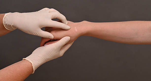
Medical professional examining a patient's wrist for ganglion cyst treatment

Treatment of Ganglion Cyst

Treatment of Ganglion Cyst The treatment of ganglion cyst depends on symptoms, cyst size, and how much it interferes with daily life. Many ganglion cysts go away on their own or remain stable without causing problems. Whenever doctors need to intervene, they have a variety of non-surgical and surgical options available. Observation (Watch and Wait) If the cyst is painless and not affecting function, doctors do not need to administer any treatment. Many cysts shrink or disappear spontaneously Regular monitoring ensures it doesn’t grow or cause new symptoms Non-Surgical Treatments 1. Immobilisation Wearing a brace or splint can reduce movement and pressure, especially in the wrist May relieve discomfort and help the cyst shrink Doctors do not reccoment long-term use, due to risks of stiffness or muscle weakening 2. Aspiration (Needle Drainage) Fluid is withdrawn using a needle and syringe Steroid injection may be added to reduce recurrence, though this is debated Quick and low-risk, but recurrence rates are high (over 50%) Aspiration may provide temporary relief but does not remove the cyst wall. Surgical Removal (Excision) Surgery may be recommended when: The cyst is painful, recurrent, or restricts movement Non-surgical options fail There’s concern about nerve compression Performed under local or general anaesthetic, the procedure involves: Removing the entire cyst and its stalk, which often connects to a joint or tendon Outpatient recovery with stitches removed in 1–2 weeks Lower recurrence rate compared to aspiration, though recurrence is still possible Recovery and Self-Care After surgery or aspiration: Keep the area clean and bandaged Avoid strenuous activity for several days Gentle exercises may be prescribed to restore movement and strength Treatment of Ganglion Cyst The treatment of ganglion cyst should be tailored to each individual, balancing symptom relief with the risk of recurrence or complications. [Next: Complications of Ganglion Cyst →]

Treatment of Ganglion Cyst Read More »

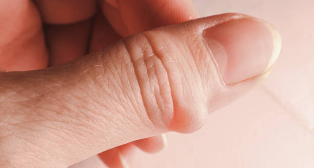
Close-up of a ganglion cyst on the thumb showing swelling and tension

Complications of Ganglion Cyst

Complications of Ganglion Cyst The complications of ganglion cyst are generally mild, but they can interfere with function or comfort if left untreated. Ganglion cysts are benign and rarely dangerous, but their size, location, or recurrence can cause difficulties over time. Pain and Nerve Compression A cyst that presses on a nearby nerve may cause: Tingling, numbness, or burning sensation Muscle weakness in the affected limb Interference with fine motor skills or grip strength This is most common when the cyst is located on the volar (palm) side of the wrist. Reduced Range of Motion Large or persistent cysts may limit movement in the wrist, fingers, or ankle May lead to stiffness, particularly after periods of rest In some people, this affects everyday tasks like typing, writing, or gripping objects Recurrence Even after removal or aspiration, cysts often return, particularly if the root (stalk) is not completely removed Surgical excision has a lower recurrence rate than aspiration but is still not guaranteed to be permanent Joint or Tendon Irritation Cysts located near tendons may cause a snapping or clicking sensation during movement Chronic pressure can lead to tendon irritation or inflammation over time Infection (Rare) Infection is very rare, but possible after aspiration or surgery Signs include redness, warmth, swelling, pus, or fever Prompt medical attention is needed for any post-procedure complications Scarring and Post-Surgical Effects After surgical removal, a scar may form at the site of the incision Some people experience persistent sensitivity or stiffness, especially if rehabilitation is limited Psychological and Cosmetic Concerns Visible cysts—especially on the hand or wrist—may cause embarrassment or self-consciousness This can affect social confidence or lead to avoidance of certain activities Complications of Ganglion Cyst Although the complications of ganglion cyst are rarely serious, monitoring and appropriate treatment help avoid persistent symptoms, discomfort, or functional problems. [Next: Back to Overview →]

Complications of Ganglion Cyst Read More »

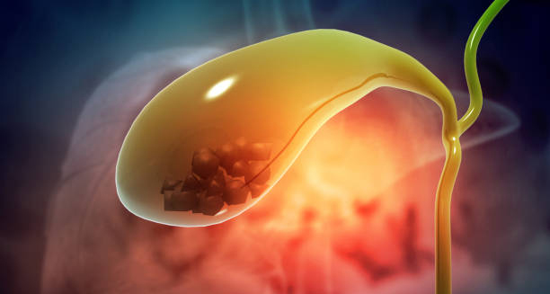
Medical illustration of gallstones inside the gallbladder

Gallstones

Gallstones These Gallstones are small, solid deposits that form in the gallbladder from substances found in bile, such as cholesterol or bilirubin. These stones can range in size from a grain of sand to a golf ball and may be present without causing symptoms for years. When they block bile flow, however, they can trigger severe pain and complications. The gallbladder is a small organ located beneath the liver. Its main role is to store and release bile—a fluid that helps digest fats. When the chemical balance of bile is disrupted, it results in the development of these stones. Therefore, causing particles to harden and form stones. Gallstones Many people with these stones experience no symptoms at all and may not even know they have them. These are known as “silent gallstones.” But when these stones obstruct the bile ducts, symptoms like upper abdominal pain, nausea, or jaundice can develop, often requiring treatment or surgery. Understanding these stones is key to recognising symptoms early, making informed dietary choices, and seeking medical care when needed. [Next: Causes of Gallstones →]

Gallstones Read More »

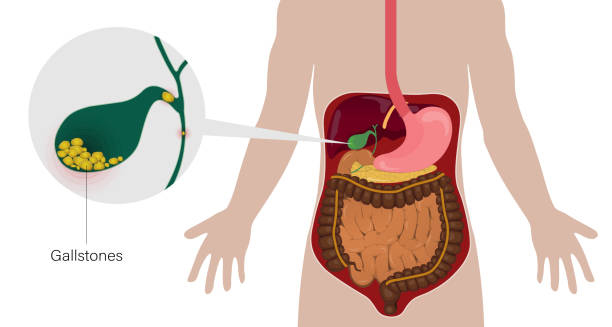
Diagram showing gallstones forming in the gallbladder and their location in the human digestive system

Causes of Gallstones

Causes of Gallstones The causes of gallstones involve an imbalance in the substances that make up bile, particularly cholesterol, bile salts, and bilirubin. Gallstones form when these substances become too concentrated and crystallise inside the gallbladder. Cholesterol Stones The most common type, especially in Western countries Caused when bile contains too much cholesterol, too little bile salts, or not enough fluid High-fat diets, obesity, and lack of physical activity can contribute to this imbalance Pigment Stones Made from excess bilirubin, a substance produced when the body breaks down red blood cells More common in people with: Liver disease Certain blood disorders like sickle cell anaemia Infections in the biliary system Sludge Formation Before actual stones develop, bile may become thick and form sludge—a precursor to gallstones. This often occurs: During rapid weight loss In people receiving intravenous feeding During pregnancy due to hormonal changes Risk Factors Gallstones are more likely in people who are: Over 40 years old Female – due to hormonal influences of oestrogen Overweight or obese Following a high-fat, low-fibre diet Living a sedentary lifestyle Other contributing factors include: Family history of gallstones Pregnancy Use of oral contraceptives or hormone replacement therapy Certain medical conditions like diabetes or Crohn’s disease Ethnicity and Geography Gallstones are more common in people of Native American, Hispanic, or South Asian descent Western dietary habits are associated with higher prevalence compared to other regions Causes of Gallstones Understanding the causes of gallstones allows at-risk individuals to take preventive steps—such as maintaining a healthy weight, eating a balanced diet, and staying active. [Next: Symptoms of Gallstones →]

Causes of Gallstones Read More »

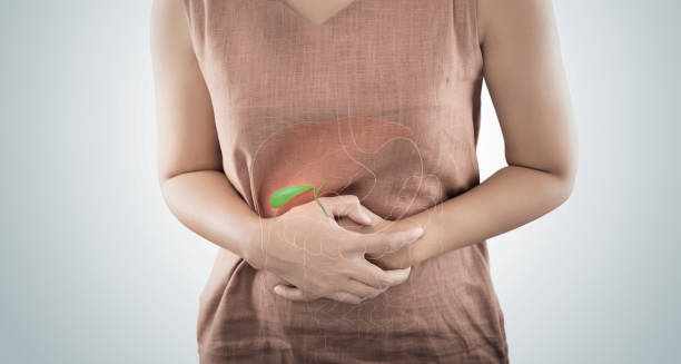
Woman clutching abdomen with a gallbladder illustration superimposed

Symptoms of Gallstones

Symptoms of Gallstones The symptoms of gallstones depend on whether the stones are causing a blockage or irritation in the gallbladder or bile ducts. Gallstones often go unnoticed, but when they obstruct the normal flow of bile, they can lead to a painful episode known as a gallbladder attack or biliary colic. Asymptomatic (Silent) Gallstones Most people with gallstones do not experience symptoms These are often discovered incidentally during scans or surgery for unrelated issues No treatment is usually required unless symptoms develop Biliary Colic (Gallbladder Attack) Sudden and intense pain in the upper right abdomen or middle of the abdomen Pain may radiate to the back or right shoulder blade Typically occurs after eating a fatty meal May last from 30 minutes to several hours Often accompanied by: Nausea or vomiting Bloating Indigestion Gallbladder attacks can happen intermittently and may return unless the underlying issue is treated. Complicated Gallstone Symptoms If gallstones lead to complications, additional symptoms may appear: Cholecystitis (Gallbladder Inflammation) Persistent pain lasting more than six hours Fever and tenderness in the upper right abdomen Nausea and vomiting Requires urgent medical treatment Choledocholithiasis (Stones in the Bile Duct) Jaundice (yellowing of the skin and eyes) Dark urine, pale stools Severe abdominal pain and fever Pancreatitis Inflammation of the pancreas caused by gallstones blocking the pancreatic duct Severe abdominal pain, nausea, and fever Requires hospital admission When to See a Doctor Seek medical attention if you experience: Repeated abdominal pain after meals Signs of infection such as fever and chills Jaundice or persistent nausea Symptoms of Gallstones Recognising the symptoms of gallstones early can help prevent complications and ensure timely treatment. [Next: Diagnosis of Gallstones→]

Symptoms of Gallstones Read More »

Doctor consulting a woman with abdominal pain

Diagnosis of Gallstones

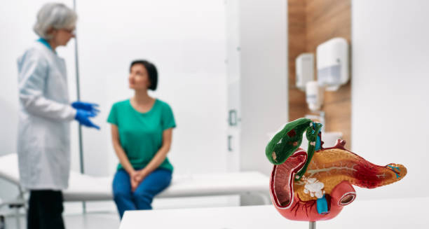
Diagnosis of Gallstones The diagnosis of gallstones typically begins with a clinical assessment based on symptoms, followed by imaging tests to confirm their presence. Doctors often find Gallstones incidentally during scans for unrelated conditions, particularly in asymptomatic individuals. In symptomatic cases, swift diagnosis is essential to prevent complications. Medical History and Physical Exam Doctors will: Ask about episodes of abdominal pain, especially after meals Look for signs of jaundice, fever, or tenderness in the upper right abdomen Discuss personal and family risk factors such as obesity, pregnancy, and gallbladder disease When doctors suspect gallstones, they usually recommend imaging tests as the next step. Ultrasound Abdominal ultrasound is the most common and accurate test for detecting gallstones It is non-invasive, quick, and painless Can identify stones in the gallbladder, inflammation, and gallbladder wall thickening Additional Imaging If doctors need further details, they may use the following: CT scan – shows complications such as infection, blocked ducts, or abscesses MRI and MRCP (Magnetic Resonance Cholangiopancreatography) – visualises bile ducts and can detect gallstones outside the gallbladder HIDA scan – assesses gallbladder function by tracking bile flow; useful in diagnosing cholecystitis Blood Tests Blood work helps assess: Liver function – elevated enzymes may suggest bile duct obstruction White blood cell count – increased levels may indicate infection Bilirubin levels – high readings are associated with blocked bile ducts These results help determine whether emergency treatment is needed. Endoscopic Tests If stones are suspected in the bile ducts: ERCP (Endoscopic Retrograde Cholangiopancreatography) may be performed Combines endoscopy and X-ray to view and remove bile duct stones Often used as both a diagnostic and therapeutic tool Diagnosis of Gallstones An accurate diagnosis of gallstones guides appropriate treatment and prevents unnecessary procedures. In cases where no symptoms are present, monitoring may be all that’s required. [Next: Treatment of Gallstones →]

Diagnosis of Gallstones Read More »

Doctor consulting a patient with a model of the gallbladder in the foreground

Treatment of Gallstones

Treatment of Gallstones The treatment of gallstones depends on whether symptoms are present and if complications have occurred. Gallstones that cause pain or digestive issues often require medical or surgical intervention, while silent gallstones may be left alone. Watchful Waiting Doctors usually do not recommend treatment for asymptomatic gallstones. Doctors may advise regular monitoring. Lifestyle changes such as a healthy, low-fat diet and weight control can reduce the risk of symptoms developing Medications Oral bile acid pills (e.g. ursodeoxycholic acid) may help dissolve small cholesterol gallstones Takes months to years to work and not effective for all types of stones Recurrence is common after stopping the medication Doctors usually reserve medication for patients who cannot undergo surgery. Surgery (Cholecystectomy) The most effective treatment for symptomatic gallstones is surgical removal of the gallbladder: Laparoscopic cholecystectomy (keyhole surgery) is the most common method Minimally invasive Short recovery time Often performed as a day procedure Doctors may need to perform Open Cholecystectomy in complex or emergency cases Removing the gallbladder does not significantly affect digestion in most people. The liver continues to produce bile, which flows directly into the intestine. ERCP and Stone Removal If gallstones lodge in the bile ducts, doctors may recommend immediate treatment: ERCP can be used to remove them without removing the gallbladder Often followed by surgery later to prevent future blockages Post-Surgical Care After gallbladder removal: Some individuals experience temporary digestive changes, such as diarrhoea or bloating A gradual return to normal diet is encouraged Most people resume regular activities within one to two weeks Treatment of Gallstones The treatment of gallstones is tailored to the severity and type of symptoms. For those with complications, timely surgery can be lifesaving. [Next: Complications of Gallstones →]

Treatment of Gallstones Read More »

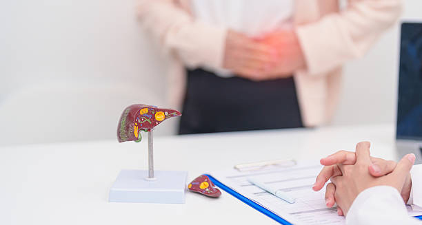
Medical consultation showing a liver and gallbladder model with a patient holding their abdomen

Complications of Gallstones

Complications of Gallstones The complications of gallstones arise when stones block bile flow or trigger inflammation. While many people live with gallstones without any problems, untreated or poorly managed cases can lead to serious health issues. Gallstones can also increase the risk of infections, organ damage, and rare cancers. Cholecystitis (Gallbladder Inflammation) Occurs when a stone blocks the cystic duct, causing the gallbladder to become inflamed Symptoms include severe upper right abdominal pain, fever, nausea, and tenderness May lead to abscesses, gallbladder rupture, or generalised infection (sepsis) if not treated Urgent surgery may be required in severe cases. Choledocholithiasis (Bile Duct Stones) Stones may migrate into the common bile duct, blocking the flow of bile Leads to jaundice, dark urine, pale stools, and abdominal pain Requires ERCP to remove the obstruction and relieve symptoms Pancreatitis Gallstones can block the pancreatic duct, leading to acute pancreatitis Symptoms include intense upper abdominal pain, vomiting, and fever Can range from mild to life-threatening and often requires hospitalisation Gallbladder Empyema Build-up of pus inside the gallbladder Results from untreated cholecystitis Increases risk of rupture and widespread infection Gallstone Ileus A rare complication where a large gallstone enters the intestine and causes a blockage Can result in severe abdominal pain, vomiting, and bowel obstruction Often requires surgery to remove the stone and relieve the blockage Gallbladder Cancer Long-term gallstone disease may slightly increase the risk of gallbladder cancer Rare, but more likely in people with chronic inflammation or porcelain gallbladder Recurrent Biliary Pain Without treatment, gallstones can cause repeated painful episodes These may become more frequent and severe over time Complications of Gallstones Recognising and managing the complications of gallstones early prevents escalation and helps preserve overall digestive health. [Next: Back to Overview →]

Complications of Gallstones Read More »

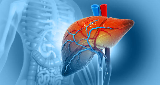
3D illustration of human liver and gallbladder with blood vessels

Gallbladder Cancer

Gallbladder Cancer The condition gallbladder cancer is a rare but aggressive form that begins in the tissues of the gallbladder. This cancer often develops silently, with few symptoms in the early stages. Making it difficult to detect until it has advanced. This delayed diagnosis contributes to its generally poor prognosis. The gallbladder is a small, pear-shaped organ located beneath the liver. Its main function is to store and release bile, which helps the body digest fats. While most gallbladder problems are due to gallstones or inflammation (cholecystitis), this disease can occasionally arise. Particularly in individuals with long-standing gallbladder disease. This cancer occurs more frequently in women and is more common in certain parts of the world. Including South America, India, and Eastern Europe. Risk factors include gallstones, chronic inflammation, obesity, age, and a history of gallbladder polyps. Gallbladder Cancer Because early symptoms are vague or absent, many cases are only discovered incidentally during surgery or imaging for other gallbladder conditions. Raising awareness about this cancer may improve early detection and support timely, effective treatment. [Next: Causes of Gallbladder Cancer →]

Gallbladder Cancer Read More »

Scroll to Top