Mental Matters

Author name: admin

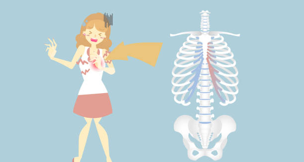
Illustration of a woman experiencing chest pain with a diagram of the ribcage highlighting costal cartilage inflammation, indicating costochondritis.

Costochondritis

Costochondritis – Overview Costochondritis is an inflammatory condition that affects the cartilage connecting the ribs to the breastbone (sternum). Leading to localised chest pain that can mimic the symptoms of a heart attack. Although alarming, this is generally harmless and not related to heart disease. It is a common cause of chest pain. Especially in younger adults and teenagers, and often resolves on its own over time. The inflammation occurs where the upper ribs attach to the sternum at the costosternal junctions. This irritation can result in sharp, stabbing, or aching pain. Which typically worsens with movement, deep breathing, or pressure applied to the chest wall. Unlike cardiac-related pain, costochondritis is usually localised and can often be reproduced by touching the affected area. Costochondritis The exact cause of this is often unknown, though it may be triggered by trauma, repetitive strain, respiratory infections, or inflammatory conditions such as arthritis. The pain may affect one or more rib joints and is usually more prominent on the left side of the chest, contributing to the anxiety it causes in patients. Costochondritis is considered a benign condition with no lasting effects. However, the chest pain can be severe and distressing, often leading to unnecessary emergency visits or cardiac evaluations. Diagnosis is based on physical examination and exclusion of more serious conditions. In summary, costochondritis is an inflammation of the cartilage in the chest wall, causing non-cardiac chest pain. It is treatable and usually self-limiting, though the symptoms can be intense and unsettling. [Next: Causes of Costochondritis →]


Costochondritis Read More »

Two workers lifting a heavy box, potentially straining their chest muscles, which can lead to costochondritis.

Causes of Costochondritis

Causes of Costochondritis Causes of costochondritis vary widely and are often idiopathic, meaning they arise without a known reason. However, several identifiable factors are known to contribute to the inflammation of the costosternal joints. Regardless of the source, costochondritis occurs when the cartilage that connects the ribs to the sternum becomes irritated or inflamed, resulting in pain and tenderness in the chest wall. Mechanical Strain One of the most common contributors is physical strain or injury to the chest wall. This may include: Heavy lifting Vigorous exercise, especially involving the upper body Prolonged or repetitive coughing Poor posture, especially over time The repetitive movement or tension on the rib joints can cause microtrauma, which leads to inflammation. Trauma or Injury | Causes of Costochondritis Direct trauma to the chest, such as from a fall, car accident, or sports injury, can irritate the cartilage between the ribs and sternum. Even minor impacts may trigger costochondritis in susceptible individuals, especially if the injury goes unnoticed. Infections Though rare, viral or bacterial respiratory infections—particularly those involving severe coughing—can inflame the rib joints. Certain pathogens may also lead to post-viral inflammation, mimicking symptoms of costochondritis. Inflammatory Conditions Autoimmune and inflammatory disorders such as: Rheumatoid arthritis Ankylosing spondylitis Psoriatic arthritis can contribute to chronic costosternal joint inflammation. In such cases, costochondritis may form part of a broader systemic condition. Other Possible Triggers | Causes of Costochondritis Allergies and asthma (due to frequent coughing) Wearing heavy backpacks Muscular imbalances or tight chest muscles It’s important to note that costochondritis is often not linked to any visible damage on imaging scans, which is why diagnosis depends largely on clinical symptoms and ruling out other causes of chest pain. In conclusion, causes of costochondritis include trauma, repetitive strain, infections, and underlying inflammatory diseases. Identifying the likely trigger helps guide effective management and reassures patients about the non-cardiac nature of the condition. [Next: Symptoms of Costochondritis →]

Causes of Costochondritis Read More »

Woman holding her chest in discomfort, indicating pain typical of costochondritis.

Symptoms of Costochondritis

Symptoms of Costochondritis Symptoms of costochondritis primarily involve pain and tenderness in the chest, particularly where the ribs meet the sternum. The pain may feel sharp, aching, or pressure-like and often leads patients to believe they are having a cardiac event. However, costochondritis pain is musculoskeletal and non-life-threatening, though it can be severe and persistent. Chest Pain The hallmark symptom is localised chest pain, which typically: Is located on one side of the chest, most commonly the left Increases with deep breaths, coughing, or sneezing Worsens with physical activity or upper body movement Is reproducible when pressure is applied to the affected rib joints This reproducibility is a key feature distinguishing costochondritis from heart-related pain. Tenderness | Symptoms of Costochondritis The chest wall is usually tender to touch over one or more costosternal junctions. Palpating these areas often intensifies the pain, confirming a musculoskeletal origin. Radiation of Pain Pain may radiate to the back or abdomen, or along the ribs. This can cause confusion with gastrointestinal or cardiac issues. However, radiation with costochondritis is usually superficial and not associated with nausea, sweating, or other cardiac warning signs. Pain Duration and Intensity Symptoms can last from a few days to several weeks or even months in chronic cases. Pain intensity may vary throughout the day and often worsens at night or after periods of physical exertion. Associated Symptoms | Symptoms of Costochondritis Some individuals may experience: Mild swelling at the affected site (rare) Tightness in the chest Anxiety due to fear of a heart condition Importantly, costochondritis does not cause fever, shortness of breath, or palpitations. If these symptoms are present, further evaluation is necessary to rule out more serious conditions. In summary, symptoms of costochondritis centre around chest pain that is sharp, localised, and reproducible on touch. While often mistaken for cardiac pain, it is a benign condition that responds well to conservative treatment. [Next: Diagnosis of Costochondritis →]

Symptoms of Costochondritis Read More »

Medical diagram of the rib cage showing inflammation in the costal cartilage, labelled as costochondritis.

Diagnosis of Costochondritis

Diagnosis of Costochondritis Diagnosis of costochondritis is based primarily on clinical evaluation, as there are no specific laboratory or imaging tests that can definitively confirm the condition. Since costochondritis shares symptoms with more serious causes of chest pain—such as heart attacks, pulmonary embolisms, or gastrointestinal issues—doctors must carefully rule out life-threatening conditions before settling on this diagnosis. Medical History The diagnostic process begins with a thorough discussion of symptoms: Onset and nature of the chest pain Triggers, such as physical activity, coughing, or breathing deeply Duration and whether the pain improves or worsens with rest Any recent trauma, infection, or repetitive strain Patients may also be asked about their medical history, including risk factors for heart disease, respiratory conditions, and inflammatory disorders. Physical Examination | Diagnosis of Costochondritis A key diagnostic feature of costochondritis is reproducible chest pain. During examination, the doctor will: Press on the chest wall at the costosternal joints Assess for tenderness and inflammation Observe the patient’s breathing and chest movement Check for signs of swelling (though rare in costochondritis) Pain that worsens with palpation of the rib junctions is a strong indicator of costochondritis. Exclusion of Other Conditions Because chest pain can signify serious issues, further tests may be conducted to rule out cardiac, respiratory, or gastrointestinal conditions: Electrocardiogram (ECG) to rule out heart attack Chest X-ray to assess lung or bone abnormalities Blood tests if an infection or inflammation is suspected Stress tests or echocardiograms, particularly in older adults or those with cardiac risk factors If tests are normal and the pain is localised and reproducible, a diagnosis of costochondritis is usually confirmed. Distinguishing from Tietze Syndrome | Diagnosis of Costochondritis Tietze syndrome is a similar condition but includes visible swelling over the rib joints and typically affects younger individuals. Unlike costochondritis, Tietze syndrome is less common and more inflammatory in nature. In conclusion, diagnosis of costochondritis involves recognising characteristic symptoms and ruling out more dangerous conditions. A careful physical examination is key to confirming this often-misunderstood cause of chest pain. [Next: Treatment of Costochondritis →]

Diagnosis of Costochondritis Read More »

Medical professional wearing gloves holding a model of the rib cage with the word “Costochondritis” overlayed.

Treatment of Costochondritis

Treatment of Costochondritis Treatment of costochondritis focuses on relieving pain and inflammation while allowing the cartilage to heal naturally. Since the condition is self-limiting in most cases, conservative therapies are often sufficient. However, persistent or severe cases of costochondritis may require more intensive intervention. Medications Nonsteroidal anti-inflammatory drugs (NSAIDs) such as ibuprofen or naproxen are first-line treatments. They help reduce inflammation and relieve pain. Paracetamol may be used for those who cannot tolerate NSAIDs. Stronger painkillers, such as codeine, may be considered for acute flare-ups but are not recommended for long-term use. Corticosteroid injections may be used in chronic or unresponsive cases to reduce inflammation at the affected joints. Physical Therapy | Treatment of Costochondritis Physiotherapy can help improve posture and reduce strain on the chest wall: Stretching exercises to relieve tightness in the chest muscles Postural correction to address ergonomic causes of costochondritis Breathing exercises to reduce tension in the rib cage Local Treatments Heat or ice packs can provide temporary relief. Apply a warm compress or cold pack for 15–20 minutes at a time to the affected area. Topical anti-inflammatories, such as diclofenac gel, may reduce localised discomfort. Activity Modification | Treatment of Costochondritis Rest and avoiding activities that exacerbate symptoms are key in the early stages. This includes: Avoiding heavy lifting or upper-body workouts Minimising repetitive chest movements Adopting ergonomic positions when sitting or working Alternative Therapies Some individuals find relief with: Acupuncture Chiropractic adjustments Massage therapy While evidence is limited, these may be useful as adjunctive therapies in chronic cases. In summary, treatment of costochondritis aims to relieve symptoms through anti-inflammatory medication, rest, and physiotherapy. Most individuals recover within weeks to a few months, particularly with early intervention and lifestyle adjustments. [Next: Complications and Recovery from Costochondritis →]

Treatment of Costochondritis Read More »

Muscular man clutching his chest with a highlighted rib area, indicating pain from costochondritis.

Complications and Recovery from Costochondritis

Complications and Recovery from Costochondritis Complications and recovery from costochondritis are generally minimal, as the condition is benign and self-limiting in most cases. However, if costochondritis is left untreated or mismanaged, it can lead to persistent pain and reduced quality of life. Fortunately, with appropriate care, recovery is usually complete and long-lasting. Potential Complications | Complications and Recovery from Costochondritis Chronic pain: Although uncommon, some individuals may experience ongoing discomfort for several months, particularly if the inflammation becomes recurrent or is aggravated by physical activity. Reduced mobility: Severe or chronic cases may cause individuals to limit movement, leading to poor posture, muscle stiffness, or deconditioning. Anxiety and misdiagnosis: The chest pain caused by costochondritis often mimics heart disease. This can lead to emotional distress, repeated emergency visits, and unnecessary cardiac tests. When to Seek Medical Attention While costochondritis is not dangerous, it’s important to consult a doctor if: The chest pain is new or severe Pain occurs with exertion or radiates to the arm or jaw There are associated symptoms such as shortness of breath, dizziness, or palpitations These may indicate a more serious cardiac or respiratory condition that requires urgent evaluation. Recovery Timeline Acute cases: Most people recover within 2 to 4 weeks with rest, NSAIDs, and activity modification. Chronic or recurrent cases: May require ongoing physiotherapy or periodic corticosteroid treatment and can last several months. Post-viral cases: Inflammation due to respiratory infections often subsides as the infection resolves, but lingering pain can persist for several weeks. Preventing Recurrence | Complications and Recovery from Costochondritis Maintain good posture and ergonomic workspaces Avoid activities that strain the chest wall Treat respiratory infections promptly Use protective gear during sports or physical labour In conclusion, complications and recovery from costochondritis are usually manageable. With early treatment and appropriate lifestyle modifications, most individuals make a full recovery and return to normal activities without long-term issues. [Next: Back to Overview →]

Complications and Recovery from Costochondritis Read More »

Illustration of a human head made of fragmented ice, symbolising cognitive and neurological decline from corticobasal degeneration.

Corticobasal Degeneration

Corticobasal Degeneration – Overview Corticobasal degeneration is a rare and progressive neurological disorder that affects movement, speech, and cognitive function. It is caused by abnormal deposits of tau protein in certain areas of the brain, leading to nerve cell deterioration. This degeneration typically begins in adults aged between 50 and 70 and gradually worsens over time, affecting one side of the body more than the other in the early stages. This condition falls under the category of atypical parkinsonian syndromes, as it shares some similarities with Parkinson’s disease. However, it presents with a broader and more complex set of symptoms, including stiffness, clumsiness, tremors, and cognitive decline. Over time, individuals may also develop difficulty speaking, swallowing, and performing coordinated movements. Corticobasal Degeneration – Overview The disease is named after the two main brain areas it affects: the cerebral cortex, which is responsible for higher brain functions such as thinking and planning, and the basal ganglia, which helps control voluntary motor movement. Damage to these regions results in a unique combination of motor, sensory, and cognitive symptoms that make diagnosis particularly challenging. Although the condition is rare, it is often misdiagnosed in its early stages because its symptoms overlap with more common conditions like Parkinson’s disease, Alzheimer’s disease, and frontotemporal dementia. There is currently no cure, but symptom management and supportive therapies can help improve quality of life. In summary, this degeneration is a progressive, degenerative brain disorder marked by asymmetric motor problems, cognitive impairment, and speech difficulties. Early diagnosis and supportive care are essential for maintaining dignity and comfort as the disease progresses. [Next: Causes of Corticobasal Degeneration →]

Corticobasal Degeneration Read More »

Illustration of a brain with virus-like particles, symbolising unknown and neurological causes of corticobasal degeneration.

Causes of Corticobasal Degeneration

Causes of Corticobasal Degeneration Causes of corticobasal degeneration are not fully understood. However, the condition is strongly associated with the accumulation of abnormal tau protein in the brain. This protein, which normally supports the internal structure of nerve cells. Therefore, becomes misfolded and clumps together, leading to cell dysfunction and death. As corticobasal degeneration progresses, these tau deposits spread through the cerebral cortex and basal ganglia, disrupting motor and cognitive function. Causes of Corticobasal Degeneration Unlike some neurodegenerative disorders that are linked to clear genetic mutations. Most cases of corticobasal degeneration are sporadic, meaning they arise with no known family history or inherited pattern. Although rare familial cases have been reported, no single gene has been consistently associated with the condition. This suggests a complex interplay between genetic predisposition and environmental factors. The build-up of tau protein places corticobasal degeneration within a group of diseases known as tauopathies, which also includes Alzheimer’s disease, progressive supranuclear palsy (PSP), and frontotemporal dementia (FTD). Each of these conditions is characterised by the presence of tau pathology, but the distribution and effects on the brain differ significantly. Some researchers have explored potential environmental or lifestyle risk factors, such as head trauma, exposure to toxins, or infections, but no conclusive link has been established. There is also no evidence that corticobasal degeneration is contagious or caused by an infection. Causes of Corticobasal Degeneration The disease affects certain areas of the brain more severely, including the posterior frontal lobe and parietal lobe, which are involved in movement and spatial awareness. This explains many of the hallmark symptoms such as clumsiness, stiffness, and coordination problems. In summary, causes of corticobasal degeneration involve the abnormal accumulation of tau protein in specific brain regions. While the exact triggers remain unknown, research continues into the genetic and environmental factors that may contribute to its onset. Understanding these mechanisms is crucial for developing future treatments. [Next: Symptoms of Corticobasal Degeneration →] https://mentalmatters.co.za/corticobasal-degeneration/

Causes of Corticobasal Degeneration Read More »

Elderly man sitting on a couch, holding and examining his hand, indicating motor symptoms of corticobasal degeneration.

Symptoms of Corticobasal Degeneration

Symptoms of Corticobasal Degeneration Symptoms of corticobasal degeneration can vary widely among individuals and tend to progress gradually. Early signs are often subtle and may be mistaken for other neurological disorders. As corticobasal degeneration advances, signs typically become more pronounced and disabling, affecting both physical movement and cognitive function. Motor Symptoms | Symptoms of Corticobasal Degeneration One of the earliest and most distinctive symptoms is asymmetrical motor dysfunction, where one side of the body becomes noticeably weaker or stiffer than the other. Common motor symptoms include: Muscle stiffness (rigidity) Tremors or involuntary shaking Slowness of movement (bradykinesia) Poor coordination or clumsiness Dystonia, involving abnormal postures or muscle contractions Apraxia, the inability to perform learned movements despite having the desire and physical ability Individuals may struggle with simple tasks like buttoning a shirt or using cutlery, even when muscle strength appears normal. These problems are often misdiagnosed as functional or psychiatric issues in the early stages. Cognitive and Behavioural Symptoms As the disease progresses, cognitive decline becomes more apparent. Symptoms include: Memory loss Difficulty planning or organising Impaired judgement Language difficulties, such as hesitant speech or difficulty finding words Irritability, apathy, or depression Behavioural changes may resemble those seen in frontotemporal dementia, particularly in the late stages. Speech and Swallowing Difficulties with speech (dysarthria) and swallowing (dysphagia) can also emerge, leading to nutritional issues and increased risk of choking. Voice may become soft, slurred, or monotone. Sensory Disturbances | Symptoms of Corticobasal Degeneration Some people experience numbness or the feeling of a “foreign limb,” where the affected limb feels disconnected or acts involuntarily—a phenomenon called alien limb syndrome. In conclusion, symptoms of corticobasal degeneration are diverse and evolve over time, typically affecting movement first and cognition later. The combination of motor and mental symptoms often makes daily life increasingly challenging, requiring a comprehensive care approach. [Next: Diagnosis of Corticobasal Degeneration →] https://mentalmatters.co.za/corticobasal-degeneration

Symptoms of Corticobasal Degeneration Read More »

Illustration of a brain with visible atrophy and lesions, symbolising neurological damage from corticobasal degeneration.

Diagnosis of Corticobasal Degeneration

Diagnosis of Corticobasal Degeneration Diagnosis of corticobasal degeneration is often difficult, especially in the early stages, due to the overlap of symptoms with other neurological disorders. There is no single test that can definitively confirm corticobasal degeneration during life. Instead, diagnosis is made through a combination of clinical assessments, neurological examinations, and imaging studies that help rule out other possible conditions. Medical History and Clinical Assessment The process typically begins with a thorough evaluation of: Onset and progression of symptoms Degree of asymmetry in movement difficulties Cognitive and behavioural changes Speech and swallowing problems Doctors will look for the combination of movement and cognitive symptoms that are characteristic of corticobasal degeneration, especially unilateral stiffness, apraxia, and speech changes. Neurological Examination | Diagnosis of Corticobasal Degeneration A neurologist will assess: Muscle tone and reflexes Coordination and balance Ability to perform learned movements Memory, attention, and language function Findings such as limb rigidity, difficulty performing simple tasks despite adequate strength, and alien limb syndrome are suggestive of corticobasal degeneration. Brain Imaging MRI scans may show shrinkage (atrophy) in specific brain areas such as the parietal lobe or frontal cortex, often more pronounced on one side. PET or SPECT scans can assess metabolic activity and highlight regions of the brain that are underactive due to nerve cell damage. These imaging results support the diagnosis and help differentiate corticobasal degeneration from Parkinson’s disease, Alzheimer’s, or progressive supranuclear palsy. Neuropsychological Testing This type of testing evaluates memory, attention, problem-solving, and language skills. It helps identify cognitive decline patterns typical of corticobasal degeneration. Exclusion of Other Conditions | Diagnosis of Corticobasal Degeneration Because symptoms overlap with other disorders, conditions such as Parkinson’s disease, frontotemporal dementia, or stroke must be ruled out. Blood tests and, in rare cases, lumbar puncture may be conducted to exclude other causes. In summary, diagnosis of corticobasal degeneration relies on clinical expertise, symptom observation, and supportive imaging. Due to the lack of a specific test, ongoing reassessment is often necessary to confirm the condition over time. [Next: Treatment of Corticobasal Degeneration →] https://mentalmatters.co.za/corticobasal-degeneration

Diagnosis of Corticobasal Degeneration Read More »

Scroll to Top