Mental Matters

Author name: admin

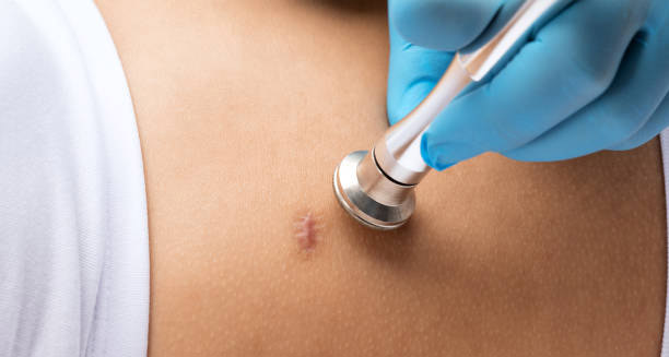
Doctor examining a small keloid scar on the chest with a stethoscope during treatment consultation

Treatment of Keloid Scars

Treating keloid scars can be challenging. These scars tend to return and often resist standard therapies. They form when the body produces too much collagen after a wound has healed. As a result, treating keloids often requires a combination of approaches. No single treatment works for everyone. The choice depends on the size, location, severity, and symptoms of the scar. Skin type and medical history also play a role. Treatment usually focuses on three goals: reducing scar size, easing discomfort, and preventing recurrence. In some cases, therapy also helps patients cope with emotional distress caused by visible or painful scars. 1. Corticosteroid Injections One of the most common treatments for keloids is corticosteroid injections. These are injected directly into the scar every 3 to 4 weeks. The steroids reduce inflammation and slow collagen production. Over time, the scar becomes flatter, softer, and less itchy. Triamcinolone acetonide is the steroid most often used. While it is usually effective, it can cause side effects such as skin thinning, color changes, or injection-site pain. Because keloids can return after injections stop, doctors often combine this therapy with other treatments. 2. Silicone Gel Sheets and Ointments Silicone-based products are a gentle, non-invasive option. These include gel sheets, self-drying gels, and ointments. They help hydrate the scar and reduce collagen overgrowth. Silicone therapy works best on early-stage or new keloids. Patients often need to wear silicone sheets for 12 to 24 hours a day for several months. These products are safe for most people, including children and those with sensitive skin. 3. Cryotherapy Cryotherapy uses liquid nitrogen to freeze and destroy scar tissue. This method is especially helpful for small, firm keloids. Over time, it can reduce the height and firmness of the scar. When combined with corticosteroid injections, cryotherapy can yield even better results. However, it may cause skin discoloration, particularly in people with darker skin. It’s also not suitable for large or highly vascular scars. 4. Laser Therapy Laser treatment is increasingly popular. It improves the texture, color, and thickness of keloid scars. Pulsed dye lasers (PDL) are most commonly used. They target blood vessels in the scar to reduce redness and swelling. Laser therapy works best when paired with other treatments like steroids or topical creams. It is safe but often requires multiple sessions. Although not a cure, laser therapy can greatly improve the scar’s appearance and reduce symptoms like itching. 5. Surgical Excision Surgery may be an option for large or painful keloids that don’t respond to other treatments. It involves cutting out the scar tissue, usually under local or general anesthesia. However, surgery alone has a high risk of recurrence—up to 70%. It often triggers the same healing response that caused the original keloid. That’s why surgery is almost always combined with follow-up therapies such as steroid injections, silicone sheets, pressure garments, or radiation. 6. Radiation Therapy Radiation therapy is sometimes used after surgical removal of a keloid. Low-dose radiation helps kill leftover fibroblast cells that might cause the scar to grow back. While effective, radiation carries long-term risks. It is used with caution, especially in younger patients or near vital organs. Still, when carefully planned, it can significantly reduce the chances of recurrence. 7. Pressure Therapy Pressure therapy involves wearing compression garments or bandages to flatten the keloid. It is especially effective on earlobe keloids and is often used after surgery or ear piercing. To be successful, pressure must be applied consistently for 12 to 18 hours a day over several months. This treatment is non-invasive and affordable, but it can be uncomfortable, which may affect compliance. 8. 5-Fluorouracil (5-FU) 5-FU is a chemotherapy drug that can also treat keloids. It is either injected directly into the scar or applied as a cream. It helps reduce collagen production and scar size. Doctors often use 5-FU together with corticosteroids. While effective, it may cause mild pain, redness, or peeling at the treatment site. 9. Natural and Alternative Therapies Some natural remedies are used to manage keloids, though scientific proof is limited. These include: Patients should consult a doctor before trying any natural treatment, especially on sensitive or broken skin. 10. Psychological Support Keloids can have a strong emotional impact. People with visible scars often feel anxious, embarrassed, or depressed. This is especially true for scars on the face, chest, or other exposed areas. Counselling, support groups, or mental health therapy can help. A holistic approach—addressing both the physical and emotional effects—is essential for long-term wellbeing. Final Thoughts Treating keloid scars takes time and a personalised strategy. While no cure exists, combining medical treatments, surgical options, and self-care can lead to meaningful improvements. Early intervention matters. People prone to keloids should avoid skin trauma and follow medical advice to prevent new scars. With new treatments and better awareness, patients now have more options than ever to manage this difficult condition. [Next: Complications of Keloid Scars →]
Treatment of Keloid Scars Read More »

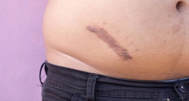
Dark raised keloid scar on lower abdomen showing post-inflammatory pigmentation

Complications of Keloid Scars

The complications of keloid scars can extend far beyond cosmetic concerns, significantly impacting an individual’s physical comfort, emotional wellbeing, and quality of life. Keloids are not harmful in a medical sense, but they often cause real problems. The complications of keloid scars can be long-lasting and upsetting, especially when they affect movement, confidence, or come back after treatment. Since keloids do not shrink on their own and are hard to treat, knowing what problems they cause is important for full care. Even though they are not cancer, keloids often behave in unpredictable ways. They may grow too much, cause pain or itching, and often return after treatment. These things make healing harder and leave many patients unhappy with the results. 1. Physical Discomfort and Functional Limits One of the most common complications of keloid scars is physical discomfort. Many people feel ongoing itching, pain, or tenderness. These symptoms get worse when the keloid is in a spot that rubs against clothing or moves often. If keloids form near joints like the elbows, knees, or shoulders, they may make it hard to move. They pull on the skin or press on tissues, causing stiffness. Larger keloids on the chest or neck can make it hard to turn the head, breathe deeply, or sleep. Keloids on the feet or hands can cause pain while walking, using tools, or wearing shoes. These small daily problems can greatly reduce a person’s ability to function. Often, they are overlooked but still cause a major loss of comfort and freedom. 2. High Chance of Coming Back Another major complication of keloid scars is how often they return. This can happen even after treatment seems to work. In fact, up to 70% of keloids may come back after surgery. Often, the new scar grows larger or feels worse than before. This happens because keloids form from an extreme healing response. The body sends too many healing cells, which create thick scar tissue. Even helpful treatments—like surgery or injections—can cause new trauma and spark this response again. As a result, patients often feel frustrated. After spending time and money on treatment, they face the same problem again, sometimes even worse. This can lead to disappointment and emotional stress. 3. Skin Color Changes and Visible Scars Keloids often cause changes in skin color. This is more common in people with darker skin tones. The scars may look red, purple, brown, or much darker than nearby skin. Even if the keloid flattens after treatment, the color may not return to normal. Keloids also tend to grow beyond the wound edges. This makes them more visible, especially on the face, ears, chest, or neck. Over time, the scar may become even more noticeable and change a person’s appearance. Many patients say the look of the scar bothers them more than the pain. It affects how they feel about themselves and how they interact with others. The complications of keloid scars often go beyond the skin—they affect how people feel inside. 4. Mental and Emotional Effects of Keloid Scars Keloid scars can deeply affect mental health, especially when they are easy to see. People may feel shy, anxious, or sad about how they look. This is especially true for teens and young adults who already feel pressure about appearance. Studies show that large or painful keloids can lead to depression or anxiety. Some people may even develop body image disorders. In certain cultures, scars are linked with illness or shame, which can lead to stigma or isolation. Living with keloids for years and trying many treatments can wear people down. They may lose hope or feel tired of trying. Financial costs and repeated doctor visits can also add to the mental burden. 5. Problems Caused by Treatment Treating keloids is not always safe or simple. Many methods come with risks. These may include: Strangely, the very treatments used to fix keloids can sometimes cause them to grow again. That’s why doctors must weigh the pros and cons carefully. People with a family history or a strong tendency to form keloids need extra care when choosing treatment. 6. Slower Wound Healing People who get keloids often heal more slowly than others. Even small injuries—like a bug bite, pimple, or vaccine shot—can take longer to go away. Worse, these small wounds may turn into new keloids. This slow healing keeps the skin in a state of stress. It also makes surgeries riskier. Doctors must work gently and plan carefully to prevent more scarring. To help, they might use pressure garments or silicone sheets after surgery. These steps raise the cost and difficulty of care but are needed to avoid more problems. 7. Impact on Daily and Work Life Keloid scars often appear on parts of the body that are hard to hide. When they form on the face, hands, or neck, they can affect daily life and job options. Some people avoid social events, job interviews, or public roles. For those with physical jobs or uniforms, like athletes or health workers, keloids may rub and hurt. This makes tasks harder and lowers work performance. In these cases, complications of keloid scars become more than a beauty issue—they affect daily life and income. This wide effect shows why keloids must be treated as serious health issues, not just skin problems. Final Thoughts on Complications of Keloid Scars The complications of keloid scars go far beyond looks. They can affect how a person moves, feels, and lives. Even though they are not dangerous by themselves, keloids can cause serious problems. The best care plan includes more than skin treatments. It should also support mental health and give clear guidance to patients. By understanding all the risks, people can make better choices about how to treat and manage their keloids. Knowledge and awareness help reduce the pain, stress, and burden of this long-lasting condition. With the right care, people can regain control of their health, confidence, and comfort. [Next: Back to Overview →]

Complications of Keloid Scars Read More »

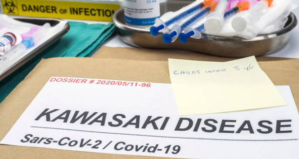
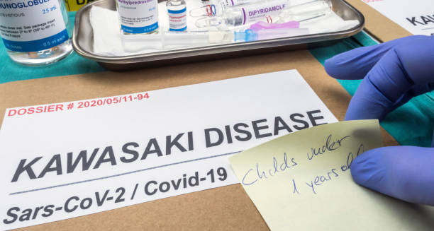
Medical dossier labelled Kawasaki Disease linked to Covid-19 with treatment vials and handwritten note

Overview of Kawasaki Disease

Kawasaki disease is a rare but serious condition that primarily affects children under the age of five. It causes swelling in blood vessels throughout the body. Doctors call this vasculitis. No one knows the exact cause yet, but the effects can be very serious—especially for the heart. If doctors don’t find and treat Kawasaki disease quickly, it can lead to heart problems. These include inflamed heart muscles, artery swelling, and even heart attacks in young children. Thankfully, with proper treatment, most children recover fully. Still, it remains one of the top causes of acquired heart disease in children living in developed countries. The illness usually develops in phases. It starts with a lasting fever and includes symptoms like a rash, red eyes, swollen hands and feet, cracked lips, and a red, bumpy tongue. These signs look like many common viral infections, which makes it hard to spot in the early days. That’s why knowing the signs and acting quickly are key to preventing long-term problems. 1. Epidemiology and Demographics Kawasaki disease mostly affects children between six months and five years old. It is seen more often in boys than girls. Children with East Asian backgrounds—especially from Japan—have the highest rates. However, the disease can appear in all parts of the world, including North America, Europe, and Africa. In some places, more cases happen during winter and early spring. This seasonal pattern may hint at a trigger. Even so, Kawasaki disease is not contagious. It cannot spread from one child to another. 2. Clinical Presentation and Stages Kawasaki disease moves through three main stages. a) Acute Phase (Weeks 1–2):This stage starts with a high fever over 39°C that lasts more than five days. Other key signs include: The child may seem very cranky and may have belly pain or sore joints. b) Subacute Phase (Weeks 2–4):Once the fever goes away, other problems become more visible. These may include: During this time, the risk of heart problems grows. So, close monitoring is very important. c) Convalescent Phase (Weeks 4–6):In this stage, most symptoms begin to fade. The child starts to feel better and gains energy. Blood test results slowly return to normal. But the blood vessels may still be healing. That’s why regular check-ups are still needed during this time. 3. Aetiology and Risk Factors Doctors still don’t know the exact overview of Kawasaki disease and its cause. Most believe the immune system overreacts to an infection. This infection could be from a virus or bacteria. Children with certain genes may respond too strongly, leading to swelling and fever. There may not be just one germ to blame. Instead, many common germs could be involved in triggering the immune system in certain children. Risk factors include: Even with these factors, not every child exposed to a germ will develop the disease. 4. Prognosis and Outcomes With fast treatment, most children recover fully in six to eight weeks. The outlook is very good. But if Kawasaki disease is not treated, about one in four children may have heart problems. These may include: To prevent these, doctors aim to start treatment within the first 10 days of fever. Children who develop heart problems may need care for the rest of their lives. 5. Differential Diagnosis At first, Kawasaki disease may look like other common illnesses in children. This makes it harder to identify. Other conditions that may look similar include: What makes Kawasaki disease stand out is a lasting fever plus four or more of the five main signs. These include the red tongue, cracked lips, rash, red eyes, and swollen hands or feet. If a child shows these signs and does not improve with fever medicine or antibiotics, doctors should consider Kawasaki disease right away. 6. Impact on Families and Healthcare Systems Having a child with Kawasaki disease can be scary and overwhelming. The symptoms appear suddenly and can be severe. Many families need to visit hospitals often. Long-term heart monitoring may also be needed. This puts emotional and financial strain on families. Because Kawasaki disease is rare, some doctors may not know the latest ways to diagnose or treat it. This can lead to delays in care. Healthcare systems need to stay informed. It is vital to train healthcare workers, even in places where the disease is not common. Quick access to blood tests, heart scans, and treatments like IVIG (intravenous immunoglobulin) is key to avoiding serious problems. Final Thoughts on the Overview of Kawasaki Disease Overview of Kawasaki disease shows us that this illness is a medical emergency. It needs to be caught early and treated right away. Even though the exact cause is still unclear, the way the disease is handled makes a big difference in how children recover. Most children do very well if they get IVIG therapy early and have their hearts monitored. But a delay in finding and treating it can lead to lasting damage. By raising awareness, more children can get help in time. Parents, doctors, and caregivers must stay alert to the warning signs—especially a lasting fever in a young child. Quick action can save lives and protect hearts. [Next: Causes of Kawasaki Disease →]

Overview of Kawasaki Disease Read More »

Medical folder labelled Kawasaki Disease with syringes and infection warning in a clinical setting

Causes of Kawasaki Disease

Understanding the causes of Kawasaki disease remains one of the most complex and elusive areas of modern paediatric medicine. Even though Kawasaki disease is well-known as an inflammatory illness, its exact cause is still unknown. What we do know is that several factors likely play a role. These include genes, infections, environmental triggers, and immune system responses. Because of this, it’s hard to find a single cause. Still, ongoing research continues to explore the most likely sources of this serious childhood condition. Doctors have searched for answers since Dr Tomisaku Kawasaki first described the illness in 1967. Over the years, many studies have suggested that the disease may result from an abnormal immune reaction. This response likely starts after a common infection or exposure to something in the environment, especially in children who are more vulnerable due to their genetics. 1. Infectious Triggers: The Leading Theory One of the strongest theories is that an unknown infection may trigger the disease. This is because many signs of Kawasaki disease are similar to common infections—like fever, swollen glands, and skin rashes. Also, more cases happen in winter and early spring. This seasonal pattern makes infection a likely cause. Experts have studied several possible germs: So far, no single virus or bacteria has been found in all cases. But many researchers believe a common, harmless germ may cause an extreme immune reaction in children with a genetic risk. 2. Genetic Susceptibility Genetics also play an important role. Kawasaki disease is more common in children from East Asian backgrounds, especially Japanese, Korean, and Chinese families—even if they live in other countries. Certain genes may make a child more likely to get the disease. These include: However, having these genes alone is not enough. The child still needs to be exposed to something—like an infection or environmental trigger—to develop Kawasaki disease. 3. Immune System Overreaction Kawasaki disease causes strong inflammation in many parts of the body. Blood vessels, especially the ones around the heart, are often affected. If not treated, this can cause serious heart problems. During the illness, the body’s immune cells become overactive. These cells release substances called cytokines, such as IL-6, TNF-alpha, and IL-1 beta. These chemicals cause the inflammation that leads to tissue damage. While the trigger might be an infection, the immune system reacts much more than it should. That’s why many experts see Kawasaki disease as a temporary autoimmune condition—where the body attacks itself. Thankfully, this immune reaction usually settles after treatment with IVIG (intravenous immunoglobulin). Most children’s immune systems go back to normal within a few weeks. 4. Environmental Factors Some researchers believe the environment may also play a part in Kawasaki disease. Although the evidence is still weak, several possible triggers have been suggested: On their own, these triggers are not enough to cause the disease. But when combined with genetic risk and other factors, they may help start the immune response. 5. Perinatal and Maternal Factors Some studies suggest that events during pregnancy might affect a child’s risk later on. Possible factors include: These may affect how the baby’s immune system develops. If the immune system becomes too reactive, it could later respond too strongly to common infections or allergens. This may partly explain why some children get Kawasaki disease while others do not. 6. Vaccines and Kawasaki Disease Some people worry that vaccines may cause Kawasaki disease. However, many studies show no proven link. Children with Kawasaki disease are still advised to get all regular vaccines. The protection they offer far outweighs any small or rare side effects. Sometimes, a child may get Kawasaki-like symptoms shortly after a vaccine. But this is a coincidence, not a cause. The reaction seen in Kawasaki disease is much stronger than what vaccines trigger. Final Thoughts The causes of Kawasaki disease are still unclear. But current evidence points to a mix of genetics, environmental exposure, and an immune overreaction to a common infection. We haven’t found a single cause, which makes prevention difficult. Still, new discoveries about genes and immune system behaviour are helping doctors find the disease faster and treat it more effectively. More research is needed to fully understand the causes of Kawasaki disease. Learning why some children get it—and others don’t—may lead to better treatments and even prevention in the future. [Next: Symptoms of Kawasaki Disease →]

Causes of Kawasaki Disease Read More »

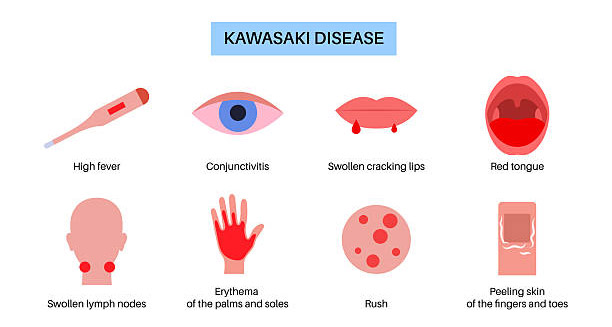
Illustration showing common symptoms of Kawasaki Disease including fever, rash, and red eyes

Symptoms of Kawasaki Disease

Kawasaki disease is characterised by a distinct set of symptoms that typically evolve in three phases: acute, subacute, and convalescent. The most common and first symptom is a high fever that doesn’t go away with regular fever medicine. But this illness also causes many other signs. These signs can affect the skin, eyes, mouth, lymph nodes, and heart. Knowing these signs well helps with early treatment, which lowers the risk of serious heart problems. Children with Kawasaki disease often look very unwell. They may feel tired, upset, and uncomfortable. Since many early signs look like viral infections, the disease can be hard to catch at first. But the mix of specific symptoms helps doctors tell it apart from other illnesses. 1. Persistent High Fever The first and most clear sign is a fever that lasts more than five days. It usually stays above 38.5°C and can even reach 40°C. Unlike other childhood fevers, this one doesn’t respond to paracetamol or ibuprofen. The child may also be very cranky and tired. Sleep problems are common. Many parents bring their child to the doctor because of this long, high fever. 2. Red Eyes (Conjunctival Injection) Another early sign is red eyes without pus. Both eyes look bloodshot, but there’s no sticky discharge like in infections. This often starts a few days after the fever. The eyes don’t usually hurt. The redness may last for several days. Although it fades on its own, it’s an important clue when seen with other signs. 3. Mouth and Tongue Changes The mouth and lips change in clear ways. Common signs include: These changes often make it hard for the child to eat or drink. The strawberry tongue is a strong sign of Kawasaki disease and appears in only a few other illnesses. 4. Rash Many children get a rash within the first few days. It can look different in each case. Some rashes are flat with raised bumps, while others look like scarlet fever or hives. It usually starts on the chest or belly and can spread to the arms, legs, or face. It might itch a little, but it doesn’t usually peel or blister at this stage. The rash helps confirm the diagnosis when seen with other signs. 5. Hand and Foot Changes The hands and feet are often affected too. Look for: These changes may cause pain. Some kids may even stop walking due to discomfort. 6. Swollen Neck Glands Swollen lymph nodes in the neck are another common sign. Usually, just one large node is involved, more than 1.5 cm wide. It’s often on one side and may hurt to touch. Though neck swelling is seen in many infections, it’s more telling when combined with other symptoms of Kawasaki disease. 7. Stomach and Gut Symptoms Some children also have stomach issues early on. These may include: These signs are not specific, which can make early diagnosis harder. Sometimes, the pain may look like appendicitis and lead to surgery by mistake. 8. Heart-Related Symptoms of Kawasaki Disease Even if heart problems don’t show at first, they often appear in the second phase of the illness. These may include: While most kids don’t show early heart symptoms, some may have chest pain, tiredness, or shortness of breath later on. These signs mean the child needs urgent heart tests. 9. Other Possible Signs Kawasaki disease can also cause: Doctors look at the full picture of symptoms. No one sign can confirm it alone. But the group of signs makes the diagnosis much clearer. 10. Phases of Symptom Progression The symptoms of Kawasaki disease appear in three main phases: a) Acute Phase (Days 1–14)High fever, red eyes, rash, mouth changes, and swollen glands are present. b) Subacute Phase (Days 15–30)Fever fades, but peeling skin, crankiness, and heart problems may begin. c) Convalescent Phase (Weeks 4–8)All visible symptoms go away. Blood test results also return to normal. However, heart monitoring is still important. Final Thoughts Spotting the symptoms of Kawasaki disease early is the key to getting treatment in time. Though each sign might look like other childhood illnesses, the full group of signs—especially if they don’t respond to usual medicine—points clearly to Kawasaki disease. The mix of long-lasting fever, red eyes, mouth changes, rash, swollen neck glands, and hand or foot symptoms should lead to urgent testing and care. Doctors, parents, and emergency teams should all stay alert—especially for children under five. [Next: Diagnosis of Kawasaki Disease →]

Symptoms of Kawasaki Disease Read More »

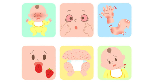
Cartoon showing visual signs used in diagnosing Kawasaki Disease in infants

Diagnosis of Kawasaki Disease

The diagnosis of Kawasaki disease is primarily clinical, based on a defined set of signs and symptoms rather than a single confirmatory test. Doctors must rely on clear signs because there is no single test to confirm the condition. This makes it harder to spot, especially since Kawasaki disease can look like common infections in children. But early diagnosis is vital. If treatment begins within the first 10 days, it can prevent serious heart problems. Doctors must watch closely for key symptoms and follow standard guidelines. These help identify the illness even when it doesn’t appear obvious at first. Even though tests and scans can support the diagnosis, the most important part is careful observation. Looking at how symptoms appear and change over time helps doctors make the right call. 1. Diagnostic Criteria Doctors around the world use a standard set of signs to diagnose Kawasaki disease. These rules come from the American Heart Association and are used in many countries. To make the diagnosis, a child must have: Sometimes, a child with all the symptoms but a shorter fever can still be diagnosed. Likewise, a child with fewer than four signs may still have Kawasaki disease if other evidence supports it. 2. Incomplete or Atypical Kawasaki Disease Not all children show the full list of symptoms. Incomplete Kawasaki disease is more common in babies under six months and older kids. Their signs might be mild or unclear. These cases are more risky because they are often diagnosed late. This can lead to missed treatments and heart problems. Doctors must stay alert. They may use extra tests and scans to confirm the condition and start treatment quickly. 3. Lab Tests to Support Diagnosis of Kawasaki Disease No single blood test can confirm the disease. Still, several tests can support the diagnosis and check how much inflammation is present: These tests are helpful, but only when used with symptoms and other findings. 4. Echocardiography Heart ultrasound (echocardiogram) is one of the most important tools for diagnosing Kawasaki disease. It is safe and painless. It helps doctors look for: Doctors should do this scan as soon as they think the child has Kawasaki disease, even before treatment begins. Follow-up scans are done at two and six weeks to check for changes. If the heart arteries can’t be seen clearly, other scans like CT or MRI may be used. 5. Conditions That Mimic Kawasaki Disease Many other illnesses can look like Kawasaki disease. This makes diagnosis harder. Some of the most common lookalikes include: Doctors take a full history and do extra tests to rule out these other problems. But if they strongly suspect Kawasaki disease, they should not wait too long for lab confirmation. Early treatment is more important, especially if the heart is already affected. 6. Specialist Help in Diagnosis of Kawasaki Disease In unclear cases, it’s smart to call in specialists. These might include: Referral is especially helpful when: In some hospitals, a team of doctors works together to spot and treat Kawasaki disease early. 7. New Tests and Research Scientists are working to find better ways to diagnose Kawasaki disease faster. Some new blood tests may help tell it apart from other illnesses. These include: These tests are still being studied and not used everywhere yet. In the future, they may help doctors find high-risk kids sooner. Researchers are also studying genes. This may lead to tools that predict which kids might get the disease before symptoms even begin. 8. Why Early Diagnosis Matters Time is critical. Giving IVIG within the first 10 days lowers the chance of heart artery problems. The sooner treatment begins, the better the outcome. Late or missed diagnoses—especially in incomplete cases—cause most of the severe heart issues. That’s why doctors should always consider Kawasaki disease if the symptoms fit, and act quickly when needed. Final Thoughts The diagnosis of Kawasaki disease takes close attention to symptoms, blood test results, and heart scans. There is no single test to confirm it. That’s why quick thinking and experience matter so much. Delays in treatment can cause lasting heart damage. But with early care, most kids recover fully. As new tools are developed, diagnosing Kawasaki disease may get easier. Until then, doctors must rely on trusted signs, careful checks, and a clear understanding of how the disease shows up in different children. [Next: Treatment of Kawasaki Disease →]

Diagnosis of Kawasaki Disease Read More »

Syringe with DNA strand and virus icon symbolising Kawasaki Disease treatment

Treatment of Kawasakvi Disease

The treatment of Kawasaki disease is a medical priority, as the condition can lead to serious complications if not addressed swiftly—particularly coronary artery aneurysms. Treating it early—within the first 10 days—lowers the chance of heart problems later on. The main goal is to calm the strong inflammation that affects the blood vessels, especially the ones that supply the heart. Other goals include lowering the fever, easing symptoms, and keeping the heart healthy in the long run. Doctors use a mix of medicines, supportive care, and regular follow-ups with heart experts. These steps have greatly improved how children recover. But early diagnosis still plays the biggest role in helping kids get better. 1. First-Line Treatment: Intravenous Immunoglobulin (IVIG) The most important treatment of Kawasaki disease is intravenous immunoglobulin (IVIG). It’s usually given as one large dose—2 grams per kilogram—over 10 to 12 hours. Doctors try to give IVIG within the first 10 days of illness. Even if given later, it can still help if the child has symptoms or inflammation. IVIG works by calming the immune system and lowering body-wide inflammation. It helps fast—over 85% of children feel better within 36 hours, and their fever goes down. This treatment also cuts the risk of heart artery problems from 25% to under 5%. If the fever doesn’t go away within 36 hours, doctors may give another dose or try different treatments. 2. Aspirin Therapy Aspirin plays a big role in treating Kawasaki disease. Though doctors usually avoid aspirin in children, it’s safe here under medical care. Aspirin helps in two ways: If the child has no signs of artery issues, aspirin may be stopped after 6–8 weeks. But if aneurysms are present, they may need to stay on aspirin longer or even add other blood-thinning medicines. 3. Corticosteroids in Treatment of Kawasaki Disease Doctors now use steroids in some cases to improve recovery. Corticosteroids like prednisolone or methylprednisolone are helpful when: Steroids slow down the immune system’s attack and ease swelling in the vessels. When given early with IVIG, they can lower the chances of heart artery problems. 4. Handling IVIG-Resistant Kawasaki Disease Around 10–15% of children don’t respond to the first IVIG treatment. These children need extra help. Treatment may include: These children need close heart checks, as they have a higher risk of artery problems. 5. Supportive Care Besides the main treatments, supportive care makes a big difference. This includes: Most kids stay in the hospital for 3–7 days, depending on how sick they are and how fast they recover. 6. Heart Monitoring and Follow-Up Because the disease can harm the heart, doctors keep checking the heart for weeks or months afterward. This includes: Kids with big aneurysms may need checkups even as teens or adults. They also need heart-healthy habits and may take medicine long-term to protect their hearts. 7. Exercise and Activity Most children can go back to playing and school once the fever is gone and they stop high-dose aspirin. But those with artery problems must avoid heavy exercise for a while. Doctors will guide the family based on the child’s heart condition. 8. Vaccine Timing After Treatment of Kawasaki Disease Kids who get IVIG need to wait before getting live vaccines like MMR or chickenpox. The delay is about 11 months. This is because IVIG can affect how well vaccines work. Other vaccines can usually stay on schedule. Talk to your child’s doctor to plan the right timing. 9. Helping Parents Understand and Cope Having a child with Kawasaki disease is stressful. Parents need clear support. Doctors should give: Both paediatricians and heart doctors work closely with families throughout recovery. Final Thoughts The treatment of Kawasaki disease has come a long way, and when started early, it works very well. IVIG and aspirin remain the most effective tools. Steroids and newer immune medicines help in harder cases. But recovery isn’t just about medicine. A full care plan—including regular heart checks, family support, and planning for the future—is key to helping children heal. Most children recover well and return to normal life, thanks to early and thorough care. [Next: Complications of Kawasaki Disease →]

Treatment of Kawasakvi Disease Read More »

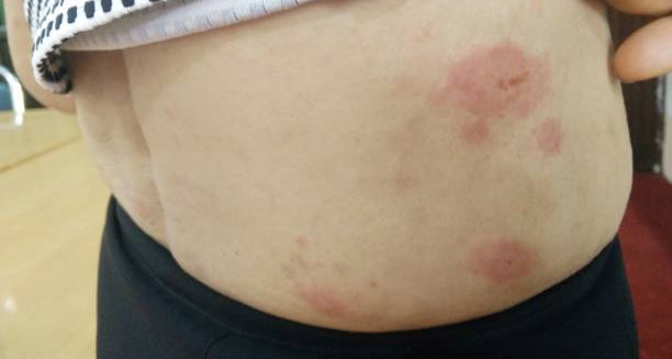
Red circular skin rashes on the abdomen indicating complications of Kawasaki Disease

Complications of Kawasaki Disease

The complications of Kawasaki disease can be significant and, in some cases, life-threatening if the condition is not identified and treated promptly. Most children get better if treated early. Still, Kawasaki disease remains a top cause of heart problems in kids in developed countries. It can harm the coronary arteries—the ones that bring blood to the heart. That’s why regular check-ups are very important for months or even years after diagnosis. Doctors use IVIG and aspirin to reduce risks. These treatments help a lot. But if doctors diagnose the disease late or if the child doesn’t respond well, serious problems can still happen. These issues usually fall into two groups: heart-related or not. 1. Coronary Artery Aneurysms (CAAs) One of the most serious issues is coronary artery aneurysms. These are bulges in the coronary arteries caused by swelling and damage. They often show up 1 to 4 weeks after the disease starts. They’re more common in kids who don’t get treatment quickly or don’t respond to it. Small aneurysms (under 5 mm) may go away on their own.Medium ones (5–8 mm) might also shrink, but doctors need to watch them over time.Large aneurysms (over 8 mm) are more dangerous. They can lead to clots, narrowed arteries, or even breaks in the artery wall. These raise the risk of heart attacks, especially in older kids. Regular heart scans and blood-thinning medicine are often needed. 2. Myocarditis and Pericarditis Some children with Kawasaki disease get myocarditis. This means the heart muscle becomes swollen, which can lower heart function. Signs include fast breathing, tiredness, poor feeding, and low blood pressure. Others may get pericarditis, or swelling around the heart. This can cause chest pain, fussiness, or fluid around the heart. Treatment usually helps these problems go away, but they can get worse without care. 3. Valvular Heart Disease The disease can also affect heart valves. This often causes mitral regurgitation, where one valve leaks a little. It’s usually mild and goes away, but doctors may need to keep an eye on it. In rare cases, surgery is needed. 4. Arrhythmias and Conduction Problems Some children develop abnormal heart rhythms. These can include: These are usually short-term and don’t cause symptoms. Still, doctors may check with ECGs, especially if kids feel dizzy or faint. 5. Thrombosis and Heart Attacks Large aneurysms can lead to blood clots. If a clot blocks an artery, it can cause a heart attack. This is rare in children but very serious. Kids may show signs like sudden tiredness, chest pain, throwing up, or acting restless. Quick treatment with blood thinners or surgery is key in these cases. 6. Peripheral Artery Aneurysms Sometimes, aneurysms show up in other arteries, like in the arms or legs. These are not as dangerous as heart-related ones but still need checking. Doctors often find them using ultrasound. 7. Recurrence of Kawasaki Disease Kawasaki disease can come back, though it’s not common. It happens in about 1–3% of kids. The second time may be harder to notice and could bring more heart risks. Parents should know it can return and seek help fast if symptoms appear again. 8. Hearing Loss Some studies show that a few children may lose hearing after Kawasaki disease. This may happen due to swelling or changes in blood flow to the inner ear. Kids who had a tough case might need a hearing test. 9. Neurocognitive and Emotional Effects Though Kawasaki disease mainly affects the heart, it may also impact a child’s brain and mood. Some kids may: These problems are often short-term. Still, parents and teachers should watch for any changes, especially if the child had a long illness or hospital stay. 10. Complications from Treatment The treatments for Kawasaki disease help a lot but can have side effects. Doctors monitor kids closely to keep them safe while on these medicines. 11. Long-Term Cardiac Follow-Up If a child had heart problems during Kawasaki disease, long-term care is crucial. Follow-up may include: Children with large aneurysms or past heart attacks face higher heart risks later in life. These kids need healthy diets, exercise plans, and sometimes limits on intense activity. Final Thoughts The complications of Kawasaki disease span a wide spectrum, from mild and temporary to severe and life-altering. Modern care helps reduce most serious risks. Still, it’s key to catch the disease early and follow up with care. Knowing what to watch for helps doctors support families and protect children’s hearts. With the right care, most kids fully recover and live healthy lives. [Next: Back to Overview →]

Complications of Kawasaki Disease Read More »

Close-up of hyperextended thumbs showing signs of joint hypermobility syndrome

Overview of Joint Hypermobility Syndrome

Joint hypermobility syndrome is a connective tissue disorder characterised by joints that move beyond the normal range of motion. People with this syndrome can bend their fingers, elbows, knees, or other joints unusually far without pain or damage in early life. However, over time, this extreme flexibility can cause joint pain, instability, fatigue, and many musculoskeletal problems. It differs from generalised joint hypermobility, which happens in healthy individuals without symptoms. Hypermobility itself is common—especially in children, dancers, gymnasts, and those of Asian or African descent. But joint hypermobility syndrome refers to cases where hypermobility causes chronic pain, stiffness, and symptoms that disrupt daily life. It is a heritable connective tissue disorder and may overlap with or resemble conditions like Ehlers-Danlos syndrome (hypermobility type). Collagen, a key protein in ligaments, tendons, and skin, plays a big role in joint hypermobility. In those with the syndrome, collagen is often weaker or more elastic than normal. This causes joints to stretch too far. This elasticity may affect not only joints but also skin, blood vessels, and internal organs. So, the syndrome is a systemic condition, not just a musculoskeletal issue. Epidemiology and Terminology of Joint Hypermobility Syndrome The prevalence of joint hypermobility syndrome varies greatly based on ethnicity, age, sex, and diagnostic methods. It appears more in females than males. Many cases begin in childhood or adolescence, but some are diagnosed only in adulthood when symptoms worsen. Generalised joint hypermobility affects 10–30% of people, but only some develop chronic symptoms of the syndrome. Genetic and cultural factors influence how common it is. For example, children of Indian, Chinese, or African heritage often have more flexible joints, usually without problems. Awareness has increased, so diagnosis has improved. Rheumatologists, physiotherapists, and paediatricians now better recognise unexplained pain, fatigue, or recurring injuries in young patients, which may once have been mistaken for growing pains or psychological issues. The term “Benign Joint Hypermobility Syndrome” (BJHS) was common but is now less used. In 2017, experts introduced “Hypermobility Spectrum Disorders” (HSD) to describe a range of hypermobility-related problems. Hypermobile Ehlers-Danlos Syndrome (hEDS) sits at the severe end of this spectrum. This change shows that hypermobility conditions exist on a continuum. Many people have chronic joint pain, dislocations, or soft tissue injuries with hypermobility but do not meet criteria for hEDS. They usually get diagnosed with joint hypermobility syndrome or HSD. Despite terminology differences, the main idea remains: joint hypermobility with lasting symptoms needs careful diagnosis, treatment, and lifestyle changes. Common Signs and Impact of Joint Hypermobility Syndrome Besides flexible joints, people with joint hypermobility syndrome often have: Children may face trouble with handwriting, coordination, or physical education, which can cause frustration or low self-esteem. Adults may be misdiagnosed with fibromyalgia, chronic fatigue syndrome, or depression because of the chronic pain and emotional effects. The syndrome may also overlap with Postural Orthostatic Tachycardia Syndrome (POTS) and other autonomic problems, creating a complex clinical picture. This overlap calls for thorough, multidisciplinary assessments. Daily life impact ranges widely. Some have mild symptoms and live active lives. Others suffer severe pain, limited function, and frequent injuries that disrupt work, exercise, and routine tasks. Fatigue and poor sleep are common and reduce quality of life. These factors can increase anxiety and depression risk. Children may struggle academically due to exhaustion, pain, or difficulty writing or sitting still. Socially, people with visible hypermobility sometimes face disbelief. Others feel misunderstood when tests appear normal despite real symptoms. Early diagnosis, validation, and proper management can prevent disability and psychological distress for those with joint hypermobility syndrome. Multidisciplinary Care for Joint Hypermobility Syndrome Managing joint hypermobility syndrome often needs several healthcare professionals. Rheumatologists usually handle diagnosis, but physiotherapists, occupational therapists, pain specialists, and gastroenterologists may also be involved. This team approach ensures patients get care that addresses both their physical and emotional needs. Joint hypermobility syndrome is a complex and often misunderstood condition that goes beyond flexible joints. Many live a lifelong challenge managing pain, fatigue, and instability. Once thought to be “benign,” it now gains recognition as a real medical condition needing careful care. With growing awareness, more people receive early diagnosis, timely support, and tailored therapies. These steps help improve symptoms and quality of life for those affected by joint hypermobility syndrome. [Next: Causes of Joint Hypermobility Syndrome →]

Overview of Joint Hypermobility Syndrome Read More »

Illustration comparing a hypermobile foot and a normal foot in Joint Hypermobility Syndrome

Causes of Joint Hypermobility Syndrome

Joint hypermobility syndrome is primarily caused by inherited variations in connective tissue structure and function, most notably involving collagen—the primary protein responsible for the strength and elasticity of ligaments and tendons. Individuals with joint hypermobility syndrome tend to have looser, more elastic connective tissue, which permits excessive joint movement but offers less stability. While hypermobility itself may be harmless or even advantageous in specific contexts, such as gymnastics or ballet, the syndrome refers to cases where hypermobility leads to chronic symptoms like pain, fatigue, and functional impairment. The causes of joint hypermobility syndrome are complex and often linked to genetics, but other factors such as age, gender, ethnicity, and individual body makeup also influence it. Some people inherit joint laxity without symptoms. Others suffer disabling problems even with only mild flexibility. Knowing these causes helps doctors and patients detect the syndrome early and manage it well. Genetic and Collagen Factors in Joint Hypermobility Syndrome The most established cause of joint hypermobility syndrome is genetic. Many cases run in families, showing a clear genetic link. Several genes likely affect how collagen and elastin fibers form and organize in connective tissues. When these proteins are more elastic or less strong than normal, ligaments and tendons loosen. Joint hypermobility syndrome is different from genetic disorders like Ehlers-Danlos syndrome (hypermobility type), but their symptoms can overlap. Ehlers-Danlos syndromes affect collagen production. Joint hypermobility syndrome may involve milder or different mutations that cause widespread symptoms without a full disorder diagnosis. Multiple family members, especially close relatives, may show similar flexibility or joint problems. This suggests the syndrome follows an inherited pattern, possibly autosomal dominant or polygenic. However, no single gene has been pinpointed for joint hypermobility syndrome. At the molecular level, abnormalities in type I and type III collagen play a major role. These collagens provide strength to ligaments, skin, and organs. When collagen is less dense or disorganized, tissues stretch more than usual. This causes: Over time, overextension leads to joint wear, pain, and soft tissue damage. It can also cause proprioceptive problems, where the brain struggles to sense joint position, increasing injury risk. In some people, other connective tissues like skin, blood vessels, and the gut lining show subtle changes. This explains symptoms like easy bruising, digestive problems, and nervous system issues seen in joint hypermobility syndrome. How Age and Hormones Affect Causes of Joint Hypermobility Syndrome Age is another important factor. Children and teens are naturally more flexible because their bodies are still growing. Usually, flexibility decreases with age as collagen stiffens and muscles strengthen. But in joint hypermobility syndrome, excess movement may persist or worsen. Children with the syndrome often seem very “bendy.” Parents may notice tricks like bending thumbs back or unusual sitting positions. While these may seem harmless, many develop pain, fatigue, or coordination problems as they grow, especially during growth spurts. Adults may find symptoms stay the same or get worse. Aging stiffens ligaments, which can ease symptoms for some. For others, years of joint strain cause pain and instability. Gender also plays a role. Women are much more likely to have joint hypermobility syndrome than men, possibly three times more. This difference relates partly to collagen differences but also to hormones, especially estrogen. Estrogen affects tissue elasticity. Its changes during menstrual cycles, pregnancy, or menopause can worsen joint instability. Pregnancy raises relaxin, a hormone that loosens ligaments for childbirth. This can increase back pain, pelvic pain, and even risks like early labor in women with the syndrome. Understanding these hormonal effects helps doctors plan treatments with physiotherapy, hormone management, and support during key life stages. [Next: Symptoms of Joint Hypermobility Syndrome →]

Causes of Joint Hypermobility Syndrome Read More »

Scroll to Top