Mental Matters

Health Conditions

Find information on various health conditions affecting people of all ages. This category covers causes, symptoms, diagnosis, and management options for physical illnesses and chronic diseases. Get insights to help you understand different health challenges and make informed decisions about your wellbeing.

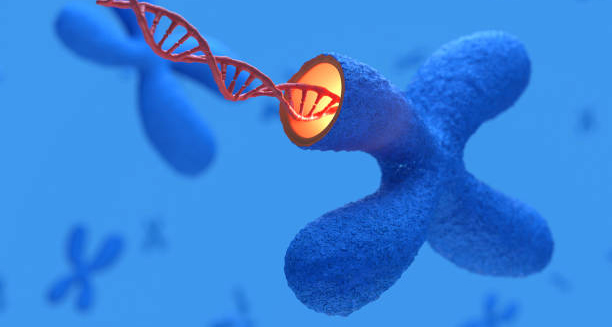
DNA strand emerging from chromosome – genetic cause of Gilbert’s Syndrome

Causes of Gilbert’s Syndrome

Causes of Gilbert’s Syndrome The causes of Gilbert’s syndrome are rooted in a genetic mutation. Furthermore, that affects how the body processes bilirubin. Gilbert’s syndrome is not caused by external factors like diet, infection, or alcohol use—rather. It is inherited and present from birth, although symptoms may not appear until adolescence or later. This inherited condition disrupts the function of an enzyme responsible for converting bilirubin into a form that the body can excrete. While it results in higher levels of unconjugated bilirubin, it does not damage the liver or lead to serious illness. The Role of Bilirubin To understand the cause of Gilbert’s syndrome, it helps to first understand bilirubin metabolism. Bilirubin is formed when: Red blood cells reach the end of their lifespan (around 120 days) Haemoglobin breaks down into heme and globin The body converts heme into unconjugated (indirect) bilirubin, which does not dissolve in water This unconjugated bilirubin travels through the bloodstream to the liver, where an enzyme called UGT1A1 converts it into a conjugated (direct), water-soluble form. The liver then excretes this bilirubin into bile, which passes it into the intestines for elimination. In people with Gilbert’s syndrome: The UGT1A1 enzyme activity is reduced by about 30–70% Less bilirubin converts into its soluble form Unconjugated bilirubin accumulates in the bloodstream, especially during periods of stress or metabolic demand Genetic Mutation in UGT1A1 A mutation in the UGT1A1 gene causes the condition by affecting production of the uridine diphosphate-glucuronosyltransferase enzyme. The most common mutation involves: The insertion of extra TA repeats in the promoter region of the gene This change reduces transcription and, therefore, enzyme levels The inheritance pattern is autosomal recessive, meaning: A person must inherit two copies of the mutated gene, one from each parent, to develop the condition Carriers with only one copy typically have normal bilirubin metabolism and no symptoms Although Gilbert’s syndrome is genetic, it does not always run clearly in families, especially if symptoms are mild and undiagnosed. Prevalence and Population Variability Gilbert’s syndrome affects 5–10% of the global population, but prevalence varies by region and ethnicity: More common in European and Asian populations Slightly less prevalent in some African and Middle Eastern groups It affects both men and women, although men are more likely to show visible jaundice, possibly due to higher bilirubin production from red cell breakdown. Triggers That Reveal the Condition The genetic mutation is present from birth, but symptoms often appear later in life due to: Puberty, which increases haemoglobin turnover Illness, such as infections that cause liver stress Fasting, which reduces bilirubin clearance Exertion or surgery, which elevate metabolic demands These events can unmask the condition in individuals who had previously never noticed symptoms. Not Caused by Liver Damage Unlike hepatitis, cirrhosis, or alcoholic liver disease, Gilbert’s syndrome: Does not cause inflammation or scarring of the liver Does not impair liver enzymes (ALT, AST) Is not related to alcohol consumption, fatty liver, or obesity This distinction is crucial to avoid misdiagnosis or unnecessary liver biopsies. Impact on Drug Metabolism Because the UGT1A1 enzyme also helps detoxify certain medications, reduced enzyme activity can affect how the body processes some drugs. Medications affected include: Irinotecan – an anti-cancer drug that requires careful dosing Atazanavir – an antiviral used in HIV treatment Paracetamol – theoretically affected but usually safe in standard doses Despite these interactions, most people with Gilbert’s syndrome can take common medications safely, especially under medical supervision. Conclusion | Causes of Gilbert’s Syndrome The causes of Gilbert’s syndrome lie entirely in a genetic mutation that reduces the body’s ability to process bilirubin. Lifestyle factors, poor diet, alcohol, and infection do not cause it, and it does not lead to liver disease. Understanding the underlying genetic mechanism helps people with the condition avoid unnecessary concern and manage their health confidently. [Next: Symptoms of Gilbert’s Syndrome→]
Causes of Gilbert’s Syndrome Read More »

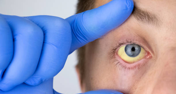
Yellowing of the eye in person with Gilbert’s Syndrome

Symptoms of Gilbert’s Syndrome

Symptoms of Gilbert’s Syndrome The symptoms of Gilbert’s syndrome are typically mild, intermittent, and often go unnoticed. In fact, many people with Gilbert’s syndrome are unaware they have the condition until it is discovered incidentally during blood tests for unrelated issues. Despite its effect on bilirubin processing, the condition does not damage the liver or other organs, and symptoms—when they do occur—are usually harmless. The hallmark of Gilbert’s syndrome is elevated levels of unconjugated bilirubin in the blood, which can lead to occasional jaundice. However, the condition may also be associated with other non-specific symptoms that can vary in severity and frequency between individuals. Jaundice: The Primary Symptom The most recognised and visible sign of Gilbert’s syndrome is jaundice—a yellowing of the skin and whites of the eyes (sclera). In most cases: The jaundice is mild and transient It occurs when bilirubin levels temporarily rise It may resolve spontaneously without any intervention The yellowing is usually most noticeable in the eyes and may not affect the skin at all in lighter-skinned individuals. In darker skin tones, jaundice may be more difficult to detect. Jaundice episodes are often triggered by: Fasting or skipping meals Dehydration Stress, both emotional and physical Viral illnesses, such as colds or flu Menstruation in some women Overexertion or intense physical activity These episodes may last for a few hours to several days, depending on the trigger and the individual’s metabolic state. Fatigue and Low Energy Many individuals with Gilbert’s syndrome report episodes of fatigue or low energy, especially during or following an episode of jaundice. While studies have not definitively linked fatigue to elevated bilirubin levels, it remains one of the most frequently mentioned symptoms among those diagnosed with the condition. Fatigue in Gilbert’s syndrome is often described as: A generalised sense of tiredness without clear cause Difficulty concentrating or mental sluggishness Feeling drained after normal daily activities It is important to note that fatigue may also be related to poor sleep, stress, or coexisting conditions, so a thorough assessment is often required. Gastrointestinal Discomfort Although Gilbert’s syndrome does not directly affect the gastrointestinal tract, some people report: Mild nausea Abdominal bloating or discomfort Loss of appetite Occasional indigestion These symptoms are generally not severe and do not require medical treatment. However, they may coincide with periods of fasting, illness, or bilirubin spikes. There is no evidence to suggest that Gilbert’s syndrome causes chronic digestive diseases, but heightened sensitivity to changes in metabolism might make the digestive tract more reactive. Dizziness and Brain Fog Some individuals with Gilbert’s syndrome mention experiencing: Light-headedness or feeling faint Brain fog, described as unclear thinking or mental sluggishness Difficulty with mental focus, especially during stressful situations or illness While these symptoms are not universally experienced, they may result from the body’s temporary adaptation to increased bilirubin or stress-related metabolic shifts. Again, these symptoms are not dangerous and often resolve quickly. Sensitivity to Medications People with Gilbert’s syndrome may be more sensitive to certain medications, especially those processed by the UGT1A1 enzyme, which is impaired in this condition. While this is not a symptom in the traditional sense, it can lead to: Nausea or adverse reactions after normal drug dosages Delayed clearance of certain medications from the body Affected drugs may include: Irinotecan (a chemotherapy drug) Atazanavir and indinavir (used for HIV) Certain anaesthetics or sedatives Though rare, patients should discuss heightened drug sensitivity with healthcare providers to avoid unexpected side effects. Anxiety or Emotional Effects The appearance of jaundice or the diagnosis itself can sometimes lead to: Health anxiety can increase when people mistake jaundice for liver disease Emotional stress during public events or work situations where visible symptoms are present Feelings of embarrassment or withdrawal, particularly among young adults Clear communication about the benign nature of Gilbert’s syndrome can alleviate these concerns and help patients feel more in control. Symptom Summary Symptom Frequency / Severity Mild jaundice Common, especially under stress Fatigue or low energy Reported by many, varies in intensity Digestive discomfort Occasional, mild and non-specific Brain fog / poor focus Sometimes reported Sensitivity to medications Rare but clinically relevant Emotional effects Common if undiagnosed or misunderstood Do Symptoms Change Over Time? For most individuals, symptoms of Gilbert’s syndrome are intermittent and manageable. The frequency and intensity may vary throughout life, often influenced by lifestyle factors such as diet, stress, and sleep patterns. In many cases, symptoms diminish with age, and people learn to recognise their own triggers and adjust their routines accordingly. When to Seek Medical Attention Gilbert’s syndrome itself does not require regular medical follow-up. However, a medical review is advised if: Jaundice becomes persistent or more severe New or unexplained symptoms develop Fatigue or digestive issues interfere with daily life Doctors prescribe a new medication, especially if it is processed by the liver Doctors do this to ensure they do not overlook any unrelated liver conditions. Symptoms of Gilbert’s Syndrome Understanding the symptoms of Gilbert’s syndrome helps individuals manage their health confidently, avoid unnecessary worry, and prevent misdiagnosis of more serious conditions. [Next: Diagnosis of Gilbert’s Syndrome →]

Symptoms of Gilbert’s Syndrome Read More »

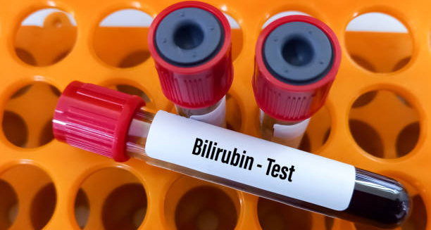
Blood sample for bilirubin test used in diagnosing Gilbert’s Syndrome

Diagnosis of Gilbert’s Syndrome

Diagnosis of Gilbert’s Syndrome The diagnosis of Gilbert’s syndrome is typically straightforward and non-invasive. Because Gilbert’s syndrome causes a harmless but noticeable increase in unconjugated bilirubin levels. Therefore, doctors often discover it by chance. Furthermore, doctors usually finds it during routine blood tests. Once doctors exclude other causes of jaundice, they can focus on diagnosing the underlying liver or biliary condition. Doctors can usually confirm the diagnosis within a few targeted assessments. Doctors diagnose most people in their teens or early adulthood. Furthermore, often during medical exams for employment, sports, or unrelated symptoms. Initial Suspicion Doctors may suspect Gilbert’s syndrome in patients who: Are otherwise healthy but show elevated bilirubin levels Experience intermittent jaundice triggered by fasting, stress, or illness Have normal liver function test results (aside from bilirubin) Show no signs of liver inflammation or damage In these cases, doctors perform further testing to confirm that the elevated bilirubin is unconjugated and not due to liver or bile duct disease. Blood Tests Blood tests are the cornerstone of diagnosis. Key indicators include: 1. Serum Bilirubin Levels Total bilirubin is elevated (usually 17–100 µmol/L) Predominantly unconjugated (indirect) bilirubin is raised Levels may fluctuate with fasting, stress, or infection 2. Liver Function Tests (LFTs) ALT, AST, ALP, and GGT are all normal, ruling out hepatitis or liver damage Albumin and prothrombin time are also within normal limits A normal liver profile with isolated unconjugated hyperbilirubinaemia is highly suggestive of Gilbert’s syndrome. Fasting or Provocation Tests In some cases, doctors may use a fasting test or a nicotinic acid challenge to confirm the diagnosis: Fasting for 24–48 hours may cause bilirubin levels to rise Doctors rarely perform these tests now because they are uncomfortable and unnecessary in most cases Modern diagnosis relies more on pattern recognition than provocative testing. Genetic Testing Genetic testing for the UGT1A1 mutation can confirm the diagnosis definitively. However: It is not routinely required unless diagnosis is uncertain Doctors may use this test for patients undergoing chemotherapy or antiviral treatment, where monitoring bilirubin metabolism is critical The most common mutation involves the UGT1A1*28 allele, which results in reduced transcription of the enzyme responsible for bilirubin conjugation. Differential Diagnosis Before confirming Gilbert’s syndrome, doctors must exclude other conditions that cause elevated bilirubin, such as: Haemolytic anaemia (excessive red blood cell breakdown) Hepatitis or liver disease Biliary obstruction Crigler-Najjar syndrome (a rare, more severe inherited condition) These conditions are usually accompanied by abnormal liver function tests or additional symptoms such as fatigue, dark urine, or pale stools. Imaging and Further Tests Doctors typically do not order imaging tests like ultrasound or CT scans unless they suspect other liver abnormalities. Gilbert’s syndrome does not cause structural changes in the liver or bile ducts. Further testing may include: Full blood count (to rule out anaemia) Reticulocyte count (to assess red blood cell turnover) Lactate dehydrogenase (LDH) and haptoglobin (to exclude haemolysis) These tests help confirm that the liver is functioning normally and that the elevated bilirubin is isolated. Communicating the Diagnosis One of the most important aspects of diagnosis is patient education. Individuals should be reassured that: The condition is completely benign There is no risk of progression to liver disease No ongoing treatment or restriction is required Recognising triggers can help manage visible symptoms Providing a written explanation or including the diagnosis in medical records can help avoid unnecessary investigations in the future. When to Refer to a Specialist Referral to a hepatologist or gastroenterologist is rarely necessary unless: Liver enzymes are abnormal Jaundice is persistent or unexplained Additional symptoms suggest another underlying condition In most cases, the diagnosis is managed entirely in primary care with simple testing and reassurance. Conclusion | Diagnosis of Gilbert’s Syndrome The diagnosis of Gilbert’s syndrome is based on a clear clinical pattern supported by blood tests. Once doctors rule out other causes of jaundice, they usually do not need to take further action. Timely diagnosis avoids unnecessary worry, reduces unnecessary investigations, and empowers individuals to manage the condition with confidence. [Next: Treatment of Gilbert’s Syndrome→]

Diagnosis of Gilbert’s Syndrome Read More »

Woman sleeping peacefully in bed as part of Gilbert’s Syndrome management

Treatment of Gilbert’s Syndrome

Treatment of Gilbert’s Syndrome The treatment of Gilbert’s syndrome is typically unnecessary because the condition is harmless and causes no long-term damage. Gilbert’s syndrome is a mild, inherited disorder of bilirubin metabolism that results in intermittent, non-dangerous episodes of jaundice. Most people with the condition require no medication, no dietary restrictions, and no lifestyle limitations. Instead, education and reassurance form the foundation of effective management. However, treatment in the broader sense includes identifying triggers, avoiding unnecessary medical interventions, and managing symptoms when they occur. In certain scenarios—such as when taking specific medications or experiencing severe fatigue—medical support may be helpful, although active treatment is rarely needed. No Cure Required Gilbert’s syndrome is not considered a disease in the traditional sense. It: In the vast majority of cases, once the diagnosis is confirmed, no active treatment is recommended or necessary. Most individuals continue with their lives without any negative health impact. Managing Triggers Although the condition is benign, people with Gilbert’s syndrome can reduce episodes of jaundice and related symptoms by managing known triggers. These include: 1. Avoid Fasting or Skipping Meals Prolonged fasting increases bilirubin levels Eating small, regular meals helps maintain stable liver metabolism 2. Stay Hydrated Dehydration can raise bilirubin levels and trigger fatigue Regular fluid intake supports optimal liver function 3. Manage Physical Stress Overexertion, particularly without rest, may bring on symptoms Balance intense activity with adequate recovery 4. Minimise Emotional Stress Stress has a direct impact on metabolism and bilirubin levels Techniques like mindfulness, exercise, and healthy sleep patterns are beneficial 5. Avoid Alcohol Excess While moderate alcohol is usually well tolerated, binge drinking may overload the liver Those with sensitive systems may benefit from limiting alcohol intake These simple lifestyle measures often reduce symptom frequency and duration, helping individuals feel more in control of their condition. Fatigue Management Some people with Gilbert’s syndrome report ongoing fatigue, though the link between bilirubin and tiredness remains debated. In such cases: Sleep hygiene and routine exercise can improve energy levels Avoiding overwork and late nights may reduce symptom flare-ups Cognitive behavioural therapy (CBT) or support groups may help if fatigue impacts quality of life If fatigue is severe or persistent, other causes such as anaemia, thyroid disease, or sleep disorders should be excluded before attributing it entirely to Gilbert’s syndrome. Nutritional Considerations There is no special diet required for those with the condition. However, general healthy eating habits support optimal liver function and overall wellbeing: Balanced meals with lean proteins, whole grains, fruits, and vegetables Avoid excessive refined sugar, salt, and saturated fats Limit caffeine and alcohol in periods of stress or illness There is no evidence that supplements, herbs, or detox diets improve bilirubin clearance in people with Gilbert’s syndrome. Medications to Use With Caution Although most medications are safe for individuals with Gilbert’s syndrome, caution is required with certain drugs that rely on the UGT1A1 enzyme for metabolism. These include: 1. Irinotecan (a chemotherapy agent) Can lead to toxic side effects in people with reduced UGT1A1 activity Dosing may require adjustment or alternative therapies 2. Atazanavir and Indinavir (used in HIV treatment) Can further elevate bilirubin and may cause visible jaundice This is cosmetic rather than harmful but should be monitored 3. Paracetamol (acetaminophen) Generally safe at recommended doses Excessive or prolonged use should be avoided, particularly during fasting Doctors should be informed of the diagnosis, especially before prescribing medications metabolised by the liver. Pharmacogenetic testing may be advised in high-risk patients. Psychological and Emotional Support Though Gilbert’s syndrome is harmless, its appearance—especially jaundice—can lead to anxiety, particularly in younger individuals or those unaware of their diagnosis. Support includes: Clear explanation of the condition’s benign nature Written documentation to prevent repeated investigations Counselling or support groups for reassurance if symptoms cause emotional distress Some people benefit from speaking to others with the condition, especially during diagnosis and adjustment. Rare Situations for Medical Intervention Treatment beyond lifestyle adjustments may be considered in rare situations, such as: Frequent, troubling jaundice interfering with daily life Reactions to prescribed medications Coexisting health conditions complicating bilirubin metabolism In such cases, specialist referral to a hepatologist or geneticist may be appropriate. However, active treatment beyond education and symptom management is extremely rare. Summary of Non-Pharmacological Strategies Strategy Purpose Regular meals Prevent bilirubin spikes Adequate hydration Support liver detoxification Balanced exercise Promote overall metabolic health Sleep and stress management Reduce symptom episodes Informing healthcare providers Avoid drug-related complications Conclusion | Treatment of Gilbert’s Syndrome The treatment of Gilbert’s syndrome involves education, reassurance, and sensible lifestyle measures. Medication and intervention are almost never required. With an understanding of triggers and the reassurance that the condition is benign, individuals with Gilbert’s syndrome can lead healthy, unrestricted lives. [Next: Complications of Gilbert’s Syndrome →]

Treatment of Gilbert’s Syndrome Read More »

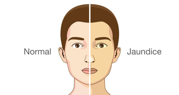
Comparison illustration of normal vs jaundiced skin tone in Gilbert’s Syndrome

Complications of Gilbert’s Syndrome

Complications of Gilbert’s Syndrome The complications of Gilbert’s syndrome are virtually non-existent in most individuals. Gilbert’s syndrome is a benign condition, and despite its impact on bilirubin metabolism, it does not lead to liver damage, chronic disease, or a shortened lifespan. However, recognising certain implications—particularly regarding drug metabolism and quality of life—can help individuals avoid unnecessary stress or misunderstanding. Most people diagnosed with Gilbert’s syndrome live full, healthy lives. That said, there are a few rare or indirect complications worth being aware of, especially in contexts like medication use or coexisting conditions. 1. Medication-Related Complications The most clinically relevant complication arises when people with Gilbert’s syndrome are prescribed drugs that depend on the UGT1A1 enzyme for detoxification. Because enzyme activity is reduced, these medications may accumulate in the body, potentially leading to: Increased side effects Delayed drug clearance Elevated bilirubin levels, causing cosmetic jaundice Medications of concern include: Irinotecan – a chemotherapy drug, known to cause diarrhoea, neutropenia, or even severe toxicity in susceptible individuals Atazanavir – used in HIV treatment, may worsen jaundice but not liver damage Certain anaesthetics and statins – usually safe but worth monitoring if prolonged use is needed Preventive strategies: Inform all healthcare providers of the diagnosis Conduct liver function monitoring during long-term treatment Consider alternative medications if side effects are significant 2. Emotional and Social Impact For many people, the most tangible complication is psychological rather than physical. These include: Worry about having liver disease Fear of long-term complications due to miscommunication or internet misinformation Embarrassment about visible jaundice, especially in public or professional settings These concerns are valid but manageable with education. Clear, consistent information from medical professionals can prevent unnecessary anxiety. 3. Misdiagnosis and Over-Investigation A frequent complication in healthcare systems is: Repeated or unnecessary testing for liver disease Referrals to specialists when not required Cost and inconvenience for patients and providers When Gilbert’s syndrome is not recognised, it can lead to misdiagnosis, unnecessary liver biopsies, or even restrictive lifestyle recommendations. This is particularly important in contexts like: Insurance or employment medicals Military or aviation screening Blood donation assessments Documenting the diagnosis helps reduce unnecessary concern and inappropriate testing. 4. Quality of Life Concerns Though the condition itself is benign, the perception of illness can affect daily life. Individuals may experience: Changes in self-image due to visible jaundice Over-cautious behaviour, avoiding activities or foods without need Reluctance to exercise or travel, fearing symptom flare-ups These lifestyle limitations are usually self-imposed and based on misunderstanding. In reality, people with Gilbert’s syndrome can: Travel freely Exercise at any intensity Eat a varied, unrestricted diet Take most medications under medical guidance Education is the most effective remedy for these lifestyle complications. 5. Coexisting Conditions In rare cases, Gilbert’s syndrome may coexist with other liver or metabolic disorders, such as: Haemolytic anaemia Hepatitis Crigler-Najjar syndrome (type II) – a more severe form of bilirubin-processing disorder While Gilbert’s syndrome does not cause these conditions, its presence may confuse the clinical picture. That’s why new or worsening symptoms always warrant a full evaluation. 6. Persistent Fatigue Though evidence is mixed, some individuals with Gilbert’s syndrome report persistent fatigue, even between jaundice episodes. This fatigue may: Reduce motivation or productivity Interfere with daily routines Trigger concern about more serious illness Fatigue can often be managed through: Structured routines Improved sleep hygiene Stress reduction Light-to-moderate exercise Where persistent, medical evaluation is advised to exclude unrelated causes. Summary of Potential Complications Complication Impact Medication sensitivity Increased side effects Emotional stress / anxiety Fear of illness or social concern Misdiagnosis or over-testing Unnecessary procedures or costs Fatigue Lifestyle impact, requires evaluation Coexisting conditions Diagnostic confusion if present Conclusion | Complications of Gilbert’s Syndrome The complications of Gilbert’s syndrome are rare and generally non-serious. Most people experience minimal interference with daily life and can safely manage any symptoms with education and awareness. The key to avoiding complications lies in early diagnosis, informed medical care, and understanding that Gilbert’s syndrome is a benign and manageable condition. [Next: Back to Overview →]

Complications of Gilbert’s Syndrome Read More »

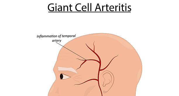
Medical illustration showing inflammation of the temporal artery in Giant Cell Arteritis

Giant Cell Arteritis

Giant Cell Arteritis Giant cell arteritis is a serious inflammatory condition that affects the blood vessels, particularly the large and medium-sized arteries of the head and neck. Also known as temporal arteritis, giant cell arteritis occurs when the immune system mistakenly attacks the artery walls, leading to inflammation, swelling, and damage to the vessel lining. This condition most commonly involves the temporal arteries, which run along the sides of the head, but it can affect other arteries as well, including the aorta. The most significant danger of untreated giant cell arteritis is sudden vision loss, which can occur if the arteries supplying the eyes become inflamed or blocked. Prompt diagnosis and treatment are essential to prevent irreversible complications. The condition predominantly affects individuals over the age of 50 and is more common in women than in men. Despite its name, giant cell arteritis does not refer to a physically enlarged artery but rather to the giant cells—a type of immune cell—seen under the microscope in biopsied tissue. These cells contribute to the aggressive inflammation that characterises the disease. Who Is Affected? Giant cell arteritis primarily affects: People over the age of 50, with incidence increasing with age Women, who are two to three times more likely than men to develop it Individuals of Northern European descent, though it can occur in any ethnic group People with a history of polymyalgia rheumatica (PMR)—an inflammatory disorder that frequently coexists with giant cell arteritis PMR and GCA are thought to be related inflammatory diseases, with up to 50% of patients with one developing symptoms of the other. Key Features and Impact Giant cell arteritis can present with a range of symptoms that may develop suddenly or gradually. The inflammation causes narrowing or blockage of affected arteries, which can severely reduce blood flow and oxygen supply to key organs and tissues. In the short term, the condition can cause: Severe headaches Jaw pain when chewing Scalp tenderness Double vision or sudden blindness In the long term, if untreated, it may lead to: Permanent vision loss Aneurysm formation, particularly in the aorta Stroke, if arteries supplying the brain are affected Because of the potentially devastating consequences, early identification and treatment are vital. How It Develops The exact cause of giant cell arteritis is not fully understood, but it is believed to be an autoimmune reaction—a condition in which the body’s immune system attacks its own tissues. In GCA, immune cells mistakenly target the lining of arteries, particularly those with elastic components like the temporal artery. This immune attack causes inflammation and the formation of granulomas, which often contain multinucleated giant cells—hence the name. These granulomas thicken the arterial walls, narrow the lumen (internal space), and reduce or block blood flow. Is It Common? Giant cell arteritis is the most common form of systemic vasculitis in adults over 50. Its prevalence varies by region and ethnicity, with higher rates reported in Scandinavian and North American populations. In the UK and many parts of Europe, the estimated annual incidence is around 15 to 30 cases per 100,000 people over age 50. Early Symptoms to Watch For Symptoms of GCA can overlap with many other conditions, making diagnosis a challenge. The most common early signs include: New-onset headaches, especially around the temples Tender scalp, making it painful to brush hair or wear a hat Jaw claudication—pain or fatigue in the jaw muscles while chewing Vision changes, such as blurred or double vision General fatigue, low-grade fever, and unintended weight loss Pain and stiffness in the shoulders or hips, suggesting coexisting PMR Some individuals may have subtle or vague symptoms, such as general malaise, which delays diagnosis. Why Vision Loss Happens One of the most feared complications of giant cell arteritis is permanent vision loss, which occurs in up to 20% of untreated cases. This usually happens when inflammation affects the posterior ciliary arteries that supply the optic nerve. Reduced blood flow leads to ischaemia (oxygen deprivation) and irreversible optic nerve damage. Visual symptoms may include: Sudden, painless loss of vision in one eye Transient blurred vision or episodes of visual blackouts (amaurosis fugax) Double vision due to muscle involvement Immediate medical intervention can sometimes save vision in the other eye, but damage to the first is usually irreversible if treatment is delayed. Diagnosis and Outlook Early diagnosis and treatment with high-dose corticosteroids can dramatically reduce the risk of complications. Once inflammation is controlled, medication is tapered over time, but careful monitoring is required to prevent relapse. Modern treatment approaches also include the use of steroid-sparing agents, such as tocilizumab, which targets specific inflammatory pathways and reduces long-term dependence on steroids. Giant Cell Arteritis With appropriate treatment, most people experience symptom relief and stabilisation. However, some patients may have relapses, requiring long-term management. Giant cell arteritis is a medical emergency when vision is threatened, and anyone over 50 experiencing new headaches, jaw pain, or visual symptoms should seek immediate evaluation. [Next: Causes of Giant Cell Arteritis →]

Giant Cell Arteritis Read More »

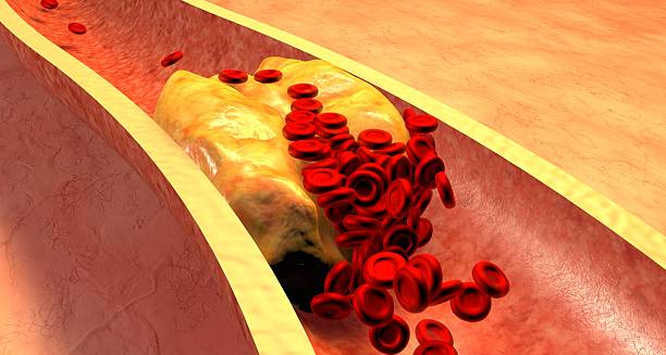
Illustration of blocked artery showing blood cells and plaque buildup, symbolising causes of Giant Cell Arteritis

Causes of Giant Cell Arteritis

Causes of Giant Cell Arteritis The causes of giant cell arteritis are still being researched. However, current evidence suggests that a combination of genetic, environmental, and immunological factors is involved. Doctors consider giant cell arteritis an autoimmune condition in which the immune system mistakenly attacks artery walls, causing inflammation and narrowing of the vessels. Understanding the underlying mechanisms helps in identifying at-risk individuals and developing better treatments. The condition is most common in older adults, especially women over 50, but doctors still do not fully understand what triggers the disease. Immune System Dysfunction The leading theory is that giant cell arteritis arises from immune dysregulation, where immune cells misidentify healthy tissue—in this case, artery walls—as harmful. This sets off a chronic inflammatory response. Key immune cells involved include: CD4+ T lymphocytes Macrophages Giant cells are large immune cells formed when multiple macrophages fuse together These cells infiltrate the tunica media and adventitia (layers of the arterial wall), forming granulomas and releasing inflammatory cytokines. The inflammation leads to: Thickening of the artery wall Narrowing or obstruction of the blood vessel Potential tissue damage due to reduced blood flow This explains the classic symptoms of ischaemia, such as headache, jaw pain, and vision disturbances. Genetic Predisposition There is a strong genetic component to giant cell arteritis. Studies have found that people with certain variations in the HLA (human leukocyte antigen) system, particularly HLA-DRB1*04, are more likely to develop the condition. While having these genes does not guarantee disease, it increases susceptibility, particularly when combined with environmental or age-related factors. Family history may also influence the risk, although doctors do not consider GCA a hereditary disease in the traditional sense. Ageing and Vascular Changes Ageing is the strongest risk factor for developing giant cell arteritis. The vast majority of cases occur in people over 50, and risk increases steadily with age. Several theories explain this age-related risk: Ageing causes degeneration of vascular structures, making them more vulnerable to inflammation The immune system becomes more prone to autoimmune activation Arterial elasticity declines, contributing to vascular stress and damage The intersection of these factors creates a favourable environment for GCA to develop. Environmental Triggers Doctors suspect that certain environmental factors may trigger the initial immune response in individuals who are predisposed. These may include: 1. Infections Viral or bacterial infections have long been hypothesised as potential triggers. Though no single pathogen has been definitively identified, several candidates include: Parvovirus B19 Varicella zoster virus (VZV) Chlamydia pneumoniae These pathogens may provoke an immune response that cross-reacts with arterial tissues, leading to autoimmunity. Some studies have found fragments of viral DNA in the arteries of patients with GCA, although evidence remains inconclusive. 2. Seasonal Variations Epidemiological data suggest a seasonal pattern to disease onset, with more diagnoses occurring during colder months. This may point to a link with seasonal infections or reduced sunlight exposure, although the exact relationship is unclear. 3. Geographic Trends Giant cell arteritis is more common in certain regions, particularly: Northern Europe and Scandinavia North America Parts of Australia and New Zealand This geographic distribution supports both genetic and environmental influences, including climate, infection exposure, and possibly vitamin D levels. Coexisting Conditions Giant cell arteritis often occurs in conjunction with polymyalgia rheumatica (PMR), an inflammatory condition that causes stiffness and pain in the shoulders and hips. Up to 50% of patients with GCA also have symptoms of PMR. This association suggests shared inflammatory mechanisms, possibly involving overlapping genetic susceptibility and immune pathways. Gender Differences Giant cell arteritis affects women more often than men, with a female-to-male ratio of approximately 2:1 to 3:1. The reasons for this are not fully understood but may involve: Hormonal influences on immune regulation Differences in vascular structure or immune gene expression Postmenopausal women are particularly at risk, further implicating hormonal changes as a contributing factor. Conclusion While the exact causes of giant cell arteritis remain under investigation, current research points to a multifactorial process involving: Immune system malfunction Genetic predisposition Age-related vascular changes Possible environmental triggers Causes of Giant Cell Arteritis Identifying and understanding these factors is crucial for early detection and better management of this potentially sight-threatening disease. [Next: Symptoms of Giant Cell Arteritis→]

Causes of Giant Cell Arteritis Read More »

Woman with grey hair touching her temples in pain, showing symptoms of Giant Cell Arteritis

Symptoms of Giant Cell Arteritis

Symptoms of Giant Cell Arteritis The symptoms of giant cell arteritis can be wide-ranging and often develop gradually, though in some cases, they appear suddenly. Giant cell arteritis is an inflammatory condition that primarily affects the large and medium arteries of the head and neck, especially the temporal arteries. Recognising the early signs of this condition is essential, as delayed diagnosis can result in serious complications, including permanent vision loss or stroke. Because the symptoms can mimic other conditions—such as tension headaches, dental problems, or sinus infections—early identification can be challenging. However, there are hallmark symptoms that should prompt immediate medical evaluation, particularly in people over the age of 50. Classic Symptoms Giant cell arteritis has a set of “classic” symptoms that often occur together or in close succession: 1. New-Onset Headache The most common symptom, affecting around two-thirds of patients Typically located at the temples, but may affect other parts of the head Often throbbing or aching, sometimes worse at night or with touch Unlike migraines or cluster headaches, GCA headaches are persistent and unresponsive to typical over-the-counter medication Patients frequently describe the pain as different from any headache they’ve had before, which should raise clinical suspicion. 2. Scalp Tenderness Pain or discomfort when brushing hair or wearing a hat The scalp may feel sore or bruised without visible signs Sensitive to light pressure, even from lying on a pillow This symptom is particularly associated with inflammation of the superficial temporal arteries. 3. Jaw Claudication Pain or cramping in the jaw when chewing, talking, or yawning Caused by reduced blood flow to the muscles of mastication Typically improves with rest Jaw claudication is highly suggestive of GCA and is rare in other conditions, making it a valuable diagnostic clue. 4. Visual Disturbances Vision symptoms can be subtle or sudden: Blurred or double vision Partial or complete loss of vision in one eye Transient blackouts of vision (amaurosis fugax) These signs often precede irreversible blindness. Prompt treatment can protect the second eye and prevent stroke. 5. Systemic Symptoms Inflammation in GCA often causes generalised symptoms such as: Low-grade fever Unexplained fatigue Night sweats Loss of appetite Weight loss These symptoms overlap with infections and malignancies, so they must be evaluated in context. Polymyalgia Rheumatica (PMR) About 40–50% of individuals with giant cell arteritis also develop polymyalgia rheumatica, a related inflammatory condition that causes: Pain and stiffness in the shoulders and hips Difficulty raising the arms or getting out of bed Symptoms worse in the morning and improving throughout the day PMR can precede, accompany, or follow the onset of GCA and may respond dramatically to corticosteroid treatment. Less Common Symptoms While the classic features are well recognised, other symptoms may also be present: Throat pain or discomfort when swallowing Earache or tinnitus Dizziness or balance issues Tongue pain, numbness, or rarely, tongue necrosis Chest pain or shortness of breath (if large arteries like the aorta are involved) These signs suggest extra-cranial arterial involvement and may indicate systemic vasculitis. Atypical Presentations In some individuals, especially those with diabetes or pre-existing cardiovascular disease, symptoms may be subtle or atypical. Headaches may be minimal, and the first sign could be vision loss or stroke. For this reason, doctors must maintain a high index of suspicion in older patients presenting with: Unexplained inflammatory markers (e.g., raised ESR or CRP) Vague symptoms like fatigue or malaise Visual complaints, even if transient Symptom Progression Without treatment, symptoms of GCA usually worsen over time, with increasing inflammation and vascular damage. Once blood flow is significantly reduced, complications can set in quickly. Vision loss from anterior ischaemic optic neuropathy (AION) can develop over hours to days, making early intervention essential. Psychological Impact The sudden onset of severe symptoms—especially vision problems—can cause significant anxiety and distress. Patients may experience: Fear of going blind Sleep disturbances Reduced independence Social withdrawal Support from medical teams, counsellors, and patient support groups can make a significant difference in coping and recovery. Summary of Key Symptoms Symptom Description Temporal headache Persistent, new in onset, throbbing Scalp tenderness Pain with light touch, brushing hair Jaw claudication Cramping jaw pain while chewing Visual disturbances Blurred vision, partial or complete loss Polymyalgia rheumatica Shoulder/hip stiffness, especially mornings Systemic symptoms Fatigue, fever, weight loss Symptoms of Giant Cell Arteritis Recognising the symptoms of giant cell arteritis can help prevent permanent disability and promote faster recovery through early treatment. Anyone over 50 with new or unusual headaches, jaw pain, or visual complaints should seek urgent medical advice. [Next: Diagnosis of Giant Cell Arteritis →]

Symptoms of Giant Cell Arteritis Read More »

Blood test sample labelled Anti C1q Antibody held by gloved hand

Diagnosis of Giant Cell Arteritis

Diagnosis of Giant Cell Arteritis The diagnosis of giant cell arteritis requires a combination of clinical evaluation, blood tests, imaging, and—when needed—biopsy. Because giant cell arteritis can cause irreversible complications such as vision loss or stroke, early and accurate diagnosis is vital. Treatment is often initiated based on strong clinical suspicion even before confirmation, due to the high risk of delay. This condition most commonly affects people over the age of 50, and the diagnostic process often begins when a patient presents with symptoms such as new-onset headache, jaw pain, or visual disturbances. Clinical Evaluation A detailed history and physical examination are the first steps. Key features that suggest GCA include: New, persistent headaches in people over 50 Jaw claudication Tenderness or thickening of the temporal arteries Scalp sensitivity Visual symptoms, such as blurred or double vision Symptoms of polymyalgia rheumatica On examination, doctors may find: Thick, tender, or pulseless temporal arteries Asymmetry in arterial pulses Bruits (abnormal whooshing sounds) over large vessels Signs of ischaemia, such as pale retina or afferent pupillary defect Blood Tests Although not diagnostic on their own, blood tests provide crucial supporting evidence. 1. Erythrocyte Sedimentation Rate (ESR) Often markedly elevated (typically over 50 mm/hr) Reflects systemic inflammation 2. C-Reactive Protein (CRP) Sensitive marker for inflammation May be raised even if ESR is normal 3. Full Blood Count May show normochromic, normocytic anaemia Thrombocytosis (elevated platelet count) is also common These results support a clinical diagnosis and help rule out other causes. Temporal Artery Biopsy The gold standard for diagnosis is a temporal artery biopsy, which involves removing a small section of artery from the temple for microscopic examination. Key findings include: Granulomatous inflammation Multinucleated giant cells Intimal thickening and vessel wall disruption While this is a highly specific test, false negatives can occur due to “skip lesions”, where inflammation affects segments of the artery unevenly. To reduce this risk, longer biopsy samples (2–3 cm) are preferred. Biopsy should be performed within 1–2 weeks of starting steroids, as treatment can begin to reduce inflammatory changes quickly. Imaging Studies Modern imaging techniques have become valuable in both diagnosing and monitoring giant cell arteritis, especially in cases where biopsy is not possible. 1. Ultrasound (Colour Doppler) Can detect the “halo sign”—a dark, hypoechoic ring around the artery indicating inflammation Non-invasive, fast, and increasingly used as a first-line test 2. MRI or CT Angiography Used to visualise larger arteries, such as the aorta or subclavian arteries Helps assess extent of vessel involvement and detect complications like aneurysms 3. FDG-PET Scan Useful in systemic or atypical cases Detects increased metabolic activity in inflamed arteries Expensive and less widely available Diagnostic Criteria The American College of Rheumatology (ACR) criteria for diagnosis include: Age over 50 New-onset localised headache Tenderness or decreased pulse in temporal artery ESR > 50 mm/hr Abnormal biopsy Meeting three or more of these criteria yields high diagnostic accuracy, though real-world diagnosis often relies on a combination of clinical judgement and test results. Differential Diagnosis Conditions that can mimic GCA include: Tension or migraine headaches Dental infections Trigeminal neuralgia Carotid artery dissection Meningitis or subarachnoid haemorrhage These possibilities must be ruled out, especially when symptoms are non-specific or when typical GCA features are absent. Initiating Treatment Before Confirmation Due to the risk of permanent vision loss, treatment with high-dose corticosteroids should begin immediately if GCA is strongly suspected—even before biopsy or imaging confirmation. Diagnosis of Giant Cell Arteritis Delaying treatment can result in devastating consequences, and early steroid use does not invalidate a biopsy for at least 7–10 days. [Next: Treatment of Giant Cell Arteritis→]

Diagnosis of Giant Cell Arteritis Read More »

Doctor giving medication and water to elderly patient in hospital bed

Treatment of Giant Cell Arteritis

Treatment of Giant Cell Arteritis The treatment of giant cell arteritis is aimed at reducing arterial inflammation, preventing irreversible complications such as vision loss and stroke, and minimising long-term treatment side effects. Because giant cell arteritis is a medical emergency when visual symptoms appear, treatment typically begins immediately, even before the diagnosis is confirmed. Corticosteroids are the mainstay of treatment, but newer therapies now offer more options for long-term disease control and steroid reduction. With prompt and appropriate management, most people can achieve disease remission and avoid the most serious consequences. Immediate Treatment Goals The urgent priorities in treating GCA are: Preserving vision in patients with visual symptoms Preventing stroke or other vascular complications Controlling systemic inflammation Alleviating symptoms such as headache, jaw pain, and fatigue Doctors typically begin treatment on the same day they suspect the diagnosis, especially if the patient presents with: Visual disturbances New-onset headache with elevated inflammatory markers Jaw claudication Corticosteroids: First-Line Therapy 1. High-Dose Prednisolone For patients without vision symptoms: 40–60 mg daily orally For patients with visual symptoms or transient loss: 500–1000 mg of intravenous methylprednisolone daily for 3 days, followed by high-dose oral steroids Corticosteroids typically produce a rapid clinical response, often within 24–72 hours. Inflammatory markers such as ESR and CRP begin to normalise, and systemic symptoms improve. However, the treatment does not cure the disease outright. Doctors use a slow tapering regimen, often lasting 12–18 months or longer, to prevent relapse. 2. Steroid Tapering Once symptoms and lab markers are stable for several weeks: Doctors gradually reduce the dose—for example, by 10 mg every 2 weeks until reaching 20 mg, then taper more slowly Doctors must individualise tapering based on symptom recurrence and inflammatory markers Relapses may occur and require dose adjustment Sudden reduction in steroid dose or rapid withdrawal can lead to flare-ups and increase risk of complications. Managing Steroid Side Effects Long-term steroid use can cause numerous side effects: Weight gain, fluid retention, and mood changes High blood pressure and diabetes Osteoporosis and fractures Increased infection risk Cataracts and glaucoma To counter these risks: Prescribe calcium and vitamin D supplements Consider bisphosphonates for bone protection Monitor blood glucose and blood pressure Schedule regular eye exams Encourage lifestyle changes such as low-sodium diets and physical activity Steroid-Sparing Therapy To reduce long-term steroid exposure, doctors increasingly use immunosuppressive or biologic agents, particularly in: Patients with frequent relapses Those with significant steroid side effects Individuals at high risk of complications from steroids 1. Tocilizumab (Actemra) An interleukin-6 (IL-6) receptor antagonist Administered weekly or monthly (subcutaneous or intravenous) Helps induce remission and allows faster steroid tapering Proven efficacy in clinical trials Tocilizumab is now widely accepted as a first-line steroid-sparing agent in GCA and may be used from the start in selected patients. 2. Methotrexate Taken weekly, often as a tablet or injection Modestly effective at reducing relapse and steroid dose Considered for patients intolerant to tocilizumab or with mild-to-moderate disease Doctors rarely use agents like azathioprine, leflunomide, or cyclophosphamide, reserving them for refractory or atypical cases. Monitoring and Follow-Up Regular follow-up is essential during treatment: Inflammatory markers (ESR and CRP) are checked every few weeks Blood pressure, glucose, and bone health are monitored regularly Eye exams are scheduled, particularly if vision symptoms were present Imaging (e.g. ultrasound or MRI) may be repeated if large-vessel involvement is suspected Doctors define relapse as the recurrence of symptoms and/or a rise in inflammatory markers. Doctors treat it by increasing the steroid dose and reassessing the treatment plan. Patient Education and Lifestyle Support Helping Patients Take Control of GCA Supporting patients is very important when dealing with a long-term illness like Giant Cell Arteritis (GCA). Here are some simple ways to help: Treatment in Different Patient Groups 1. Older Adults 2. People with Polymyalgia Rheumatica (PMR) 3. Large-Vessel GCA They must also keep an eye out for serious problems like aneurysms or vessel damage. This form may need treatment for a longer time. Doctors use scans like CT, MRI, or PET-CT to check blood vessels. Treatment of Giant Cell Arteritis The treatment of giant cell arteritis requires a multidisciplinary, patient-centred approach. With timely intervention and modern therapies, most individuals achieve suspension and retain their quality of life. [Next: Complications of Giant Cell Arteritis →]

Treatment of Giant Cell Arteritis Read More »

Scroll to Top