Mental Matters

Author name: admin

Cartoon of a man sweating and holding his head in pain, indicating symptoms of Japanese encephalitis

Symptoms of Japanese Encephalitis

Japanese encephalitis is a viral brain infection that often presents a deceptive clinical picture, with the majority of infected individuals exhibiting no symptoms at all. In fact, up to 99% of people infected with the Japanese encephalitis virus (JEV) show no symptoms or only mild flu-like signs. But when the virus reaches the brain, symptoms can worsen quickly and become life-threatening. Knowing the symptoms of Japanese encephalitis helps people act early and get medical help in time. The symptoms of Japanese encephalitis range from mild to severe. In some, the disease starts with general signs and quickly turns into a brain infection. The virus can cause seizures, coma, and death if not treated properly. The Incubation Period and Asymptomatic Cases After a mosquito bite, the incubation period lasts 5 to 15 days. During this time, the virus multiplies in the body without causing symptoms. Most people remain unaware of the infection. In some, however, the virus crosses into the brain, leading to clinical illness. In more than 99% of cases, people experience only mild symptoms such as: These mild symptoms often resemble the common flu and usually go away on their own. Still, vulnerable people—especially children and those with weak immune systems—face a higher risk of complications. Prodromal Phase: Early Symptoms of Japanese Encephalitis In the small number of people who develop symptoms, Japanese encephalitis begins with a prodromal phase lasting 2 to 5 days. Early warning signs include: These signs can be mistaken for malaria, dengue, or the flu. However, Japanese encephalitis becomes clearer when neurological signs develop shortly afterward. Acute Encephalitic Phase Once the virus enters the brain, it triggers the acute encephalitic phase. This happens in about 1 in 250 infected people. Symptoms may appear within hours or days after the fever begins. Key signs during this stage include: a. Changes in Mental Status b. Neurological Symptoms Some patients may show Parkinson-like features such as facial stiffness, tremors, or slow movements. c. Speech and Behavior ChangesPeople may speak slowly, mumble, or stop speaking altogether. Children may become aggressive, irritable, or withdrawn. Recovery and Late Symptoms of Japanese Encephalitis If the patient survives the acute phase, symptoms may ease over a few weeks. However, full recovery is rare. Many people live with long-term effects such as: As many as 30–50% of survivors develop permanent disabilities. Recovery often needs months of physical, speech, or mental health therapy. Symptoms in Children vs Adults Children are more likely to show early warning signs such as: Adults may instead display mood changes or confusion first. Sadly, children also face a greater chance of lasting brain damage, especially when treatment is delayed. Unusual Presentations of Japanese Encephalitis Some people may have rare or unexpected symptoms, which can delay diagnosis. These include: Doctors in high-risk areas must stay alert to these rare forms—especially during mosquito season. Why Knowing the Symptoms of Japanese Encephalitis Matters There is no specific medicine to cure Japanese encephalitis. Treatment focuses on easing symptoms and preventing complications. This is why recognising early signs—especially neurological ones—is so important. Prompt medical care can save lives and limit brain damage. Raising awareness also helps. Families should know that symptoms like fever with confusion, seizures, or stiff neck mean urgent care is needed—especially in young children. Conclusion The symptoms of Japanese encephalitis range from mild or invisible to sudden and severe brain inflammation. Understanding this range helps with early detection and life-saving care. In areas where the disease is common, doctors should always consider Japanese encephalitis when patients show signs of brain infection—especially during mosquito season. Education, fast action, and vaccines are key to reducing the harm caused by this serious but preventable illness. [Next: Diagnosis of Japanese Encephalitis →]

Symptoms of Japanese Encephalitis Read More »

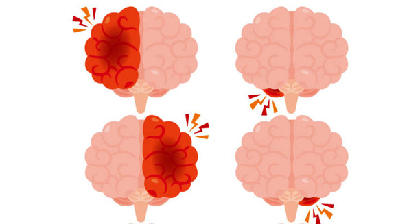
Illustration of four brains showing areas of inflammation, used to depict diagnosis of Japanese encephalitis

Diagnosis of Japanese Encephalitis

Japanese encephalitis is a challenging condition to diagnose, especially in its early stages when symptoms are vague and often resemble other common infections. As a viral disease that targets the brain, it needs quick and accurate diagnosis. This can help improve outcomes and reduce the risk of long-term brain damage. Doctors rely on clinical signs, a history of exposure, and lab tests to identify the virus. Diagnosis can be complex, especially in areas with limited resources. But early detection is vital for patient care and public health planning. There is no cure for Japanese encephalitis. So, the main goal of diagnosis is to confirm the illness, rule out similar diseases, and begin supportive treatment. It also helps track outbreaks and guide vaccine efforts in high-risk areas. Clinical Symptoms and Early Clues The first step in diagnosing Japanese encephalitis is a thorough clinical check. Doctors assess the patient’s symptoms, such as high fever, headache, confusion, seizures, and nerve problems. These signs often get worse over a few days. If coma or paralysis sets in, doctors suspect a serious brain infection. However, many other illnesses can look similar. Malaria, meningitis, and other viral infections can cause the same symptoms. So, Japanese encephalitis is usually a diagnosis of exclusion, especially in regions with many infectious diseases. A recent stay in rural or mosquito-prone areas can provide a strong clue. In children, sudden seizures and confusion without injury should raise concern. Role of Exposure and Travel History Doctors must review the patient’s exposure history to assess the risk of infection. These questions are key: These details help doctors decide whether testing is needed. In areas where the virus is common, all unexplained brain infections should be checked for Japanese encephalitis—especially in people who are unvaccinated. Testing for Japanese Encephalitis Lab tests are essential for diagnosis. The most used test is called MAC-ELISA, which looks for IgM antibodies in the blood or spinal fluid. These antibodies appear 4–8 days after symptoms begin and peak in the second week. MAC-ELISA is: But it also has limits: So, test results must be considered along with clinical signs and exposure history. Other Tests and Imaging PCR testing can detect the virus’s genetic material in blood or spinal fluid. It works best early in the illness, before symptoms of brain infection appear. PCR is accurate but costly and often only found in research labs or major cities. Doctors may also perform a lumbar puncture to check the spinal fluid. Common findings include: Though these results are not specific, they support the diagnosis if JEV antibodies or RNA are present. Brain scans can also help. MRI often shows damage in areas like the thalamus, brainstem, or basal ganglia. CT scans may show swelling or localized damage. While not diagnostic, imaging helps rule out other diseases and supports clinical suspicion. Excluding Other Illnesses Many diseases mimic Japanese encephalitis. Doctors must rule out: Comparing symptoms, lab data, and exposure helps doctors avoid misdiagnosis. Reporting and Public Health Impact Japanese encephalitis is often a notifiable disease. Confirmed cases must be reported to health authorities. This helps: Public health labs confirm diagnoses and gather data to prevent future infections. Conclusion The diagnosis of Japanese encephalitis depends on careful evaluation of symptoms, history, and lab tests. Because the early signs are vague, doctors must stay alert—especially in unvaccinated patients returning from high-risk areas. While tests like MAC-ELISA or PCR are key, the full picture—including brain scans and spinal fluid analysis—is essential. In areas with few resources, clinical signs and exposure history often guide the diagnosis. Finding the illness early helps not only the patient but the wider community. Accurate and fast diagnosis supports public health goals, slows outbreaks, and strengthens vaccine efforts against this severe yet preventable disease. [Next: Treatment of Japanese Encephalitis →]

Diagnosis of Japanese Encephalitis Read More »

Close-up of vaccine vials and syringe labeled Japanese Encephalitis for treatment and prevention

Treatment of Japanese Encephalitis

Japanese encephalitis is a potentially life-threatening condition caused by the Japanese encephalitis virus (JEV), which is transmitted by mosquitoes. While most cases are asymptomatic, a small number develop severe neurological disease that needs urgent medical attention. Since there is no specific antiviral drug for this illness, the treatment of Japanese encephalitis focuses on supportive care. The aim is to manage symptoms, avoid complications, and help the patient recover. Quick action increases the chances of survival and lowers the risk of long-term damage. In areas where the virus spreads often, treatment of Japanese encephalitis also helps reduce death rates and ease pressure on hospitals. Knowing what treatment options exist for both the early and recovery phases helps doctors, families, and decision-makers plan better. Hospital Support is Key to the Treatment of Japanese Encephalitis Because a virus causes Japanese encephalitis, antibiotics do not work. So far, no antiviral drug has shown consistent success in trials. This makes supportive hospital care the main method of treatment. Doctors monitor vital signs, manage each symptom, and work to stop new problems from appearing. a. First Steps to Stabilise the Patient Care begins with keeping the airway, breathing, and blood flow stable. Many patients need: b. Monitoring Brain Health The virus can cause swelling in the brain. So, doctors watch for: If brain pressure rises, staff may raise the bed head, use diuretics like mannitol, or start a breathing machine to ease the load on the brain. Seizure Control and Managing Unconsciousness Seizures often happen, especially in children. Treating them fast protects the brain. a. First-Line Seizure Treatment Doctors use diazepam or lorazepam right away. If that fails, they may give phenytoin, valproic acid, or phenobarbital. b. Long-Term Seizure Plans Some people develop long-term epilepsy after the illness. These patients may need daily seizure medicine. A neurologist usually guides this, using EEG scans as needed. For patients who fall into a coma, care gets more complex. They may need: Nurses also work to stop pressure sores, blood clots, and infections. Broader Recovery in the Treatment of Japanese Encephalitis Some people face movement issues like tremors or rigid muscles. Paralysis may also appear. Though no drug reverses these directly, some treatments help reduce symptoms: Recovery varies, but early rehab usually helps. Fever and vomiting often cause weight loss and low fluids. So, doctors focus on keeping energy and water levels stable: Rehabilitation and Future Care Even when patients recover physically, many still struggle with thinking or mood. Problems may include memory loss, speech trouble, or depression. So, a full rehab plan includes: The treatment of Japanese encephalitis also requires planning for life after hospital. Before sending patients home, doctors check their physical, mental, and emotional state. They build a custom care plan, which may include: New Drugs and Public Health Support Doctors continue to study medicines like ribavirin, interferon-alpha, minocycline, and IVIG. However, none are yet part of standard care. In poor regions, hospitals may not have machines or ICU beds. Here, basic care is vital. Community health workers help by: Public health teams also work after diagnosis to stop more infections. They may: These steps help reduce future outbreaks and save lives. Final Thoughts on the Treatment of Japanese Encephalitis The treatment of Japanese encephalitis focuses on support and symptom control. There is no cure yet, but good hospital care, seizure control, and rehab can greatly improve outcomes. Quick action and complete rehab help many regain function and live independently. At the same time, public health efforts are just as important. Fighting mosquitoes and increasing vaccination can reduce the spread of the virus, especially where hospitals lack resources. As travel grows and climates change, the treatment of Japanese encephalitis must remain a public health priority. [Next: Complications of Japanese Encephalitis →]

Treatment of Japanese Encephalitis Read More »

Illustration showing complications of Japanese Encephalitis including fever, vomiting, seizures, and neurological symptoms

Complications of Japanese Encephalitis

Japanese encephalitis is a serious neuroinvasive disease that can lead to a broad spectrum of complications, especially in individuals who develop severe encephalitic illness. Although most people infected with the virus do not show symptoms, those who do often face grave outcomes. About 20–30% of symptomatic cases result in death, while 30–50% of survivors live with long-term disabilities. These statistics show just how devastating the complications of Japanese encephalitis can be for patients and healthcare systems alike. The effects of this mosquito-borne illness do not end with the initial infection. Many survivors face ongoing issues that impact their brain, mood, and ability to function daily. In young children, these effects can slow growth, school progress, and overall well-being. Learning about these complications helps doctors, families, and health planners prepare better care and support. Neurological Effects in the Complications of Japanese Encephalitis The most common complications of Japanese encephalitis involve the nervous system. The virus inflames parts of the brain like the thalamus, brainstem, basal ganglia, and spinal cord. When these areas are damaged, patients can develop serious neurological problems. Motor Problems:Many survivors have trouble moving. They may experience: These issues can make walking or self-care difficult. Some people need physiotherapy for life or devices like crutches or wheelchairs. Seizures and Epilepsy:Seizures are another common issue. Patients may have: Treatment includes anti-seizure medication and regular check-ups with a neurologist. Cranial Nerve Issues:If the virus affects cranial nerves, it can lead to: These complications often require speech therapy or even surgery. Cognitive and Psychological Complications of Japanese Encephalitis Another group of complications of Japanese encephalitis involves mental functions. Damage to the brain can impair thinking and learning, especially in children. Cognitive Challenges:Survivors might face: In children, these issues often lead to poor school performance. Adults may find it hard to return to work, especially if their job involves planning or decision-making. Emotional and Behavioural Changes:The illness and its effects can also impact mood and behaviour. Survivors may develop: These symptoms often need therapy, medication, or both. Support for families is also important, as caregivers can struggle to manage behavioural changes at home. Senses, Speech, and Development in Japanese Encephalitis Survivors Speech and Swallowing Problems:Damage to the brain’s speech centres or throat muscles may cause: Speech therapy helps many children and adults improve these skills. Some may need communication devices. Hearing and Vision Loss:A few survivors lose hearing or vision. This might be due to nerve damage or brain processing issues. They may experience: Hearing aids or glasses often help. It’s vital to test these senses during recovery, especially in school-aged children. Fatigue and Sleep Problems:Even after recovery, many people feel tired or struggle to sleep. This may result from: Managing sleep and energy levels includes lifestyle changes and, in some cases, medication. Broader Effects and Prevention of Japanese Encephalitis Complications Delayed Development in Children:Young children who survive may experience delays in: They benefit from early screening and tailored care. A team of doctors, therapists, and educators can help improve outcomes. Impact on Families and Finances:Families often face high emotional and financial costs. Ongoing care for a disabled child or adult can lead to: Support like financial aid, caregiver training, and respite care can ease the burden. Risk of Death:About one in three severe cases results in death. Death often happens within the first week due to: In very severe cases, palliative care can help keep the patient comfortable. Families also need support through grief and loss. Prevention and Early Action:Many complications of Japanese encephalitis can be reduced by: Ongoing care includes regular check-ups, therapy, school or job support, and counselling. Outlook for Survivors of Japanese Encephalitis The complications of Japanese encephalitis are serious and long-lasting. From physical and mental issues to emotional and financial struggles, the effects can touch every part of life. But with early care and proper support, many survivors improve. Raising awareness, boosting access to care, and strengthening public health efforts—especially in high-risk areas—can help reduce the damage this illness causes. Vaccination, mosquito control, and health education remain the most powerful tools to prevent these life-changing complications. [End of Article – Back to Overview]

Complications of Japanese Encephalitis Read More »

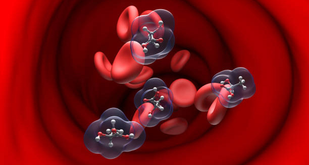
3D molecular representation of isovaleric acid molecules in the bloodstream, linked to Isovaleric Acidaemia

Isovaleric Acidaemia

Isovaleric acidaemia is a rare but serious inherited metabolic disorder that affects the body’s ability to process certain proteins, particularly the amino acid leucine. Isovaleric acidaemia belongs to a group of disorders called organic acidemias. These disorders cause toxic organic acids to build up in the blood. In isovaleric acidaemia, the body lacks an enzyme called isovaleryl-CoA dehydrogenase. This enzyme breaks down isovaleric acid, a byproduct of leucine metabolism. When the enzyme is missing or not working properly, isovaleric acid builds up to harmful levels. This buildup leads to many health problems, some of which can be life-threatening if left untreated. The condition follows an autosomal recessive inheritance pattern. This means both parents must carry a faulty copy of the IVD gene for their child to be affected. Although it is very rare, with about 1 in 250,000 live births worldwide, isovaleric acidaemia occurs in many ethnic groups. It is more common in some regions due to genetic founder effects. Thanks to newborn screening programs in many countries, early diagnosis and treatment are now possible. This has helped improve long-term outcomes. Clinical Forms of Isovaleric Acidaemia Isovaleric acidaemia shows up mainly in two forms: acute neonatal onset and chronic intermittent. The acute neonatal form appears within the first few days of life. It can quickly cause a severe metabolic crisis. Symptoms include vomiting, lethargy, seizures, and sometimes coma. Without prompt treatment, this form can be fatal. The chronic intermittent form usually starts later in infancy or early childhood. It often occurs during times of metabolic stress like infections or fasting. People with this form may seem healthy between episodes but can suddenly develop severe symptoms needing emergency care. One unique feature of isovaleric acidaemia is a “sweaty feet” smell. This odor comes from the buildup of isovaleric acid in body fluids. Although this smell can help with diagnosis, it does not always appear. Therefore, doctors should not rely on it alone. More important are biochemical tests like blood ammonia levels, urine organic acids, and acylcarnitine profiles. These tests confirm the diagnosis and guide life-saving treatment. Managing Isovaleric Acidaemia Treatment focuses on lowering the production and buildup of toxic substances like isovaleric acid. This involves a diet low in leucine, special amino acid-free formulas, and supplements such as carnitine and glycine. These supplements help the body remove harmful compounds. During metabolic crises, patients often need hospital care. This care includes intravenous fluids, sugar (dextrose), and other emergency treatments to stabilise the patient. Long-term care requires regular diet monitoring and metabolic check-ups. Usually, a metabolic specialist and dietitian oversee this care. Like many rare diseases, awareness, early diagnosis, and proactive care are key to avoiding problems and improving life quality. Advances in genetic testing and metabolic medicine help diagnose the disorder earlier, even in newborns without symptoms. However, challenges remain, especially in places where newborn screening is not widely available or where special metabolic care is limited. Overview of This Article This article will explore all aspects of isovaleric acidaemia in detail. The next sections will cover: Although isovaleric acidaemia is rare and not widely known, this article aims to inform families, caregivers, doctors, and the public with clear, honest, and caring information. Raising awareness helps reduce delays in diagnosis, encourages early treatment, and improves the lives of those living with this complex metabolic disorder. [Next: Causes of Isovaleric Acidaemia →]

Isovaleric Acidaemia Read More »

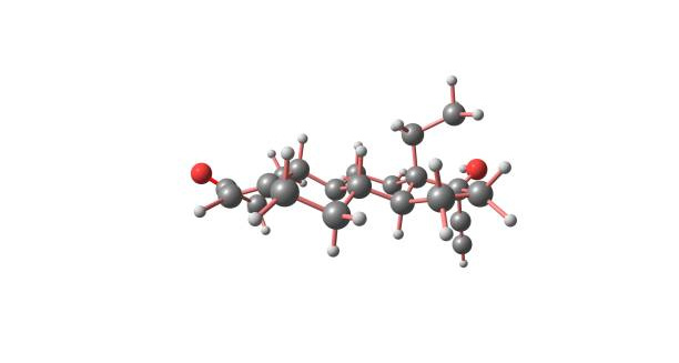
Molecular structure associated with leucine metabolism linked to causes of Isovaleric Acidaemia

Causes of Isovaleric Acidaemia

Isovaleric acidaemia is caused by a genetic mutation that disrupts how the body breaks down leucine, an essential amino acid found in protein-rich foods. This inherited metabolic disorder results from a deficiency in an enzyme needed to safely process leucine. Specifically, mutations in the IVD gene affect the enzyme isovaleryl-CoA dehydrogenase, causing toxic substances like isovaleric acid to build up. These toxins lead to the symptoms and complications of the disease. This section explains the genetic basis, inheritance pattern, biochemical effects, and environmental triggers behind isovaleric acidaemia. 1. Genetic Mutation in the IVD Gene The root cause lies in mutations of the IVD gene on chromosome 15. This gene provides instructions to make isovaleryl-CoA dehydrogenase, a mitochondrial enzyme crucial for breaking down isovaleryl-CoA, a leucine metabolite. Mutations may cause: Over 60 mutations have been found, including missense, nonsense, and frameshift types. The mutation type often influences whether symptoms appear early and severe or later and milder. Even siblings with the same mutation can show different symptom severity, indicating other genetic or environmental influences. 2. Autosomal Recessive Inheritance Isovaleric acidaemia follows an autosomal recessive pattern. A child must inherit two faulty copies of the IVD gene—one from each parent—to develop the disease. Parents who carry one defective gene usually show no symptoms. For two carrier parents: This inheritance explains why the disorder can appear unexpectedly. Carrier testing and genetic counselling help families understand their risks. 3. Disruption of Leucine Metabolism Leucine is a branched-chain amino acid in many protein foods like meat, dairy, and nuts. The body breaks it down in steps, one involving isovaleryl-CoA dehydrogenase. When this enzyme is deficient: 4. Triggers and Environmental Stressors Though genetic, isovaleric acidaemia symptoms often flare during external stress: These triggers pose higher risk for infants and young children, requiring careful management. 5. Genotype–Phenotype Correlation Research shows some link between mutation type and disease severity: Still, symptom severity can vary even with the same mutation, influenced by other genes, metabolism, and environment. This variability highlights the need for personalized care. 6. Carrier Frequency and Ethnic Variability Isovaleric acidaemia is rare but carrier rates differ by population. Some communities show higher carrier frequencies due to founder effects, such as: Knowing these patterns helps tailor screening and education efforts. 7. Prenatal and Preimplantation Genetic Diagnosis Couples with known mutations can use genetic counselling for reproductive planning. Options include: These methods enable early detection and informed decision-making. Conclusion Isovaleric acidaemia results from mutations in the IVD gene causing enzyme deficiency and toxic buildup during leucine breakdown. It follows an autosomal recessive pattern, with symptoms often triggered by metabolic stress, especially in infants. Though rare, the condition can be managed well through early diagnosis, genetic counselling, dietary control, and medical care. Understanding its genetic and biochemical roots paves the way for better outcomes and support for affected families. [Next: Symptoms of Isovaleric Acidaemia →]

Causes of Isovaleric Acidaemia Read More »

Crying infant showing symptoms of Isovaleric Acidaemia in early infancy

Symptoms of Isovaleric Acidaemia

The symptoms of isovaleric acidaemia vary widely depending on the form and severity. Some people show life-threatening signs within days of birth, while others stay symptom-free for months or years. Stress, illness, or diet may trigger a metabolic crisis. Early recognition of symptoms is crucial to avoid serious brain damage, coma, or death. This section covers the full range of symptoms—from subtle signs to severe acute episodes. Isovaleric acidaemia presents mainly in two forms: the acute neonatal type and the chronic intermittent type. Each form has distinct symptoms, but some signs overlap. 1. Symptoms in the Acute Neonatal Form The acute neonatal form usually appears between 3 and 14 days after birth. Babies often seem healthy at first but worsen quickly when they start feeding and processing protein. Without fast treatment, they risk coma and death. Common signs include: If untreated, this form can be fatal. Survivors may face intellectual or developmental delays depending on how long and severe the crisis was. 2. Symptoms in the Chronic Intermittent Form This form appears later, often in infancy or childhood. Individuals may seem healthy between episodes but fall ill when stressed by infection, fasting, or high protein intake. Common symptoms include: This form is often mistaken for gastrointestinal illness or behavioural problems until proper testing is done. 3. Subtle and Non-Specific Symptoms Milder cases, especially those diagnosed early through screening and on treatment, may have subtle symptoms: These signs may not cause immediate alarm but show underlying metabolic imbalance. Regular follow-up and blood tests remain important. 4. Symptoms During Metabolic Crises A metabolic crisis is life-threatening and requires urgent hospital care. Symptoms develop quickly and include: Emergency treatment with glucose, fluids, acidosis correction, and metabolite reduction is critical. Families learn to spot early warning signs and seek immediate care. 5. Differences in Presentation Between Age Groups Newborns face the highest risk due to immature metabolism and immunity. Older children usually have episodic symptoms, which can be confused with common illnesses. Adults with late diagnosis may experience chronic fatigue, brain fog, or protein intolerance. Conclusion Symptoms of isovaleric acidaemia range from severe neonatal crises to mild, chronic issues in older individuals. Early signs like vomiting, lethargy, seizures, and poor feeding need urgent attention in newborns. In children, failure to thrive and behavioural changes may signal chronic intermittent disease. Awareness of these symptoms helps ensure prompt diagnosis and treatment. Newborn screening and clinical vigilance save lives and improve long-term outcomes. [Next: Diagnosis of Isovaleric Acidaemia →]

Symptoms of Isovaleric Acidaemia Read More »

Medical scientists performing laboratory tests for Isovaleric Acidaemia diagnosis

Diagnosis of Isovaleric Acidaemia

Diagnosing isovaleric acidaemia can be challenging, particularly due to its rarity and the non-specific nature of many early symptoms. The condition often looks like common illnesses such as sepsis, stomach infections, or brain disorders in newborns and young children. However, as awareness grows and newborn screening expands, detecting isovaleric acidaemia early has become easier. This section explains how doctors diagnose this condition through clinical signs, lab tests, genetic checks, and newborn screening. Early diagnosis helps prevent metabolic crises and allows timely treatment, which improves long-term health. Clinical Suspicion and History Taking Doctors first suspect isovaleric acidaemia when infants show symptoms like poor feeding, tiredness, vomiting, seizures, or unexplained acid buildup in the blood. A “sweaty feet” smell is also a key clue. Family history matters too. If siblings have the condition or parents are related, the chance of inherited metabolic disease rises. Symptoms that start after eating protein may also hint at this disorder. Because these signs overlap with infections or birth injuries, doctors may not think of isovaleric acidaemia at first. But when blood tests show metabolic acidosis with a high anion gap, ketosis, and low blood sugar, metabolic disorders must be considered. Biochemical Testing After suspicion, lab tests confirm the diagnosis by measuring toxic substances from broken-down leucine. a. Plasma Acylcarnitine ProfileThis blood test checks for high levels of isovalerylcarnitine (C5), which is a clear sign of isovaleric acidaemia. It is often done on dried blood spots during newborn screening. b. Urine Organic Acid AnalysisTesting urine shows raised amounts of isovalerylglycine, isovaleric acid, and related compounds, which strongly indicate the condition. c. Blood Ammonia and LactateHigh ammonia and lactate levels may appear during crises. These markers help doctors understand the crisis severity and plan treatment. d. Arterial Blood Gases and Anion GapMetabolic acidosis with a raised anion gap means organic acids build up in blood, prompting more metabolic tests. e. Serum Glucose and KetonesLow blood sugar and ketones may also appear during stress, supporting the diagnosis. Genetic Testing Definitive diagnosis comes from finding mutations in the IVD gene on chromosome 15. This gene controls the enzyme that breaks down isovaleryl-CoA. Genetic tests confirm the diagnosis and allow screening of family members and prenatal testing. Some populations have common mutations, which makes testing easier. Newborn Screening Many countries test newborns for isovaleric acidaemia using tandem mass spectrometry on dried blood spots. This test detects high C5 acylcarnitines within days after birth. Early screening helps by: However, screening can give false positives or negatives, especially in mild cases or premature babies. Abnormal results need further biochemical and genetic checks. Differential Diagnosis Symptoms and lab results in isovaleric acidaemia can resemble other metabolic diseases like: Doctors use biochemical markers and genetic tests to tell these apart. For example, isovalerylglycine in urine points to isovaleric acidaemia, while other acids suggest different disorders. Prenatal Diagnosis Families with a history of isovaleric acidaemia may choose prenatal testing by chorionic villus sampling or amniocentesis. These tests check the fetus for defective IVD genes using: These help families prepare for medical care and early treatment after birth. Enzyme Assays Although rare now, doctors can measure isovaleryl-CoA dehydrogenase activity in cells from skin or blood. Low or absent activity confirms the diagnosis. This method is mostly used in research or where genetic testing is unavailable. Family Testing and Cascade Screening After diagnosis, doctors recommend testing siblings and relatives. Finding carriers or affected people early helps start care and lower risks. Sometimes siblings have mild or no symptoms but still carry the condition. Conclusion Diagnosing isovaleric acidaemia takes clinical awareness, lab tests, genetic analysis, and newborn screening. Though early symptoms may mimic common newborn illnesses, signs like the sweaty feet smell, metabolic acidosis, and high C5 acylcarnitines guide doctors to the right diagnosis. Timely diagnosis saves lives by enabling quick treatment and preventing brain damage. As newborn screening spreads, early detection and care improve, giving hope to families facing this rare but serious disorder. [Next: Treatment of Isovaleric Acidaemia →]

Diagnosis of Isovaleric Acidaemia Read More »

Nurse administering IV treatment to patient with Isovaleric Acidaemia

Treatment of Isovaleric Acidaemia

Isovaleric acidaemia is a lifelong inherited disorder that needs ongoing care to stop toxic substances from building up in the body. Treatment varies for each person. Doctors consider how severe the condition is, the patient’s age, medical history, and whether they were diagnosed early through newborn screening or after symptoms started. The main goals of treatment are to lower isovaleric acid levels, support the body’s metabolism, and prevent metabolic crises. With the right care, many people with this condition can live fairly normal lives, especially if diagnosed early and helped by a team of specialists. This section covers emergency care during metabolic crises and long-term steps to keep metabolism stable and avoid problems. Emergency Care During a Metabolic Crisis When someone with isovaleric acidaemia has a crisis—often caused by infection, stress, fasting, or eating too much protein—they need urgent medical help. The treatment aims to: a. Intravenous Glucose and FluidsHigh-dose sugar solutions are given through a vein to stop the body from breaking down proteins and fats for energy. This stops harmful substances from forming. Doctors also fix dehydration, acid buildup, and imbalances in salts and minerals. b. Protein RestrictionDuring the crisis, protein is stopped temporarily to prevent more toxin build-up. After the patient stabilises, doctors carefully reintroduce protein under close supervision. c. Ammonia ScavengersIf blood ammonia is high, medicines like sodium benzoate help remove nitrogen and protect the brain. d. Carnitine and Glycine Supplements These supplements reduce toxins and help balance metabolism. They can be given by mouth or through a vein depending on how severe the crisis is. Long-Term Diet Management A key part of managing isovaleric acidaemia is a low-protein diet, especially low in leucine. Leucine is the amino acid that causes toxic buildup. a. Leucine-Restricted DietLeucine is in many protein-rich foods like meat, dairy, eggs, beans, nuts, and some grains. A dietitian with metabolic expertise creates a diet that provides enough nutrition while limiting leucine. This includes: Monitoring growth and nutrition is important, especially for children. b. Medical Foods and SupplementsSpecial formulas help people get nutrition without leucine. These may include amino acid blends without leucine, extra calories from sugars, and vitamins and minerals. Long-Term Use of Carnitine and Glycine L-carnitine and glycine are usually taken daily for life. They help clear toxins and support energy production in cells. Doctors adjust doses based on weight, age, how often crises occur, and lab tests like blood carnitine levels. Regular tests ensure the treatment stays safe and works well. Monitoring and Follow-Up People with IVA need lifelong check-ups, including: Regular visits help catch health changes before crises happen. Illness and Infection Plans Illnesses often trigger crises in IVA. To prevent this, many patients follow a “sick day” plan that includes: Caregivers learn to watch for signs like loss of appetite, vomiting, or strange behaviour, and act fast to avoid hospital stays. Emergency Preparedness Because crises can be life-threatening, patients carry an emergency letter that explains their condition and needed treatments. Wearing a medical alert bracelet is also recommended. This helps any doctor give the right care, even if they don’t know the patient. Liver Transplant (Rare Cases) In very rare cases, when crises keep happening despite good care, a liver transplant might be an option. The new liver can provide the missing enzyme and improve health. But this is a last resort because transplants have risks. Psychological and Educational Support Living with a rare metabolic disease is tough emotionally and socially. Children may face: Psychological help, school counselling, and support groups improve quality of life. Teaching classmates and teachers about IVA helps reduce stigma. Conclusion Treating isovaleric acidaemia is complex but effective. Early diagnosis, personalised diet plans, supplements, and emergency care help patients live well. The key is close monitoring, family education, and quick action at signs of illness. New treatments, like gene therapy, hold promise for the future. For now, careful diet control and medical support remain the best ways to manage this rare but treatable disorder. [Next: Complications of Isovaleric Acidaemia →]

Treatment of Isovaleric Acidaemia Read More »

Woman sitting tired in a chair possibly depicting complications of isovaleric acidaemia

Complications of Isovaleric Acidaemia

Isovaleric acidaemia (IVA) is a serious inherited metabolic disorder that, if left unmanaged or poorly treated, can lead to a range of short-term and long-term complications. This condition affects how the body breaks down leucine. When that process fails, toxic substances like isovaleric acid build up. These toxins can harm many parts of the body. Early diagnosis and treatment reduce the risks, but people with IVA still face several health problems throughout their lives. This section explains the most common, severe, and often hidden complications of isovaleric acidaemia. These include metabolic, neurological, physical, emotional, and social effects. Knowing about these risks helps doctors, caregivers, and patients take early action, leading to better health outcomes. Acute Metabolic Crises The most urgent danger in isovaleric acidaemia is a metabolic crisis. These happen when stress, infections, fasting, or too much protein causes toxin levels to rise quickly. Common warning signs include: These episodes are medical emergencies. Without fast treatment, they can lead to organ failure, brain swelling, or death. Babies and young children face the highest risk, but crises can occur at any age during times of stress. Prevention is key. A strict diet, early illness care, and regular check-ups can help stop these crises. Neurological Effects of Isovaleric Acidaemia IVA can also affect the brain, especially during metabolic crises. High levels of ammonia and toxic acids can damage brain cells. Some possible effects include: The more crises a person has, and the later the diagnosis, the higher the risk of brain damage. Early testing in newborns and quick treatment can lower the risk of mental or developmental problems. Growth and Nutrition Problems People with isovaleric acidaemia often follow a low-protein diet. If not planned well, this can lead to poor growth and nutrition. Issues may include: Too little protein or food can also trigger the body to break down its own muscle. This releases even more toxins. Diet plans should be adjusted often by a specialist to keep growth on track and avoid malnutrition in Irritable Bowel Syndrome patients. Heart-Related Risks Although rare, some people with IVA develop heart problems. This can happen after long exposure to toxins that affect how the heart gets energy. Possible heart issues include: For people with moderate to severe IVA, regular heart tests like ECGs and ultrasounds can help catch problems early. If needed, doctors can start treatment right away. Liver and Kidney Function The liver and kidneys work hard to remove harmful substances from the body. In people with IVA, these organs may become stressed, especially after repeated crises. Possible problems: Most people do not reach full liver or kidney failure. But it is important to keep checking how these organs work, especially in adults who’ve had the condition for many years. Blood and Carnitine Levels Carnitine helps the body get rid of toxins. People with IVA often have low carnitine levels, which can lead to anaemia or tiredness. Signs and risks: L-carnitine supplements are often part of treatment. Regular blood tests help manage these risks and keep energy levels steady. Mental and Emotional Strain Living with isovaleric acidaemia can be mentally and emotionally hard. People often feel burdened by strict diets and the constant risk of emergency. Common struggles include: Some children may also show behavioural problems. These can come from brain effects or frustration about food limits. Counselling, support groups, and talking with others can help people cope and feel more supported. Learning and School Difficulties IVA can affect how well children learn and get along at school, especially if their brain was affected early in life. Possible school issues: Schools can help by creating learning plans just for that child. Teachers, psychologists, and special education teams should all work together to support the student. Isovaleric Acidaemia and Pregnancy As people with IVA grow older, pregnancy becomes a concern. Many women with IVA can become pregnant, but pregnancy puts extra stress on the body. Some pregnancy risks include: Before pregnancy, women should get genetic counselling. A high-risk obstetrician and metabolic doctor should monitor closely. With careful planning, pregnancy can go well for both the mother and the baby. Missed or Late Diagnosis One of the most dangerous complications of isovaleric acidaemia is not being diagnosed in time. This happens more often in places without newborn screening. IVA can look like other conditions, such as: Doctors, especially in newborn intensive care units, need to be aware of IVA. Early testing and fast action can save lives and prevent long-term damage. Conclusion The complications of isovaleric acidaemia can be serious. They range from sudden health emergencies to lifelong physical, mental, and social challenges. But many of these risks can be managed or even avoided. The best approach includes early testing, careful diet planning, emotional support, and regular check-ups. With a full care team in place, people with IVA can live healthy, active lives. New advances in screening and treatment give hope for an even better future. [Next: Back to Overview →]

Complications of Isovaleric Acidaemia Read More »

Scroll to Top